


方案详情
文
The molecular origins of second-order nonlinear effects in type I collagen fibrils have been identified with sumfrequency
generation vibrational spectroscopy. The dominant contributing molecular groups are: 1), the methylene groups associated
with a Fermi resonance between the fundamental symmetric stretch and the bending overtone of methylene; and 2), the
carbonyl and peptide groups associated with the amide I band. The noncentrosymmetrically aligned methylene groups are
characterized by a distinctive tilt relative to the axis perpendicular to the main axis of the collagen fiber, a conformation producing
a strong achiral contribution to the second-order nonlinear effect. In contrast, the stretching vibration of the carbonyl
groups associated with the amide I band results in a strong chiral contribution to the optical second-order nonlinear effect. The
length scale of these chiral effects ranges from the molecular to the supramolecular.
方案详情

Biophysical JournalVolume 93 December 2007 4433-44444433 Rocha-Mendoza et al.4434 Sum Frequency Vibrational Spectroscopy: The Molecular Origins of theOptical Second-Order Nonlinearity of Collagen Israel Rocha-Mendoza,* Diego R. Yankelevich,*Mingshi Wang,* Karen M. Reiser, Curt W. Frank,* and Andre Knoesen* *Department of Electrical and Computer Engineering, tDepartment of Neurological Surgery, University of California, Davis, California;and *Department of Chemical Engineering, Stanford University, Palo Alto, California ABSTRACT The molecular origins of second-order nonlinear effects in type l collagen fibrils have been identified with sum-frequency generation vibrational spectroscopy. The dominant contributing molecular groups are: 1), the methylene groups asso-ciated with a Fermi resonance between the fundamental symmetric stretch and the bending overtone of methylene; and 2), thecarbonyl and peptide groups associated with the amide I band. The noncentrosymmetrically aligned methylene groups arecharacterized by a distinctive tilt relative to the axis perpendicular to the main axis of the collagen fiber, a conformation pro-ducing a strong achiral contribution to the second-order nonlinear effect. In contrast, the stretching vibration of the carbonylgroups associated with the amideI band results in a strong chiral contribution to the optical second-order nonlinear effect. Thelength scale of these chiral effects ranges from the molecular to the supramolecular. The collagens are a superfamily of extracellular matrix pro-teins that play a crucial role throughout the lifespan of allmetazoan lifeforms, from early growth and development throughmaintenance of homeostasis. All collagens are composed ofthree polypeptide chains, called a-chains, that fold togetherto form the characteristic triple helix. Each a-chain is coiledinto a left-handed helix that winds around a common axis toform a right-handed triple helix (Fig. 1). The collagen mole-cule may be either a homotrimer (three identical a-chains) ora heterotrimer (a mixture of two or three genetically distinctchains). More than 27 collagen types and 42 distinct a-chainshave been identified in vertebrates, as well as 15 additionalproteins with collagenlike domains (1). All members of thecollagen superfamily share certain characteristics, necessi-tated by the coiled-coil structure of the triple helix. Glycine,the smallest amino acid, must occupy every third position toallow packing of the coiled-coil structure. Hydroxyprolinefrequently occupies the third position in the Gly-X-Y triplet,providing stability to the helix, while proline is often in theX position (2-6). Each twist is approximately three-amino-acids in length. These tight twists do not result in sterichindrance due to the location of glycine, the smallest aminoacid, at the center of the superhelix, a conformation thatallows the bulky side chains of amino acids at the X and Ypositions to point outward. The three a-chains are connectedby hydrogen bonds between the NH of the glycine and theCO of the proline in the X position of a neighboring a-chain.The pyrrolidine rings of proline and hydroxyproline impose ( Submitted April 24, 2007, and accepted for publication August 14,2007. ) ( Address reprint r equests t o A ndre K n oesen, Tel.: 53 0 -758-9165; E-mail: knoesen@ece.ucdavis.edu. Israel Rocha-Mendoza’s present a d dress is Cardiff U n iversity, School o f Biosciences, Cardiff, Wales, UK.Editor: Enrico Gratton. ) additional steric restrictions, causing each a-chain to foldinto a left-handed helix while simultaneously twisting togetheralong a common axis to form a right-handed superhelix. Atthe supramolecular level, collagen molecules are organizedinto complex assemblies of fibrils or networks related to theirtissue functions; structural features of these assemblies, inconjunction with other posttranslational modifications, serveas the basis for grouping the different collagen types into nine“families”(1). The fibril-forming collagens represent themost abundant group, consisting of collagen types I, II, ⅢII, V,XI, XXIV, and XXVII. In this article, we focus on type Icollagen, the predominant member of this group. Type I collagen is characterized by a complex hierarchicalorganization, in which discrete structural features are dis-cernible at scales of organization extending over many ordersof magnitude (7). The process of fibril formation begins assoon as procollagen, the precursor of collagen, undergoesenzymatic cleavage of its nonhelical propeptide extensions atits C-and N-termini, shortly after it is secreted by the cell. It isenergetically favorable for the collagen molecules to line upnext to each other, with each molecule offset by approxi-mately one-quarter its length from neighboring molecules.This offset of 67 nm is known as the D-period (8). Micro-fibrils, consisting of pentameric assemblies of aligned colla-gen molecules,represent the first supramolecular structures.The fibers have a mean diameter between 0.5 and 3 microm-eters and can be up to several hundred micrometers long.These structures are stabilized over time by enzymaticallymediated intermolecular covalent crosslinks. In many tissues,microfibrils interact to form architecturally complex struc-tures. Although fibrillar collagen has been extensively studiedfor decades, the molecular mechanisms underlying manystructural and functional properties have been remarkablydifficult to elucidate, perhaps due in part to the complexity ofits organization. The electro-optical properties of collagen, FIGURE 1The triple helical structure for a collagen molecule. The amino-acid sequence of a single a-chain is Gly-X-Y, where the smallest amino-acidglycine (Gly) and X and Y are frequently proline (Pro) and hydroxyproline(Hyp), respectively. The sequence shown is (Gly-Pro-Hyp)3. The small Glyamino acid is always in the center of the superhelix and the bulky pyrrolidinerings of Pro and Hyp point outward. The noncentrosymmetric orientation ofthe methylene groups in the rings produces a achiral nonlinear effect. Thethree a-chains (denoted with blue, green, and magenta) are connected byhydrogen bonds between the NH of the glycine and the CO of the proline inthe X position of a neighboringa-chain, indicated by dashed lines in thefigure,and create a helical ladder in the middle of the triple helix that gives riseto a supramolecular nonlinear chiral effect. Figure reproduced by permissionof the publisher (Taylor & Francis Ltd., http://www.informaworld.com) ofRamachandran et al. (3). such as piezoelectricity and second harmonic generation, haveproven to be particularly elusive with respect to identifyingproximal molecular mechanisms. Piezo-electricity and second harmonic generation arepresent only in materials characterized by the absence of acenter of symmetry on a microscopic scale that extends overmacroscopic distances. Noncentrosymmetry in nonlinearoptical organic media can be satisfied in at least three waysFirst, noncentrosymmetric alignment of dipoles produces alarge second-order hyperpolarizability, an effect we refer toas the “achiral”contribution. Second, the chirality of helicalmacromolecules, which are by definition noncentrosymmetric,can produce a significant nonlinear chiral contribution (9). Acharacteristic of the chiral contribution is that a fundamentalinput beam polarized parallel to the plane of incidence,produces a second harmonic radiation polarized orthogonal tothe input polarization. Third, the intrinsic asymmetry present atany interface can produce surface effects; however, this effect istypically negligible in macromolecular organic structures. Themolecular origins of chirality in second-order nonlinear effectshave been recently reviewed (10). Freund and co-workers have performed an extensive in-vestigation of the polarity of collagen with SHG (11-15).The cylindrical symmetrical arrangement of collagen mol-ecules about the long axis present in collagen fibril impliesan effective C.. symmetry when an average is taken over thedimensions of the wavelength of light. Freund confirmed thatthe C.. symmetry produces an achiral effect that generatesthe largest second harmonic radiation polarized along thesymmetry axis for a fundamental beam polarized parallel tothe same axis. They concluded that the polarity in type Icollagen is associated with two structures: a network of veryfine polar filaments permeating the tendon and a small numberof polar surface regions that may represent dense accumu-lations of the filaments. All of the filamentous structureshave the same directionality. While they could not determinethe molecular origins of the polarity, they argued that struc-tural considerations suggested that it was unlikely to arisefrom the collagen triple helix itself. First, covalent bonds thatcould potentially contribute to polarity, such as C=O, N-H,and C-H bonds are almost all perpendicular to the main axis.Second, the dipolar effects produced by C-N bonds wouldcancel each other out. The contribution of bound water wasexcluded because SHG produced in wet and dry samples didnot differ remarkably. They concluded that the “polar struc-tures ofconnective tissue are likely to be composed of othersubstances. .The contribution of supramolecular helicity to nonlinearchiral effects has been an area of recent interest, followingthe report that the helical arrangement in helicene is thesource of a supramolecular nonlinear chirality (9). Campag-nola and co-workers speculated that the SHG in collagencould be enhanced by chiral contributions (16). While eacha-chain is expected to produce a chiral second-harmonic con-tribution, the cooperative effect of the triple-helical supra-molecular structure of collagen should produce an even largerchiral nonlinear effect. No conclusive experimental evidencewas presented in support of this claim at that time. Recently,Plotnikiv et al. (17) supported this claim by concluding thatSHG signal arises from the pitch angle of a single collagenglycine-proline helix. While this conclusion was justified bytheoretical fittings of SHG polarization anisotropy signal offibrillar collagen, there is no identification of the moleculargroups contributing to the chiral and achiral second-orderoptical nonlinearity. Pena and co-workers showed that thechiral enhancement in the optical nonlinearity of collagen issmall (less than a factor of 2) by performing SHG experimentson thin layers of collagen molecules isotropically oriented ona substrate but were not organized into fibrils (18). It wasconcluded that the large SHG signals observed in connectivetissue is due to the high density and microscopic crystallineorder in collagen fibrils. Williams and co-workers concludedthat the SHG is produced by the outer shell of a collagen fibriland within a <50 nm region, instead of from the bulk of thefibril by measuring ratios of forward and backward scatteringof SHG from fibrils (19). Sum-frequency generation vibrational (SFG-V) spectros-copy represents an attractive approach for identifying themolecular groups responsible for second-order nonlinear op-tical effects (20-22). Initially, applications of SFG-V focusedon relatively small molecules oriented at interfaces; themethod is increasingly being applied to more complicatedmacromolecules such as proteins (23-27). Helical proteins areof particular interest, since they are inherently noncentrosym-metric and possess electric-dipole-allowed second-order non-linearity (28,29). For example, our group has identified theachiral molecular origins of second-order nonlinear effects inpoly-a-benzyl-L-glutamate (PBLG), a synthetic a-helicalprotein, self-assembled film (25). In this article, we use sum-frequency generation vibrational(SFG-V) spectroscopy to identify the molecular structuresthat give rise to both the chiral and achiral optical second-order nonlinear effects in native Type I collagen fibrils. Insum-frequency generation a sample is irradiated with twobeams of coherent light at different angles of incidence, one ata frequency in the visible range, one at a frequency in theinfrared range. Within the sample two photons (one from eachbeam) combine to form a single photon at a third frequency,which is dependent on the second-order optical nonlinearitytensor elements (@sF =Wvis +wrR). In SFG-V spectroscopy,wr is tuned while keeping ωvis fixed, and near some mo-lecular vibration resonances the output signal is increasedthrough resonant enhancement, producing the SFG-V spec-tra. A vibrational transition will only be observable in a SFspectrum if it is both Raman- and IR-active, which alsoimplies that centrosymmetric molecular transitions will not bedetected (20). Near a molecular vibration transition (ω,),provided the input frequencies are not close to an electronicresonance of the medium, each molecular hyperpolarizabilitytensor element pii'k’, expressed in the molecule-fixedcoordinate system (i',j',k’ =a, b,c), is proportional to afactor that depends on the Raman transition dipole momentand the IR transition dipole moment associated with thatvibration transition (21). In the limit of weak coupling be-tween the microscopic nonlinear molecular units, the macro-scopic second-order susceptibil(2)ity tensor elements are XikNZinkRjk,ij’k Bi in the laboratory coordinate system (i,j,k=x,y,z), where N is the number density of molecules whichcontribute to the SFG signal, Rikiik are elements of the Eulerrotation matrix, Bij'k is the hyperpolarizability tensor in themolecular coordinates, and the subscripts ijk are associatedwith the sum-frequency,visible, and IR electric fields, respec-tively. The output intensity is Isr oXeff Ivis’ I and Xeff =e(@sf)·L(@sf2:e(Wvis)·L(@vis),e@R)·L(@R】,where e(w) is the unit polarization vector and L(w) theFresnel factor atω (30). By controlling the input polarizationsand detecting a specific output polarization, specific tensorelements can be accessed, from which the orientation ofmolecular groups can be deduced. The coordinate system shown in Fig.2 is used for ourSFG-V transmission measurements. The main axis of the FIGURE 2 SSFG in the transmission mode. In the figure, ω1,2, and ωsFare the visible, infrared and sum-frequency beams, respectively. Thecollagen fibrils are oriented along the x axis. collagen fiber is oriented along the x axis and the z axis. Thez axis is perpendicular to the sample in the plane of inci-dence. A uniform orientation of the collagen fiber parallel tothe x axis is important to avoid misinterpreting any aniso-tropic effects as chiral contributions (31). The notations Pand S refer to beam polarizations parallel and perpendicularto the plane of incidence,respectively. With the main axis ofthe collagen molecules along the x axis, the 27 Xijk tensor ele-ments can be divided into elements that contribute to eitherachiral or chiral effects. The susceptibility tensor elementsthat contribute to chiral effects are those pertaining to or-thogonal components,e.g.,Xijk (i(jk)(10,18,22,28,32).The remaining elements contribute to the more commonsecond-order nonlinear effects, the achiral effects. Experi-mentally one can access the chiral and/or achiral tensor ele-ments by setting different input (visible and IR) beampolarizations and analyzing the SFG intensity. Table 1 showsthe tensor elements accessed by the different input/outputpolarization combinations according to the coordinate system TABLE 1 Macroscopic second-order susceptibility tensorelements of collagen for the different input/output polarizationcombinations; the main axis of the collagen fiber is orientedalong the x axis I/O polarization configurations Chiral (SF, VIS, IR) Achiral elements elements PPP Xxxx, Xxxz, Xxzx, Xxzz Xzxx, Xzxz: Xzzx, Xzzz SSP Xyyx, Xyyz SPS Xyxy, Xyzy SSS Xyyy PSS Xxyy, Xzyy SPP (chiral) Xyxx, Xyzz Xyzx,Xyxz PSP (chiral) Xxyx, Xzyz Xxyz乙 Xzyx PPS (chiral) Xxxy, Xzzy Xxzy, Xzxy shown in Fig. 2. The PPP (with e(wsf), evis, and en allP-polarized), SSP, PSP, and SPP configurations couple toIR-vibrations molecules within the plane of incidence thathave components normal and/or parallel to the collagen fibrilaxes. The SSS,PPS, SPS, and PSS configurations couple tothe IR-vibrations of molecules perpendicular to the plane ofincidence. Only achiral elements are accessed by the input/output beam polarization combinations configurations PPP,SSP, SPS, SSS, and PSS. Both chiral and achiral elementsare accessed by the SPP, PSP, and PPS configurations. Inthese configurations the achiral contributions are zero undercertain conditions. One example is the isotropic orientationof chiral molecules in a liquid (32). Another example occursin azimuthally isotropic media with the symmetry axis alongeither the x or z axis, in which case Xyxx = Xyzz = Xxyx=Xxxy=Xzzy=0 (28,33). To avoid that anisotropic effects aremisinterpreted as chiral contributions, we will use secondharmonic imaging to ensure that the sample has a uniformdistribution of fibers along a preferential axis, and will alignthis axis to the x axis in our SFG-V measurements. With thisuniformity and orientation established in our experimentalconfiguration, it can be assumed that only chiral tensor ele-ments contribute sum-frequency generated by the SPP, PSP,and PPS configurations. The polarization properties of SHG intensity from fibrillarand filament protein structures is well modeled using a C.cylindrical rod symmetry (15,17,34). This symmetry hasfour nonzero elements Xxxx, Xxyy =Xxzz, Xyxy = Xyyx =Xzxz=Xzzx, and Xyzx=-Xzyx =-Xzxy = Xyxz. Typically inSHG measurements a single beam with a wavelength far offelectronic resonances is used. Considering the particular casewhen the beam is propagating along the z direction, with alinear polarization in the xy plane, the only tensor elementsprobed by the laser beam are Xxxx and Xxyy = Xyxy. A smallratio between these two independent susceptibility tensors,Xxxx/Xxyy < 1, implies that the SHG intensity signalpolarized along the fibril axis is less when the polarizationof the fundamental electric field is parallel to the fibril thanperpendicular to it. Having a signal polarized perpendicularto the plane of incidence is an indication that a chiralcontribution is taking place and in such a case it is biggerthan the achiral contribution. This technique has been used todetermine the relative chiral and achiral contributions of col-lagen fibrils, myosin filaments and astroglial filaments (17,34). In contrast to such SHG measurements, with SFG-Vspectroscopy we were able to explicitly identify the chiraland achiral contributions of the molecular groups to thesecond-order optical nonlinearity of collagen. EXPERIMENTAL METHODS Native type I collagen fibrils, a few hundred of microns in width and ~1-cm-long, were prepared in vitro from native rat tail tendons (Sprague-Dawley).Individual tendon fascicles were removed from rat tail tendon bundles undera dissecting microscope. SFG spectroscopy measurements of collagen samples were made in transmission mode under conditions of ambienttemperature and humidity. The IR and visible light beams were overlappedspatially and temporally as shown in Fig. 3. The visible green 0.532-umbeam was produced by a frequency-doubled Nd:YAG laser with 22-pspulses at 10 Hz repetition rate (model No. PL2143C; EKSPLA, Vilnius-53,Lithuania) whereas the infrared tunable beam (2.5-8 um) was produced byan OPG/OPA/DFG unit (EKSPLA PG501VIR/DFG). For this wavelengthrange the system was delivering 60-15 uJ pulse energy while its line widthwas kept smaller than 6cm.The energy fluence in the collagen samplewas kept constant by setting the energy of the focused IR and visible beamsfrom 50 uJ and 3 uJ, respectively. The IR wavelength was stepped undercomputer control in 5 nm increments. Using a combination of a polarizer,a 入/2 plate, and a focusing lens, the visible beam was attenuated, set toP/S-polarization and focused onto the collagen sample.The IR P/S-polarizationwas set using a ZnSe IR-coated Fresnel rhomb (II-VI Corporation,Saxonburg, PA) and a ZnSe 25 cm focal length lens focused the beam tothe sample. The angle between the incident infrared and visible laser beamswas kept constant at 90°. The direction of the transmitted SFG beam wasdetermined by the momentum conservation condition. Due to the roughnessof the sample, the transmitted SFG and the pump visible beam wherestrongly scattered. The use of a telescope system allowed the SFG beam tobe collected without the necessity of tracking its direction. An analyzer was FIGURE 3 Experimental configuration for transmitted SFG spectroscopy.ω1, W2, and wsF are the visible, IR, and SFG beams, respectively. The anglebetween the IR and visible is kept at 90°with respect to each other. Theangle o between the transmitted visible and the SFG beams is determined bymomentum conservation. (In the figure, CS,L,f, F, PL,F, and PMT meansCollagen Sample, Lens, focal length, Linear Polarizer, Filters, andPhotomultiplier Tube, respectively. Dashed lines represent the scatteredωcollected by the lens whereas dotted line is the normal of the xy plane. placed before the photomultiplier tube (PMT) to characterize the polariza-tion of the SFG signal. An interference filter (Notch-Plus; Kaiser OpticalSystems, Ann Arbor, MI) was used to reject the scattered 0.532 jum beam,and sharp-cut-edge color filters (model No. J30635; Edmund Scientific,Tonawanda, NY) were used to filter spurious signals from the OPA whiletransmitting the sum-frequency signal. The signal produced in the PMT wastransformed via a 50 resistance and then preamplified using a 300 MHzquad preamplifier (model No. SR445; Stanford Research:Systems,Sunnyvale,CA). The preamplified data was averaged using a gated integrator(model No. SR250; Stanford Research Systems) where baseline subtractionwas performed to reduce the background noise floor.The averaged data wasdigitized using an A/D (IO Tech, Cleveland,OH) and sent to a PC. SHG polarization modulation imaging (35) was used to confirm that themicrofibrils were distributed evenly over the region of exposure and thatmolecular orientation was parallel to the main axis ofthe collagen fibril. ATi:Sapphire oscillator (SpectraPhysics, Tsunami, Mountain View, CA) wasused to generate linearly polarized ~200 fs pulses at a wavelength of 840nm, with a maximum energy of 5 nJ, and at a repetition rate of 79 MHz. Thelaser beam was spatially filtered and collimated.The polarization directionof the linearly polarized input beam was rotated at a 1.5 kHz rate with anelectro-optic modulator (EOM, model No. 360-80; Conoptics,Danbury,CT)and quarter-wave plate (Babinet-Soleil compensator, Karl LambrechtCorporation, Chicago, IL). The EOM has its axes oriented at 45° to thepolarization of the input beam and the quarter-wave plate has its axisoriented at 45° to that of the EOM. The quarter-wave plate converts theelliptically polarized light that exits the EOM into linearly polarized lightrotated by some angle with respect to the laser polarization direction. Thedegree of rotation is directly proportional to the applied voltage: from 0° for0 V to 180° for the full-wave voltage of the EOM. A function generator(DS345; Stanford Research Systems) provided a saw-tooth waveform at 4kHz with an amplitude that drives a high-voltage power supply (model No.302A; Conoptics,Danbury, CT) to the full-wave voltage of the EOM. Thebeam was focused onto the sample by a long working distance microscopeobjective (100X,NA=0.7, 6.35 mm working distance; Mitutoyo, Aurora,IL) to a measured beam waist ~1.5 mm. The sample is mounted on motorized x-y-z translation stages (model No. ESP300 controller, 850Fmotorized micrometers; Newport, Irvine, CA, connected to a model No.PCI-GPIB card; National Instruments, Austin, TX). The transmitted secondharmonic signal was collected using a microscope condenser (model No. 1.4Achromat; Olympus, Center Valley, PA). The second harmonic signal wasfiltered with color glass bandpass filter (model No. BG-40, Newport) with apeak transmission of ~95% (at ~415 nm) and the second harmonic signalwas detected with a photomultiplier tube (modelNo.R212UH; Hamamatsu,Bridgewater,NJ) mounted in a light-tight enclosure to minimize stray light.The polarization modulated beam produced a SHG signal with frequencycomponents at the first and second harmonic of the modulation frequency(35). Lock-in amplifiers (model No. SR830; Stanford Research Systems)were set to detect the amplitude and phase of the SHG signal at both the firstand second harmonic of the modulation frequency. The reference frequencywas the 4 kHz harmonic of the signal modulating the driving the EOM.Assuming that a collection of collagen molecules in a fibril has Csymmetry, the intensity of the first modulation harmonic is proportional tothe maximum second harmonic intensity produced in the plane of thesample, and the phase of the second harmonic of the modulated intensity isproportional to the projection of the average orientation for collagenmolecules onto the plane of the sample. The x and y outputs from the lock-inamplifier were digitized by an A/D converter (model No. PCI-6025E;National Instruments) from which the second harmonic intensity producedby collagen and average orientation of the collagen molecules were deter-mined. A second harmonic image of the orientation of collagen assembliesof a typical sample used in our experiments is shown in Fig. 4. The sampleshows good uniformity in fibril orientation over an area of 300 × 300micron which is larger than the 50-um diameter spot of the focused IR beamused in the SFG-V measurements. Unpolarized Fourier transform IR vibrational spectra (model No. 670 FT-IR; Nicolet Nexus, Waltham,MA) were taken in transmission mode. Ramanspectral measurements (model No. RFS 100/S Raman spectrophotometer;Bruker, Madison, WI) using a diode-pumped Nd:YAG laser as pump, cou-pled to a microscope (RamanScopeII; Bruker), were taken in reflection modefrom a sample mounted onto a optically thick gold-coated microscope slide. FIGURE 4 The spatial distribution of colla-gen fibrils obtained by polarization modulatedsecond harmonic imaging in a typical sampleused in the sum-frequency spectroscopy mea-surements. Collagen assemblies are selectedwith a uniform distribution of fibrils along adominant axis to minimize anisotropic contri-butions to chiral second-order nonlinear effects.The standard deviation of the distribution ofangles is 13.0°and the average is 17.5°. Vibrational IR and Raman spectra offibrillar collagen We performed our investigation in a type I collagen fibrillararray because both the macromolecular structure as well asthe supramolecular organization of collagen molecules con-tribute to the macroscopic optical nonlinearity of fibrouscollagen. This requirement significantly complicates the in-terpretation of the SFG-V spectra. Even for relatively simplemolecular structures the spectral assignments for SFG-V isless straightforward than for IR absorption and Raman spec-tra (20). Furthermore, extracting information about the orien-tation of molecules or parts of molecules from the SFG-Vspectra is challenging (36,37). In addition, the interpretationof IR absorption and Raman spectra of protein is compli-cated by overlap in the contributions produced by the manydifferent vibrational modes that can occur in these large,complex molecules. The investigation of structure at the su-pramolecular level of organization introduces an additionallevel of complexity in the interpretation of the SFG-V spec-tra. For such reasons we complement of our SFG-V spectrawith IR absorption and Raman spectra taken on the samesample, since a vibration peak is only sum-frequency-activeif it is both IR- and Raman-active. The unpolarized IR and unpolarized Raman spectra for thecollagen sample are shown in Fig. 5. The observed IR andRaman spectral features are consistent with other measure-ments of collagen in the spectral region from 1200 cm-to3400 cm. General spectral assignments are made by com-paring the polarized SFG-V spectra with unpolarized Ramanand IR spectra, and focusing on commonalities. The spectro- FIGURE 5Unpolarized Raman and FT-IR spectrum of a native RTTcollagen sample. scopic characteristics of collagen appear to be dominated bythe tripeptide sequence glycine, proline, and hydroxypoline(Gly-Pro-Hyp) (38), based on similarities between the IRspectra of polyproline and polyglycine and the spectra ofcollagen (38-41). Due to the proximity of the frequencypositions of methyl (CH) and methylene (CH2), a commonmistake is to assign the peaks of these spectral regions to thevibrational stretching and bending modes of both groups inconnective tissue (40,42-44). The overall content of methyl incollagen is very low when compared to methylene; for every1000 residues in collagen there are~1236 methylene and 230methyl groups (45,46). For this reason, collagen spectral fea-tures in the 1300-1500 cm and 2800-3000 cm-regionsare primarily due to the methylene groups of the proline sidechains where no methyl groups are present. Collagen spectracomprise seven general regions: 1. The amide A region caused by the first excited statestretching vibrations of peptide NH-groups involved ininterchain hydrogen-bonding (38,39,42). 2. The amide B that originates from an interaction betweenthe amide A band and the overtone of the amide IIvibration (47) (such an interaction is known as a Fermiresonance). 3. The methylene CH2 region. 4. The amide I region that arises mainly from the C=Ostretching vibration with a small contribution from N-Hin-plane bending (42,47). 5. The amide II region caused by an out-of-phase combi-nation of C-Nstretching and N-H bending modes(42,47). 6. The CH2 wagging and bending region. 7. The amide III region caused by a combination of N-Hbending and C-N stretching vibration mixed with the CH2wagging vibration from the glycine backbone and prolineside chains (38). Based on polarization-sensitive SFG-V measurements, we as-sign the IR band centered at 2945 cmto a Fermi resonancebetween the symmetric stretch vibration of methylene at2869 cm-and the overtone of the methylene bending modeat 1457cm-. The peak found at 1403 cm- is acharacteristicband of polyglycine peptides due to the CH2 wagging mode(41). The peak at 1338 cm is the absorption from CHwagging vibration of the imino-acid side chains (38), the peakat 1239 cm-is the amide II (38). The other IR molecularvibrations assignments are: amide A at 3326 cm-; amide Bat 3078 cm- (47); amide I at 1659 cm; and amide II at1549 cm. In the Raman spectra the dominant peaks areassigned to the Fermi resonance at 2940 cm-,the symmetricCH2 resonance at 2890cm,amide I resonance at 1670cm,the CH2 bending mode at 1450 cm, and the amide ⅢIresonance at 1272 cm. The spectral bands of amide A andamide B peaks are observable but are poorly resolved. Notethat the amide II is absent in this Raman spectra. SFG-vibrational spectra of fibrillar collagen Fig. 6 shows for the first time, to our knowledge, the SFG-Vspectra of collagen type I fibers. The IR and Raman spectraof collagen indicate that the amide I, ⅢI, A and B bands, aswell as the CH2 bending and wagging modes, are both IR-and Raman-active and all appear in Fig. 5. The amide A andB bands are present but very weak (for example, see alsoFig. 7). Freund and co-workers predicted that because ofextensive cancellation of dipoles of the C-N bonds they donot contribute to optical nonlinearity (14). This is nowexperimentally confirmed in the measured IR, Raman, andSFG-V spectra. The amide I band is only present in IR-spectra; as expected, it does not appear in the SFG-V spectra. (2) The achiral Xikelements contribute to the spectrameasured with the PPP- and SSS-configuration. The chiralelements contribute to the spectra measured with the PPS-and SPP-configuration. The dominant peaks in the achiral andt(2)he chiral spectra are distinctly different. The achiral Xiikelements produce a dominant peak in CH2 region at 2949cmof the PPP spectra and we assign it to the CH2 groupslocated in the pyrrolidine rings of proline and hydroxyprolineamino-acid residues stabilized toward the outside of thecollagen molecule by strong steric interactions. In contrast,thechiral Xijk?elements produce a dominant peak in the amideI region at 1627 cmof the PPS spectra and we assign it to ahelical ladder formed by hydrogen-bonding between thepeptide NH groups of glycine residue in one a-chain and theCO groups in the proline residue of the neighboring a-chain inthe middle of the triple helical collagen molecule. The COgroups are the main contributors to the chiral signal. Theintensity ratio of the dominant CH2 band in the achiral PPPSFG-V spectrum with respect to the dominant CO band of the FIGURE 6 SFG-V PPS,PPP,SPP, and SS spectra of collagen type Itakenin transmission mode. Wavenumbers,[cm] FIGURE 7 Comparison of the achiral SFG-V spectra PPP, SSP, and SSSin the 2800-3400 cmspectral region. chiral SFG-V spectrum, Ippp/lpps =0.8, confirms that theoptical nonlinearity of collagen is caused by both a dipolar andchirality contributions of an approximately equal order ofmagnitude. The achiral and chiral SFG-V spectrums directly confirmthat the nonlinear optical nonlinearity of collagen is causedby both a dipolar and chirality contributions of an approx-imately equal order of magnitude. In the achiral nonlinearspectra the remaining prominent bands are attributable to theamide I, amide ⅢI, and others that involve methylene groups,i.e., the bending mode, the wagging mode at 1385 cm , andfinally the amide II band. In the chiral nonlinear spectra theremaining prominent bands involve the Fermi resonance8(CH2) that involves methylene groups, i.e., the bendingmode, the wagging mode at 1385 cm, and finally theamide ⅢI band. Methylene group: the dominantachiral contribution The experimental results shown in Fig. 6 clearly indicate thatthe methylene functional group in the pyrrolidine rings ofproline and hydroxyproline amino-acid residues plays adominant role in the achiral contributions to the second-ordernonlinearity of collagen. More detailed spectral results in the2800-3400 cmregime are shown in Fig. 7. Peak-fitting inthe methylene CH2 region consistently produced three peaks.The Raman spectra show the presence of a very strong peakat 2936 cm- and two weaker peaks at 2897 cm and 2965cm. The IR spectra reveal two peaks of equal strength at2874 cm- and 2970 cm-, and a slightly stronger peak at2931 cm . PPP spectra has a very strong peak at 2949cm, a strong peak at 2994 cm, and a very weak peak at 2888 cm. We performed a detailed polarization analysis ofthe SFG signal at the 2949 cmpeak assuming symmetriesof either symmetric or asymmetric stretching CH2 modes(see below for details), and only the symmetric symmetryproduced results that are consistent with the experimentalresults. Therefore, we assign this band to a Fermi resonancebetween the symmetric stretch vibration of methylene at2888 cm-and the overtone of the methylene bending modeat 1478 cm. The broad characteristic of this band could bedue to a mixing of the Fermi resonance and the correspond-ing asymmetric stretch mode at 2994 cm. The Fermi bandalways dominates the achiral SSP, PPP, and SSS spectra inthis frequency region; a phenomenon observed also in theremain achiral spectra (SPS and PSS, not shown). Themixing of the Fermi resonance and the corresponding asym-metric stretching mode of CH2 is evident for the bandshoulder near 3000 cm- of the PPP and SSS spectra. In theSSP this shoulder is less pronounced since the tensor ele-ments Xyyz from the symmetric and asymmetric vibrationalstretching modes have opposite signs and a destructive in-terference occurs between the two contributions (48). In collagen proteins, the methylene groups of proline andhydroxyproline (with three and two CH2 molecules in theirside chains, respectively) point out from the rodlike collagentriple helix. The CH stretching spectral region of methyl andmethylene groups, between 2800 and 3000 cm, has beenextensively studied and documented in SFG-V spectroscopyof surface adsorbates (48-52). A detailed assignment of meth-ylene vibration spectral contributions from SFG-V spectra iscomplicated even when present in relatively simple organicmolecules with methylene groups identified as a major con-tributing factor (48). Following the approach by Lu and co-workers (48), the average orientation of the methylene groupsat interfaces was estimated by correlating the expected ef-fective susceptibility tensor in the laboratory system with thecorresponding SFG intensities measured at different input/output beam polarization configurations. The methyleneorientation model with respect to the helical, or fiber, axis isshown in Fig. 8. In the figure (x, y, z) and (a, b, c) denote thelaboratory-fixed and the molecular-fixed coordinate systems,respectively. The symmetric stretch vibration of CH2 consists FIGURE 8Orientation model of methylene groups in collagen. of in-phase oscillations of the hydrogen atoms along the axisof symmetry and the asymmetric vibration consists of out-of-phase oscillations of the hydrogen atom along the bond axis inthe ac plane. The laboratory and the sample coordinatesystems are considered the same with x axis along the fiberdirection. In the molecular-fixed coordinates, the c axis isalong the symmetry axis of CH2 (bisecting the two CH bondsaway from the H-atom side), the a axis is in the H-C-H planeperpendicular to c, and the b axis is orthogonal to a and c out ofthe CH2 plane. The three angles (w,0,d) are the transfor-mation Euler angles, where o denotes the twist angle alongthe molecule symmetry (c axis), 0 the polar angle between thez axis of the laboratory coordinate system and the c axis of themolecular coordinate system, and w the azimuth angle be-tween the projection of the c axis of the molecule and the x axisof the laboratory system. The molecular symmetry of a spe-cific functional group determines the nonzero elements of themolecular polarizability tensor B.. In this work we use thesymmetry of the symmetric stretch of the methylene groupbecause the overtone and the fundamental frequencies of thesymmetric CH2 stretching modes produce the Fermi reso-nance. The methylene group has symmetry with three non-vanishing tensor elements, Baac, Bbbc, and Bccc, for thesymmetric stretching mode (49,50). Based on the Eulerrotation angles (w, 0, 中), assuming a random orientationdistribution of the CH2 molecules within the molecular abplane and applying the relationship Baac + Bbbc =2Bcccbetween the nonvanishing tensor elements (48,49), theeffective macromolecular susceptibility is x2)= where tensor elements are reduced to only three independentcomponents by The three independent tensor elements depend only on thehyperpolarizability tensor element Becc, with c along themolecular axis of symmetry, and the respective polar 0 andazimuthal w average angles. The SFG intensities ratio of thetwo input/output polarization combinations SSP and SSS isproportional to the three components, Xxxx, Xyyy, and Xzzz,in the form Issp/lsss = dr, where d is a constant ofproportionality and r is given by The orientation of methylene groups was simulated usingEqs. 1 and 2 considering a Gaussian distribution for angles0 and w along the collagen fibrils. The input angles of the IRand visible beams with the z axis were -45° and 45°.respectively. Fig. 9, a and b, show the experimental andsimulated polarization state of the SFG signal, respectively,for the four possible input beam polarization aPP,aPS,aSP,and oSS. The polar-angle a is the position of the analyzermeasured with respect to the plane of incidence (i.e., the xzplane of Fig. 2). The experimental results are qualitatively ingood agreement with the simulation. If there was no tilt withrespect to the z axis (i.e., 0=0°, then Xxxx=Xyyx=Xzzx=0,Xyyy=Xxxy =Xzzy=0, and Xzzz=Xxxz=Xyyz/0), theconfigurations SSS and PPS would not excite any SFGsignal (see Table 1). The nonzero intensities obtained in ourSSS and PPS spectra indicate that on average the methylenegroups are tilted with respect to the z axis. To estimate theorientation of the symmetry axis of the methylene groups, (a) 90 FIGURE 9Polarization analysis of the Fermi resonance peak: (a) experi-mental data and (b) simulated data. In the figure: a PP (-O-),αSP (--),α PS (-●-), and a SS (-A-). The polar angle denotes the angle a of theanalyzer measured with respect to the plane of incidence. the experimental condition Issp ≥ Ippp ≥ Isss =Issp,obtained for the average maximum intensities of the polarplots (a=0 and w for P,T/2 and 3m/2 for S) shown in Fig.9 b, is the determining criteria. Fig. 10 b shows the SFG ofthe Ippp, Ipps, Issp, and Isss intensities, simulated from Eq.1 to Eq. 3, as a function of the polar angle, 0, with a fixedazimuth angle, , where a Gaussian distribution wasassumed for both the polar and the azimuth angles. Thebest fit to our experimental results was obtained for0 =14.5° and w=25°, with the respective distribution widths ofgg=20° and aw=85°. Different values for 0 and w yieldresults that are not experimentally observed, e.g., Issp =Ippp= Isss or inverted ratio for Ippp and Ipps, etc. The fittedangles indicate that the CH2 molecules that contribute to thenonlinear response have an axis of symmetry nearly normalto the fibril and in the plane of incidence. The small, butdistinctive, 14.5°tilt from the normal to the main axis isenough to obtain strongly polarized SFG signals parallel andperpendicular to the fiber axis present in Fig. 9 a in the PPPand SSS configurations. Using Eq. 3, a value of r= 2.1 isobtained demonstrating that there is a stronger molecularnonlinear response for fields oscillating along the plane ofincidence. The SHG investigation by Williams and co-workers concluded that the nonlinearity originates from theouter shell region, estimated at 50 nm, of a collagen fibril(19). In the context of their conclusion and our finding thatthe orientation of the methylene groups plays a dominantrole in the achiral second-order nonlinearity in collagenfibers, given that the spot size used in the SFG-V measure-ments average over several microfibrils that are alignedparallel to each other, it is concluded that only the methylenegroups oriented toward the outside of individual microfibrilscontribute collectively to produce a large second-orderoptical nonlinearity. Carbonyl group: the dominant chiral contribution Given the prominence of the amide I band in the chiralspectra, and its presence in the achiral spectra, the C=0 andN-H play an important role in the molecular origins ofoptical nonlinearity in collagen. Unlike the helical arrangement of C=O and N-H groups ina protein a-helix that are stabilized by intrachain hydrogenbonds, in the collagen triple helix they are stabilized by in-terchain bonding between adjacent a-chains. Even so, theat-helix and the triple-helical collagen has the helical arrange-ment of the C=0 and N-H groups in common, and have thesame hyperpolarizability tensor symmetry for amide I. In thecoordinate system shown in Fig. 2 where the fiber axis is alongthe x axis, the orientational average hyperpolarizability non-zero tensor elements of the helically arranged amide groups incollagen fiber are expected to be Xxxx Xxzz=Xxyy, Xzzx=XyyxXzxz =Xyxy, Xzyx =-Xyzx Xzxy =-Xyxz, and Xxzy =-Xxyz (27). The first four entries cause an achiral contribution, and the last three are associated with the chiral effect. The mea- sured SFG spectra of collagen in the amide I region are ingeneral consistent with this symmetry. Although the amide Ipeak dominates both the PPS and SPP nonlinear SFG-Vspectra, it is predominant in the PPS configuration wherethe SFG signal is polarized parallel to the fibrils directions(Fig.9). The C symmetry of the macromolecular effectivesusceptibility tensor of collagen fibrils suggests that the chiraltensor elements of the amide I are such that Xxzy
确定


还剩10页未读,是否继续阅读?

产品配置单

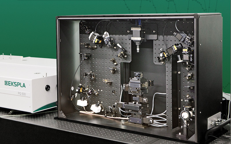
北京欧兰科技发展有限公司为您提供《胶原蛋白中光学二阶非线性的分子学来源检测方案(其它光谱仪)》,该方案主要用于其他中光学二阶非线性的分子学来源检测,参考标准--,《胶原蛋白中光学二阶非线性的分子学来源检测方案(其它光谱仪)》用到的仪器有Ekspla SFG 表面和频光谱分析系统、Ekspla CARS 相干反斯托克斯拉曼显微光谱仪、Ekspla PL2230型高能量皮秒激光器
推荐专场


激光拉曼光谱(RAMAN)
更多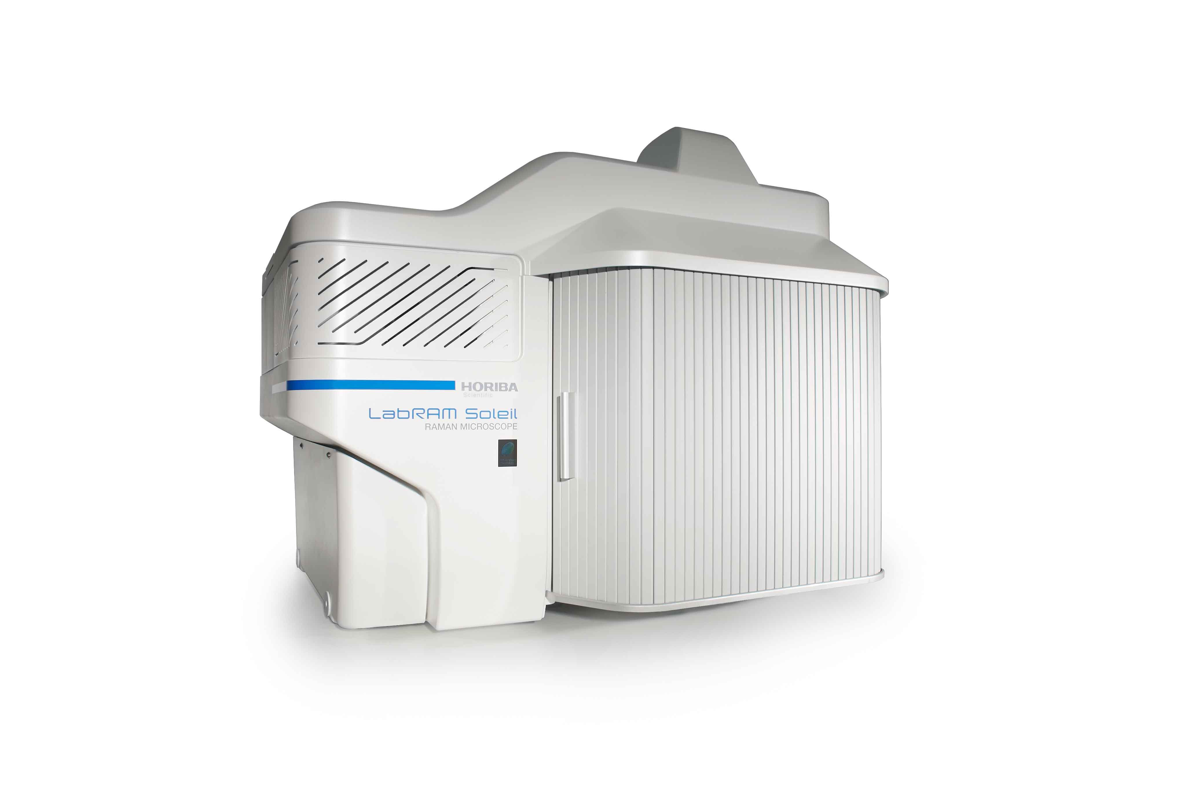


相关方案
更多
该厂商其他方案
更多