


方案详情
文
Sum frequency generation (SFG) spectroscopy using internal reflection geometry
was employed to investigate the conformational order of octadecanethiol (ODT)
monolayer on gold thin films in an electrolyte solution. This approach is convinced
to be useful to study the molecular structure on the electrode/solution interface
under electrochemical condition.
方案详情

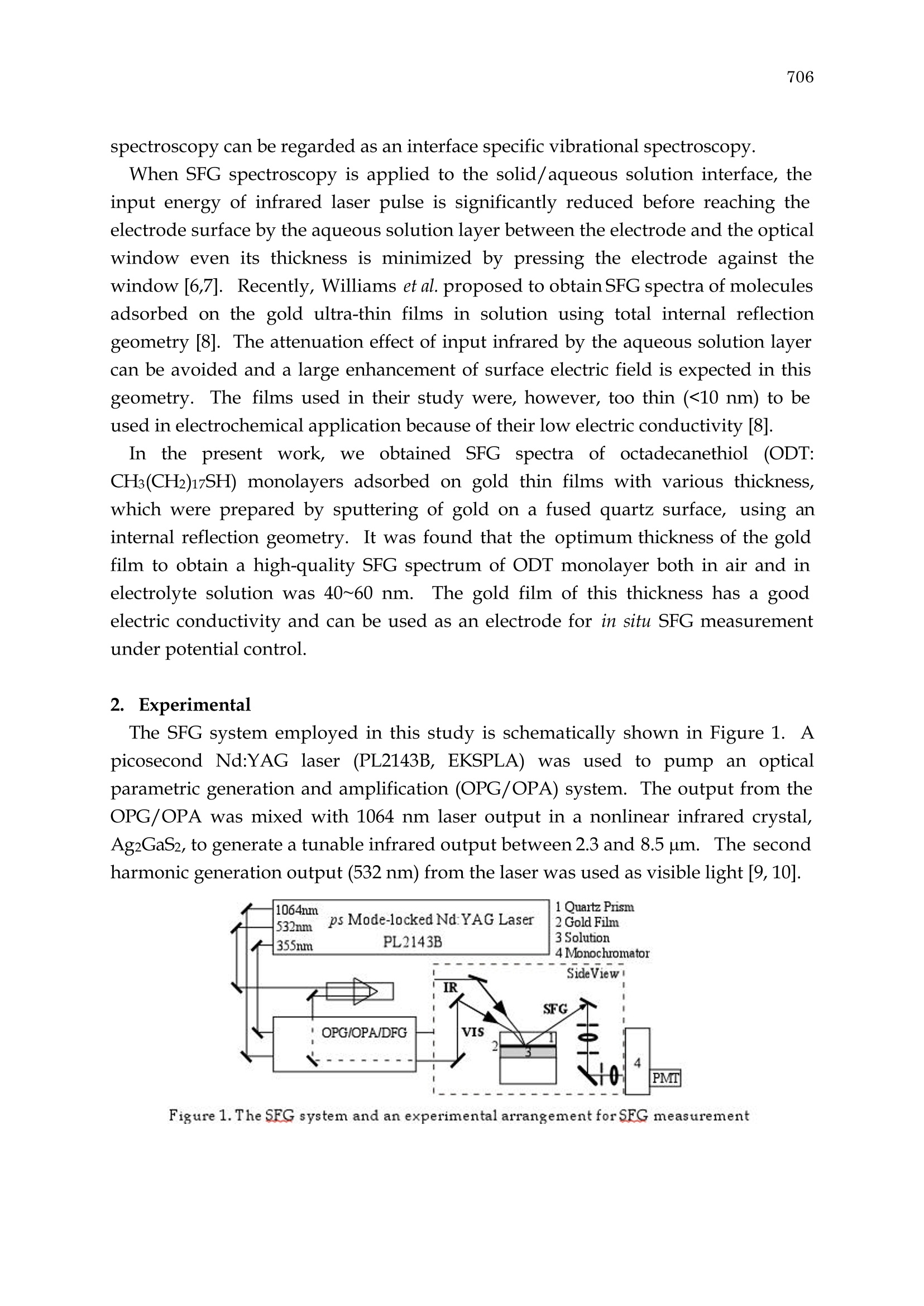
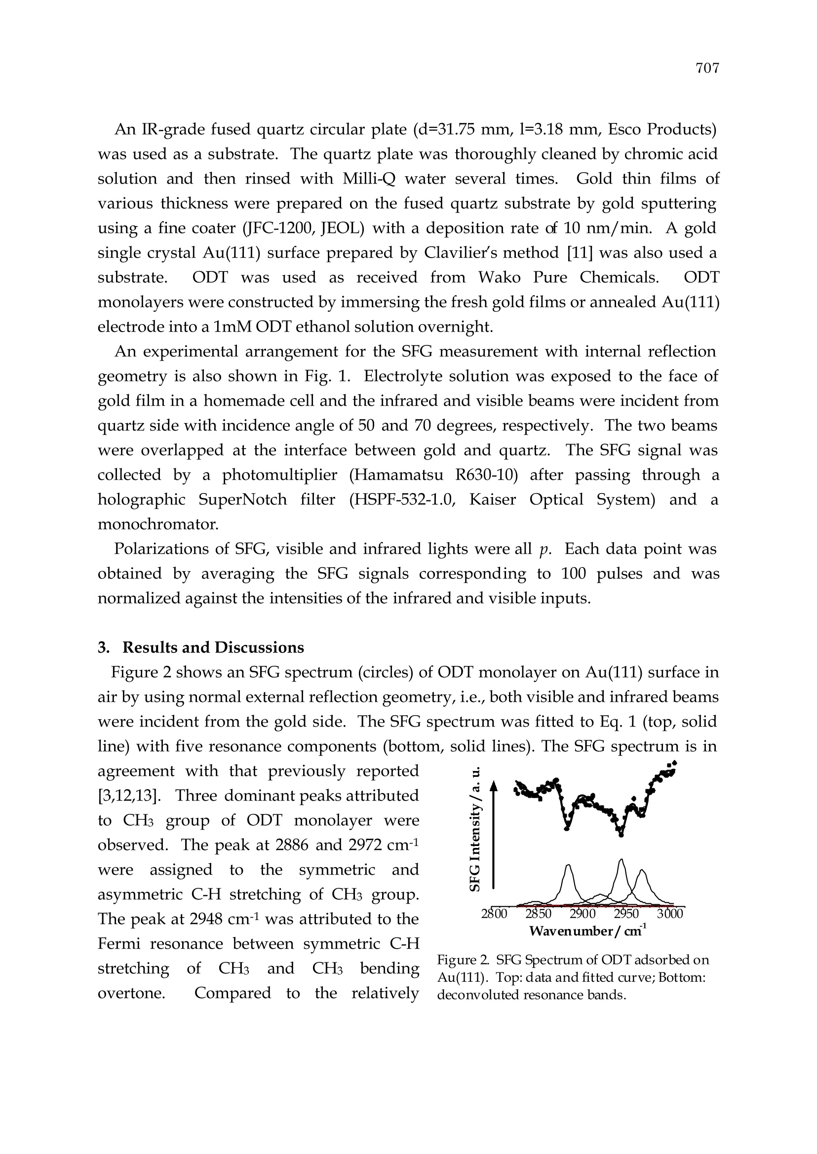
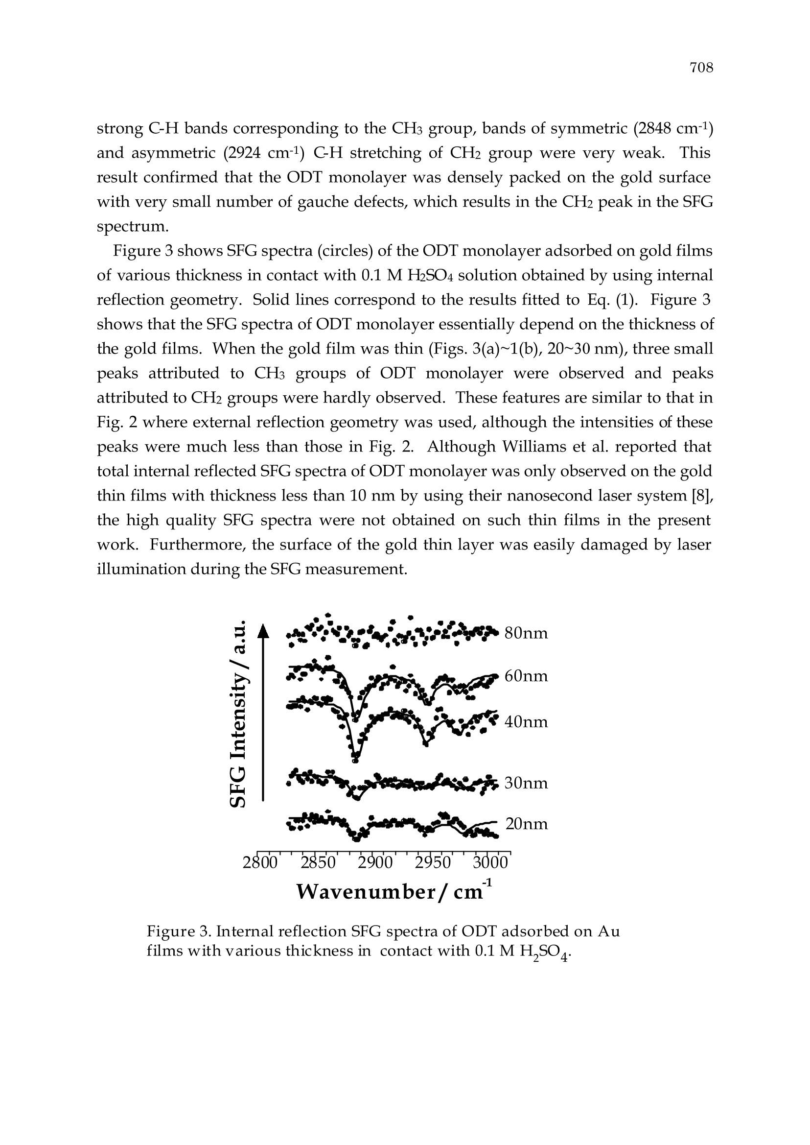
705Studies in Surface Science and Catalysis 132, 705-710Y. Iwasawa, N. Oyama and H. Kunieda (Editors)2001 Elsevier Science B. V.All rights reserved. 706 Conformational Order of Octadecanethiol (ODT) Monolayer at Gold/Solution Interface: Internal Reflection Sum Frequency Generation(SFG) Study Shen Ye, Satoshi Nihonyanagi, Ken Fujishima and Kohei Uosaki* Division of Chemistry, Graduate School of Science, Hokkaido University, Sapporo060-0810,Japan Sum frequency generation (SFG) spectroscopy using internal reflection geometrywas employed to investigate the conformational order of octadecanethiol (ODT)monolayer on gold thin films in an electrolyte solution. This approach is convincedto be useful to study the molecular structure on the electrode/solution interfaceunder electrochemical condition. 1. Introduction Sum frequency generation (SFG) spectroscopy is becoming a powerful tool in theresearch on surface science because of its high surface/interface selectivity andversatile applicability [1-6].SFG is a second-order nonlinear optical process inwhich two photons at frequencies ωi and 02 generate one photon of sum-frequencyat03=01+02. The SFG is forbidden in the bulk of a cetrosymmetric medium and isonly active on the surface or interface where the inversion symmetry is necessarilybroken. For IR-visible SFG, a pulsed visible laser at a fixed frequency ω1 and apulsed infrared laser with a tunable frequency ω2 are used. SFG signal is resonantlyenhanced when 2 matches vibrational modes (@n) on the interface as: where IsrG is the intensity of SFG signal, On An and In are the resonant frequency,strength and damping constant of the vibration modes, respectively,and xand eare the non-resonant contribution and its phase angle, respectively. Therefore, SFG spectroscopy can be regarded as an interface specific vibrational spectroscopy. When SFG spectroscopy is applied to the solid/aqueous solution interface, theinput energy of infrared laser pulse is significantly reduced before reaching theelectrode surface by the aqueous solution layer between the electrode and the opticalwindow even its thickness is minimized by pressing the electrode against thewindow [6,7]. Recently, Williams et al. proposed to obtain SFG spectra of moleculesadsorbed on the gold ultra-thin films in solution using total internal reflectiongeometry [8]. The attenuation effect of input infrared by the aqueous solution layercan be avoided and a large enhancement of surface electric field is expected in thisgeometry. The films used in their study were, however, too thin (<10 nm) to beused in electrochemical application because of their low electric conductivity [8]. In the present work,we obtained SFG spectra of octadecanethiol (ODT:CH3(CH2)17SH) monolayers adsorbed on gold thin films with various thickness,which were prepared by sputtering of gold on a fused quartz surface, using aninternal reflection geometry. It was found that the optimum thickness of the goldfilm to obtain a high-quality SFG spectrum of ODT monolayer both in air and inelectrolyte solution was 40~60 nm.The gold film of this thickness has a goodelectric conductivity and can be used as an electrode for in situ SFG measurementunder potential control. 2. Experimental The SFG system employed in this study is schematically shown in Figure1. Apicosecond Nd:YAG laser (PL2143B, EKSPLA) was used to pump an opticalparametric generation and amplification (OPG/OPA) system. The output from theOPG/OPA was mixed with 1064 nm laser output in a nonlinear infrared crystal,Ag2GaS2, to generate a tunable infrared output between 2.3 and 8.5 um. The secondharmonic generation output (532 nm) from the laser was used as visible light [9, 10]. Figure 1. The SFG system and an experimental arrangement for SFG measurement An IR-grade fused quartz circular plate (d=31.75 mm, l=3.18 mm, Esco Products)was used as a substrate. The quartz plate was thoroughly cleaned by chromic acidsolution and then rinsed with Milli-Q water several times.Gold thin films ofvarious thickness were prepared on the fused quartz substrate by gold sputteringusing a fine coater (JFC-1200, JEOL) with a deposition rate of 10 nm/min. A goldsingle crystal Au(111) surface prepared by Clavilier’s method [11] was also used asubstrate. ODT was usedas received from Wako Pure Chemicals. ODTmonolayers were constructed by immersing the fresh gold films or annealed Au(111)electrode into a 1mM ODT ethanol solution overnight. An experimental arrangement for the SFG measurement with internal reflectiongeometry is also shown in Fig. 1. Electrolyte solution was exposed to the face ofgold film in a homemade cell and the infrared and visible beams were incident fromquartz side with incidence angle of 50 and 70 degrees, respectively. The two beamswere overlapped at the interface between gold and quartz.The SFG signal wascollected by a photomultiplier (Hamamatsu R630-10) after passing through aholographic SuperNotch filter (HSPF-532-1.0, Kaiser Optical System) andamonochromator. Polarizations of SFG, visible and infrared lights were all p. Each data point wasobtained by averaging the SFG signals corresponding to 100 pulses and wasnormalized against the intensities of the infrared and visible inputs. 3. Results and Discussions Figure 2 shows an SFG spectrum (circles) of ODT monolayer on Au(111) surface inair by using normal external reflection geometry, i.e., both visible and infrared beamswere incident from the gold side. The SFG spectrum was fitted to Eq. 1 (top, solidline) with five resonance components (bottom, solid lines).The SFG spectrum is in agreement with that previously reported[3,12,13].. Three dominant peaks attributedto CH3 group of ODT monolayer wereobserved. The peak at 2886 and 2972 cm-1were: assignedto the symmetricandasymmetric C-H stretching of CH3 group.The peak at 2948 cm-1 was attributed to theFermi resonance between symmetric C-Hstretchingof CH3andCH3bendingovertone. Comparedto0tthe relatively Figure 2. SFG Spectrum of ODT adsorbed onAu(111). Top: data and fitted curve; Bottom:deconvoluted resonance bands. strong C-H bands corresponding to the CHs group, bands of symmetric (2848 cm-1)and asymmetric (2924 cm-1) C-H stretching of CH2 group were very weak.Thisresult confirmed that the ODT monolayer was densely packed on the gold surfacewith very small number of gauche defects, which results in the CH peak in the SFGspectrum. Figure 3 shows SFG spectra (circles) of the ODT monolayer adsorbed on gold filmsof various thickness in contact with 0.1 M HSO4 solution obtained by using internalreflection geometry. Solid lines correspond to the results fitted to Eq. (1). Figure 3shows that the SFG spectra of ODT monolayer essentially depend on the thickness ofthe gold films. When the gold film was thin (Figs. 3(a)~1(b), 20~30 nm), three smallpeaks attributed to CH3 groups of ODT monolayer were observed and peaksattributed to CH2 groups were hardly observed. These features are similar to that inFig. 2 where external reflection geometry was used, although the intensities of thesepeaks were much less than those in Fig. 2. Although Williams et al. reported thattotal internal reflected SFG spectra of ODT monolayer was only observed on the goldthin films with thickness less than 10 nm by using their nanosecond laser system [8],the high quality SFG spectra were not obtained on such thin films in the presentwork. Furthermore, the surface of the gold thin layer was easily damaged by laserillumination during the SFG measurement. Figure 3. Internal reflection SFG spectra of ODT adsorbed on Aufilms with various thickness in contact with 0.1 M H SO As the thickness of the gold films increased to 40-60 nm (Figs. 3(c)-1(d)), thesignal/noise (S/N) ratio of the SFG spectra improved and the three peaks attributedto CHs group were observed clearly. The gold films were more tolerant to the laserillumination. These SFG spectra were almost the same as that in Fig. 2. It shouldbe mentioned here that the gold films with the thickness of 40~60nm show a goodelectric conductivity and can be used in electrochemical measurement. In fact, thisgold film is using as the substrate for in situ SFG measurement under electrochemicalcondition. When the gold film was thicker than 80 nm, no peak was observed (Fig.3(e)). The intensity of light decreases exponentially with thickness of the gold films as: where a is the absorption coefficient, k is the imaginary part of refractive index, A iswavelength and d is the thickness of the film. The penetration depths, where lightintensity is decreased to 1/e (36.8%), for visible (532 nm) and infrared (3.4 um) wereestimated to be 17 nm and 11 nm, respectively [14]. Since the penetration depthswere much smaller than the optimum thickness for internal reflection SFGmeasurement shown in Fig. 3, the intensities of both visible and infrared lasers areexpected to be attenuated significantly by the gold thin films. A strong SFG signalfrom ODT monolayer on gold surface was, however, observed clearly as that innormal external reflection SFG spectra (Figs. 2and 3). Thus, another enhancementfactors should be considered here. It is well known that the intensity of the Ramanscattering signal for a surface adsorbed species is enhanced by 105-106 when agrained metal thin layer is used [15].The strong electromagnetic (EM) fieldassociated with the surface plasmon polariton and the collective electron resonanceare considered as major reasons for the surface enhanced Raman scattering (SERS).Similar surface enhanced phenomenon associated with surface roughness of theevaporated metal thin layer was reported for IR absorption [16]. Surface structureand morphology of the gold thin film should play an important role also in thethickness dependent SFG spectra shown in Fig.3. One should be able to reduce the light loss within the gold films by illuminatingthe visible and observing SFG output both from the water side because theabsorption of visible and SFG lights by water are negligible. This study is now inprogress. In conclusion, we have demonstrated that internal reflection SFG spectroscopy isuseful in determining conformational order of ODT monolayer on gold films inelectrolyte solution. The internal reflection SFG spectroscopy can be used as anefficient tool to study the electrochemical interface. Acknowledgment: This work was partially supported by Grant-in-Aids for ScientificResearch on Priority Area of"Electrochemistry of Ordered Interfaces" (No. 09237101)and for Encouragement of Young Scientists (No. 10740314) from the Ministry ofEducation, Science, Sports and Culture, Japan. REFERENCES 1. Y. R. Shen, Nature 337,519 (1989). 2. Y. R. Shen, Proc. Natl. Acad. Sci. USA 93,12104 (1996). 3. C. D. Bain,J. Chem. Soc. Faraday Trans. 91,1281 (1995). 4. P. B. Miranda and Y. R. Shen, J. Phys. Chem. B 103,3292 (1999). 5. D. E. Gragson and G. L. Richmond, J. Phys. Chem. B 102,3847 (1998). 6. A. Tadjeddine and A. Peremans, in Spectroscopy for Surface Science (R. J. H. Clarkand R. E. Hester,eds.), Wiley & Sons Ltd`, Chichester, UK, 1998, p.159. 7. W. Daum, K. A. Friedrich, C. Klunker, D. Knabben, U. Stimming, and H. Ibach,Appl. Phys. A. 59,553 (1994). 8C. T. Williams, Y. Yang, and C. D. Bain, Lagmuir 16,2343 (2000).9 S. Ye, S. Nihonyanagi, and K. Uosaki, Chem. Lett.,734 (2000). 10. S. Ye, T. Saito, S. Nihonyanagi, K. Uosaki, M. Paulo, D. Kim, and Y. R. Shen,submitted. 11. J. Clavilier, R. Faure, G. Guinet, and R. Durand, J. Electroanal. Chem. 107,205(1980). 12. M. A. Hines, J. A. Todd, and P. Guyot-Sionnest, Langmuir 11,493 (1995). 13. S. Ye, S. Nihonyanagi, and K. Uosaki, Nonlinear Optics, submitted. 14.D. W. Lynch and W. R. Hunter, in Handbook of Optical Constants of Solids (E. D.Palik, ed.), Academic Press, Inc.,Orlando, 1985, p. 275. 15. W. Suetaka, Surface infrared and Raman spectroscopy, methods and applications,Plenum Press, New York, 1995. 16. M. Osawa,K. Ataka, K. Yoshi, and T. Yotsuyanagi, J. Electron Spectroscopy andRelated Phenomena 64/65,371(1993).
确定
还剩4页未读,是否继续阅读?

产品配置单


北京欧兰科技发展有限公司为您提供《金/溶液界面,十八烷基硫醇,单分子层膜构象中SFG光谱,和频光谱检测方案(其它光谱仪)》,该方案主要用于其他中SFG光谱,和频光谱检测,参考标准--,《金/溶液界面,十八烷基硫醇,单分子层膜构象中SFG光谱,和频光谱检测方案(其它光谱仪)》用到的仪器有Ekspla SFG 表面和频光谱分析系统、Ekspla PL2230型高能量皮秒激光器、Ekspla CARS 相干反斯托克斯拉曼显微光谱仪
推荐专场
其它光谱仪
更多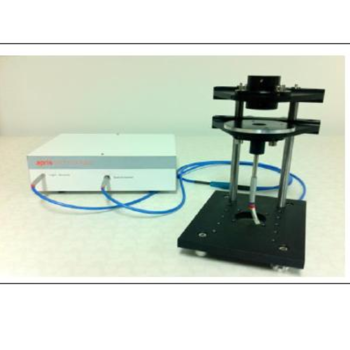


相关方案
更多
该厂商其他方案
更多