


方案详情
文
帕金森症怀疑与元素相关的神经毒性脑内多巴胺信号通路中的氧化应激有关。微量元素的同位素特征图像可以提供特定信息的作用神经元退化。详细介绍了一种利用激光的方法烧蚀电感耦合等离子体质谱(LA-ICP-MS)对神经组织中锌,铜,磷,铁进行二维成像。
方案详情

www.science.uts.edu.au Trace Element Imaging of 6-OHDAInducedParkinson's Disease in Rat Brains Using LaserAblation ICP-MS Dominic Hare1, Brian Reedy1, Michael Dawson1, Fred Fryer2, Per Svenningsson3, Rudolf Grimm4, Per Andren5,Philip Doble1 1Department of Chemistry, Materials and Forensic Science, University of Technology, Sydney, Broadway, New South Wales, Australia 2Agilent Technologies Australia, Sydney, NSW. Australia 3 Department of Pharmacology and Physiology, Karolinska Institute, Stockholm, Sweden 4 Integrated Biology Solutions Unit, Agilent Technologies Inc., Santa Clara, California, United States of America 5 Laboratory for Biological and Medical Mass Spectrometry, Uppsala University, Uppsala, Sweden ABSTRACT Parkinson's disease (PD) is suspected to be associated with neurotoxic elements thatcause oxidative stress along the dopaminergic pathway in two structures of the brainknown as the substantia nigra and the striatum. Therefore, isotope-specific mapping oftrace elements in neurological tissue may provide specific information on the role ofvarious elements in neuronal degradation. This poster details a method utilising laserablation inductively coupled plasma mass spectrometry (LA-ICP-MS) for the two-dimensional mapping of Zn, Cu, P, and Fe in neurological tissues. Rats were lesionedon one side of the brain with 6-hydroxydopamine to induce Parkinson’s disease. Theother side of the brain remained unaffected. 10 pm thick transverse sections of thebrain containing either the substantia nigra or the striatum were ablated and theelemental content measured by the ICP-MS. Two-dimensional contour maps wereconstructed that show isotopic concentration across the entire section. Resolution ofthe images was equivalent to 40 pm pixel size. Preliminary results demonstrated adecrease in zinc expression and a corresponding increase in copper expressionaround the site of the lesion in the substantia nigra sections. This is consistent withprevious reports.Increased levels of phosphorus were also noted in the lesionedhemisphere. INTRODUCTION Imaging of mammalian tissue using MALDI-MS has been described in the literature asan effective method for mapping protein expression in situ [1]. This technique,however, is not capable of identifying trace elements. Laser ablation-inductivelycoupled plasma-mass spectrometry, however, is capable of analysing solid tissuesections in a similar manner to MALDI imaging. Trace elements have long been associated with neurological disorders [2], such as theincreased oxidative stress caused by Fe in Alzheimer’s disease [3]. Previous studiesanalysing digested tissue sections have shown that the regional distribution of certainmetals in Parkinson’s disease mouse models varies significantly [4]. We introduce a new method applying a conventional laser ablation ICP-MS system tothe analysis of tissue sections, with a specific application to the study of trace elementsin 6-OHDA-induced Parkinson's disease in mice brains. MATERIALS & EQUIPMENT Figure 1: 10 um sections ofstriatal tissue 10 pm sections of Sprague-Dawley animals with6-OHDA lesions in the right hemisphere wereobtained from the Karolinska Institutee andLaboratory for Medical and Mass Spectrometry,Sweden.Samples were analysed by a NewWave UP213 Nd:YAG laser ablation unitcoupled to an Agilent Technologies 7500ceSeries ICP-MS. ACQUISITION & DATA PROCESSING Variable laser parameters examined included spot size (65-40 pm), laser power (50-100%), scan speed, intersite pause and pulse frequency. Anywhere between 1 and 13target isotopes were monitored. Data was output as single files for each x-axis line,and were combined using Visual Basic. Visualisation was performed using ENVI. Figure 1: Data analysis procedure. Each ablation line is recorded as asingle data file. Data files are combined into one ASCII file, which isimported into ENVI visualisation software. RESULTS Method 1: Initial studies used a 40 pm diameter laser pulsefiring at 10 Hz for 2 seconds. The intersite dwelltime was 5 seconds, after which the process wasrepeated on an 80 pm grid.This methodexamined primarily first row transition metals.Figure 2 shows a micrograph of brain tissue afterablation. Run time for this method was 5+ days. Figure 3 shows the image for 66Zn Figure 2: Micrograph of braintissue post-ablation. Method 2:65 pm² square laser pulses with ashorter intersite dwell time of 1second were used in a effort toreduce run time. Close gridspacing increased total ablatedmaterial with only 1pm betweencraters. Samples were run forapproximately 24 hours.Targetisotopes included non-metals like31P,shown in Figure 4. Figure 3: 66Zn using Method 1 Method 3:40 pm diameter laser pulses at 1Hz were usedi to increaseresolution while retaining shorterrun times.The scan speed wasset at 40 pm/s and a high laserpower ensured single laser shotsablated enough material. Nointersite dwell time resulted infewer isotopes being monitored.This method has, however,produced the highest resolution Figure 4: 31P using Method 2 image thus far, shown in Figure 5. Method 4: The same parameters as method3 were used with the OctopoleReaction System (ORS) enabledin order to remove possible matrix-based polyatomic interferences.He was used as the reaction gas.Figure 6 shows an 56Fe image,where after interference removal itis apparent that a much higher56Fe background is present in theglass microscope slide backing. Figure 5: 31P using Method 3 Preliminary analysis of data hasshown slight variations in tracemetal content, like Zn and Cu, inorthe substantia nigra. Additionally,striatal sections show possibleincreased phosphorylation.Further work is required to confirmthese observations. Figure 6: 56Fe using Method 4 CONCLUSIONS Laser ablation ICP-MS has proven to be a viable technique for determining thespatial distribution of trace elements, including non-metals like phosphorus inneurological tissue. This method has the potential to become a complementarytechnique to current molecular imaging procedures, specifically MALDI imaging ofpeptides and proteins. Future work will move towards reliable quantification of traceelements and further development of the analytical technology. REFERENCES [1] Stoeckli, M., Chaurand, P., Hallahan, D. E., and Caprioli, R. M. (2001) NatureMedicine 7,493-6. [2] Aschner, M., Silverman, W. F., Sekler, I., and Zatta, P. (2003) Metal lons andNeurodegenerative Disorders, 307-322. ( Qian, Z. M., and Wang, Q. (1998) Brain Research Reviews 27,25 7 -267. ) Tarohda, T., Ishida, Y., Kawai, K., Yamamoto, M., and Amano, R. (2005)Analytical and Bioanalytical Chemistry 383, 224-234.
确定
还剩1页未读,是否继续阅读?

产品配置单
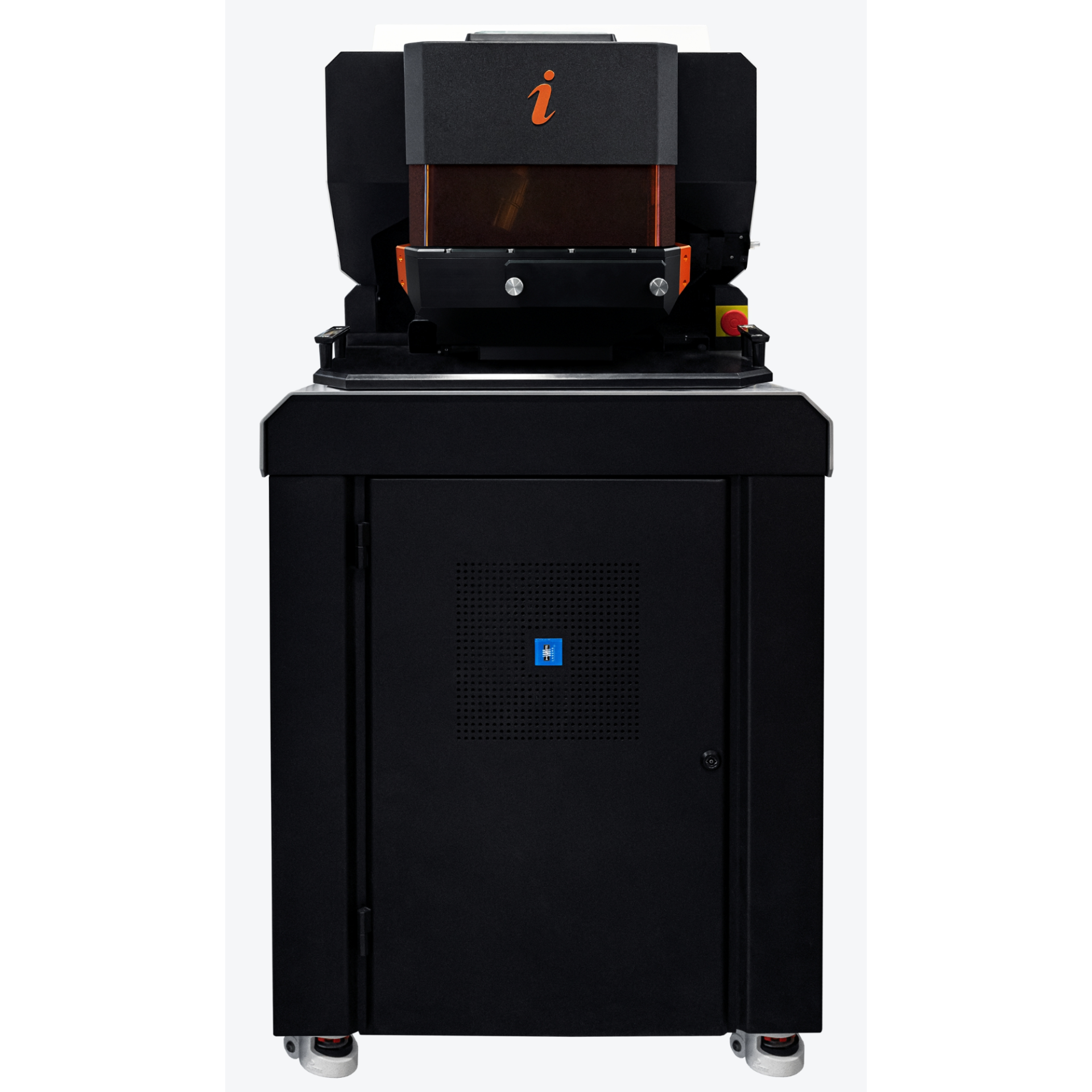
上海凯来仪器有限公司为您提供《脑部切片中微量元素 Zn Cu检测方案(激光剥蚀进样)》,该方案主要用于其他中微量元素 Zn Cu检测,参考标准--,《脑部切片中微量元素 Zn Cu检测方案(激光剥蚀进样)》用到的仪器有ESL213 灵活的激光剥蚀系统
推荐专场
相关方案
更多
该厂商其他方案
更多