


方案详情
文
肺部充填荧光微球,灌注组织后需修改常规的组织加工方法,从而保留微球发荧光特性方便显微镜下观察。福尔马林固定石蜡包埋的免疫组化法,改良后使用Histoclear II替代二甲苯。详细步骤参考本文。
方案详情

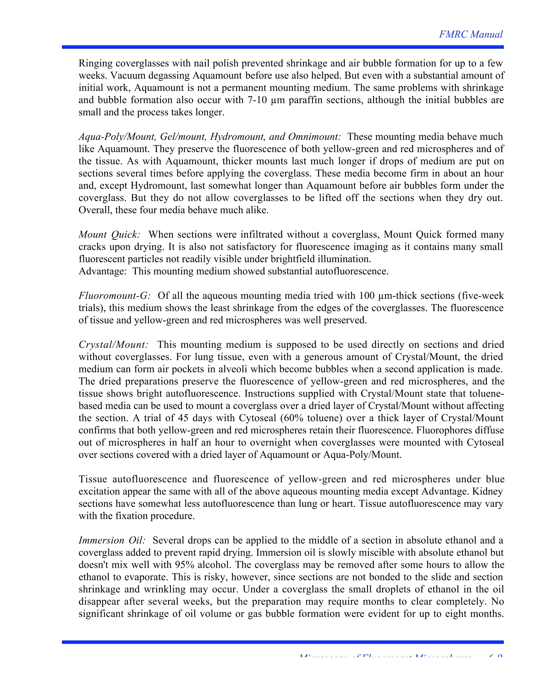
FMRC Manual Microscopic Use ofFluorescentMicrospheres Histological studies of tissues and organs perfused with fluorescent microspheres require modificationof most routine tissue processing methods in order to preserve fluorescent microspheres for microscopicvisualization. For example, formalin fixation followed by routine embedding in paraffin is not feasibleas the standard organic solvents used during tissue processing dissolve polystyrene microspheres.Several different ways of processing tissue were studied and the advantages and disadvantages of eachare evaluated. These included:1) formalin fixation followed by embedding in paraffin; 2) formalinfixation followed by embedding in glycol methacrylate;3) air-dried lung followed by Vibratomesectioning; 4) formalin fixation followed by Vibratome sectioning; 5) rapid freezing followed bycryomicrotome sectioning. Methods for staining sections and the types of mounting media that can beused to apply coverglasses to slides are also evaluated. Aldehyde fixation followed by embedment in paraffin or methacrylate produced sections of well-preserved tissue for microscopic examination. Paraffin blocks were sectioned at 7-10 um with Sturkeydisposable knives on a JB-4 microtome. Methacrylate blocks were sectioned with 12 mm widetriangular glass knives on a Sorvall (now Research & Manufacturing Co.) MT-2 microtome. The kniveswere made on a Sorvall Glass Knife Maker. Unembedded tissue was sectioned with a Vibratome 1000 modified with a 1000 Q rheostat in thecutting blade advance. This allowed a slow forward motion of the oscillating blade through thespecimen. This rheostat modification was especially useful for specimens such as airways, which aretough and difficult to cut. A faster forward blade movement for these specimens resulted in deformationof the block and uneven section thickness. Without the rheostat modification, the forward motion of therazor blade carriage had to be stopped for short time periods with the speed control knob while thevibration of the blade continued. A blade clearance angle of 15-17° worked best for dry and ethanol-dehydrated specimens while 17-20° seemed better for gelatin-embedded specimens. Sections were examined on a Leitz Diaplan compound microscope fitted with an incident mercury arclamp. Three filter sets were used: 1) blue excitation light (450-490 nm) with a suppression filter for allwavelengths below 520 nm in the observation light path (commonly used for the fluoresceinisothiocyanate (FITC) fluorophores); 2) green excitation light (530-560 nm) with a suppression filter forall wavelengths below 580 nm (commonly used for the rhodamine fluorophores); and 3) UV excitation(340-380 nm) with emission suppressed below 430 nm. The blue and UV excitation light causedmicrospheres of blue,green, yellow-green, and red emission colors to glow brightly (although most ofour perfusion protocols used yellow-green and red microspheres only). The green excitation light was11cfuseful only for the red microspheres. With blue epi-illumination, microspheres photographed wellagainst the green autofluorescence of tissue with Ektachrome 160T (tungsten) film. The UV illumination was used in combination with bright field transmitted illumination to provide better histologicalvisualization. A. Paraffin (Paraplast Plus)Embedding Paraffin sections can be cut serially and 7-10 um-thick sections provide good histological detail. Anantimedium (or transition fluid)a fluid miscible with both alcohol (used to dehydrate the tissue) andparaffin—has to be used to allow infiltration of the tissue with paraffin since alcohol is not miscible withparaffin. Hydrocarbons such as toluene and xylene have routinely been used for this purpose (Humason,1979). Such hydrocarbons are now regarded as hazardous chemicals and as fire hazards due to their highvapor pressures (Lyon, et al., 1995). More pertinent to the present study, toluene and xylene also havethe undesirable property of dissolving microspheres or removing the fluorophore from microspheres. Several companies now sell proprietary mixtures as substitutes for hydrocarbons. Several of theproprietary mixtures were evaluated-Histoclear, Histoclear II, Americlear, and Hemo-De. Of these,only Histoclear II retained the fluorescent of the microspheres in paraffin sections. The reasons for thedifferences in the properties of these four substitutes are not known since they are proprietary mixtures.A schedule for processing with Histoclear II is given in Table 1. Based on the manufacturer instructions,infiltration times were increased approximately 50% compared to the usual processing times used withtoluene. Lyon, et al. (1995) recommended butyldecanoate (CH3-(CH2)3-O-0=C-(CH2)8-CH3, sold as "Estisol220") as a substitute for toluene. They recommended clearing and infiltrating times about 4-6 timeslonger (up to 12 hours per change) than the typical toluene schedule. Paraffin is less soluble inbutyldecanoate than in toluene; at 45° C, a saturated solution of Paraplast Plus in butyldecanoate is lessthan 40% paraffin. A schedule for processing with butyldecanoate is given in Table 2. The fluorescenceof microspheres is not as intense as after processing in Histoclear II, particularly for the red Table 1. Tissue Processing with Histoclear II Antimedium Tissue blocks up to approximately 10 mm3. Formaldehyde or glutaraldehyde fixation (not evaluatedwith other fixatives). Wash in tap water, 3-4 changes of 30 minutes 2To 35% ethanol-2 changes of 15 minutes3456789 To 70% ethanol-2 changes of15 minutes To 70% ethanol-30 minutes To 95% ethanol-2 changes of 15 minutes (orovernight) To 100% ethanol- 6 changes of 15 minutes To 100% ethanol + Histoclear II, 1:1-20 minutes To Histoclear II-2 changes of30 minutes To Histoclear II - 60 minutes (probably a minimum for this medium) To 50% Paraplast Plus dissolved in Histoclear II at 45°C -60 minutes To pure Paraplast Plus at 60°C -2 changes of 60 minutes Transfer tissue to embedding mold under an infrared lamp Table 2. Tissue Processing with Butyldecanoate Antimedium Tissue blocks up to approximately 10 mm. Formaldehyde or glutaraldehyde fixation (not evaluatedwith other fixatives). 1. Wash in tap water,3-4 changes of 30 minutes 2. To 35% ethanol-2 changes of 15 minutes 3. To 70% ethanol-2 changes of 15 minutes 4. To 70% ethanol-30 minutes 5 To 95% ethanol -2 changes of 15 minutes (or overnight) 6. To 100% ethanol -6 changes of 15 minutes 7. To 100% ethanol + butyldecanoate, 1:1-30 minutes 8. To butyldecanoate-2 changes of 60 minutes 9. To butyldecanoate-overnight (alternative steps 8 & 9 -2 changes of 12+ hours, agitating occasionally) 10. To 35-40% Paraplast Plus dissolved in butyldecanoate at 45°C-60 minutes 11. To pure Paraplast Plus at 60C-2 changes of 60 minutes 12. Transfer tissue to embedding molds under an infrared lamp microspheres. Yellow-green microspheres are still bright under blue excitation but red microspheresshow fluorescence with green excitation only. Biobond tissue adhesive was used to improve adhesion of paraffin sections to glass slides. Overnightdrying after the sections are mounted on Biobond-coated slides is recommended for good adhesion. TheBiobond coat is quite clear and does not stain as a gelatin-coated slide does. B. 2-Hydroxyethylmethacrylate (Historesin) Embedding This class of methacrylate resin is also known as glycol methacrylate. The embedding procedure weused was adapted from that developed by Chappard (1985) and it has proven very useful for uniformembedment of lung tissue that contains cartilage-reinforced airways and heavily muscled arteries.Chappard (1985) suggested infiltrating dense tissue with the fully catalyzed hydroxyethylmethacrylate(GMA) monomer at -20°C so that the catalyst can diffuse fully into tissue blocks. At room temperature,the fully catalyzed mixture will polymerize in minutes but polymerization is often incomplete in thecenter of dense tissue blocks larger than approximately 6-8 mm3 as the catalyst cannot diffuse to thecenter of tissue blocks in such a short time. Tissue in catalyzed Historesin can be kept at -20°C forseveral weeks without polymerization if the monomer is changed every 24 to 48 hours. Even slightlyhigher temperatures (e.g.,-16°C) allow polymerization in less than 24 hours. Blocks with cartilage orthick-walled arteries require 14 or more days in the catalyzed monomer. A schedule for processing withHistoresin is given in Table 3. Blocks, 4 x 6 mm in size, of fixed lung, heart muscle and kidney section well at 3-5 um after 14 daysinfiltration at -20°C. Air dried lung may also be embedded in GMA with this procedure. Both yellow-green and red microspheres retain their fluorescence throughout the embedding process. Table 3. Tissue Processing for Historesin Embedding Tissue blocks 5 x 5 x 8 mm. Glutaraldehyde fixation (not evaluated with other fixatives). 1Rinse in buffer used for the glutaraldehyde-3 changes of 15 minutes23456 To 35% ethanol-2 changes of 15 minutes To 70% ethanol - 4 changes of 15 minutes To 95% ethanol-2 changes of 15 minutes To 100% ethanol- 4 changes of 15 minutes To 100% ethanol + Historesin Infiltrating Solution, 1:1- 20 min (optional) .7. To Historesin Infiltrating Solution at 40 C - overnight (vacuum degas enough of theInfiltrating Solution for step 7 and to make up the required Embedding Solution as well; lungtissue may benefit by being kept under vacuum overnight instead of in a refrigerator) 8. To Historesin Embedding Solution at -20°C - 24 hours (always handle the EmbeddingSolution on ice and return to freezer as quickly as possible) 9 To Historesin Embedding Solution at -20°C -6-13 days, changing to freshly made Solutionevery 24 to 48 hours (usually safe to leave tissue in the same solution over a week-end afterthe thirdday) 10. Transfer tissue to embedding molds on ice, fill the lower half of the mold with freshEmbedding Solution, and cover the tissue and solution with a piece of Parafilm; polymerizeovernight in a refrigerator at 4°C or keep larger molds on a bed of crushed ice 11. Attach to specimen mounts with the last of the Embedding Solution C. Vibratome Sectioning of Air-Dried Lung Unfixed lungs can be dried over a period of several days by inflating with air at a known pressure. Drylungs are sufficiently rigid to be sliced, and cubes of tissue, 10-15 mm on a side, can be cut out of theslices and fastened with a gel cyanoacrylate glue to 15x19 mm hardwood specimen mounts which fit inthe chuck of the Vibratome. Of a number of glues tried, Quick Tite Super Glue Gel was the only reallysatisfactory product. "Non-gel" glues wick into much of the dry lung and render it too hard to section. Sections were cut dry with the slowest advance of the cutting blade at a thickness of 100-150 um andpositioned without mounting medium under coverglasses on microscope slides. The coverglasses werethen attached to the slides with narrow strips of Scotch Magic Tape. D. Vibratome Sectioning of Unembedded Fixed Tissue Tissue (lung, heart, kidney) was formalin fixed and then hardened in absolute ethanol for one week totwo months. Cubes of unembedded tissue were glued to reusable, rectangular aluminum specimenmounts with Quick Tite Super Glue Gel. As absolute ethanol does not have the water to catalyze thecyanoacrylate glue, pieces were briefly wet in 95% ethanol before being glued to the aluminumspecimen mounts. The glue deteriorates in absolute ethanol so that mounted blocks cannot be storedlong term. Tissue blocks were kept wet during sectioning by periodic application of drops of ethanolwith a Pasteur pipette. A slow blade advance is required, especially through airways. Bronchi reinforced with cartilage andheavily muscled arteries will section down to 50-100 um. For bronchi, it is necessary to section throughthe same cartilage ring that is glued to the aluminum specimen mount to keep the specimen frombending in front of the knife blade. The histology of fixed lung sections is better than that of unfixed, airdried tissue. E. Vibratome Sectioning of Gelatin-Embedded (Bacto Gelatin) Fixed Tissue Embedding irregularly shaped or subdivided specimens in a supporting medium allows the productionof cohesive sections. Also, none of the specimen is rendered unsectionable by infiltrated hardened glueas happens with the unembedded material described above. Gelatin embedding does not degrade tissuehistology or the fluorescence of the microspheres, but the processing time is lengthy. Gelatin-embeddedspecimens are usually frozen before sectioning, but Igarashi,et al. (1994) recently described a methodfor serially sectioning whole rat embryos using a rotating blade machine. We adapted their procedure foruse with the oscillating-blade Vibratome. Tissue pieces were fixed in buffered formalin. To prevent crosslinking (fixing) the gelatin in theinfiltrating solutions, tissue was washed in water for at least one day with a minimum of 10-15 changesof water. Purified gelatin solutions of 15% and 30%(15 and 30 gms in 100 ml distilled water) wereprepared at 55°C. Thymol (0.1%) was added as a bacteriostatic agent. A processing schedule for gelatinembedding is given in Table 4. Blocks of gelatin with tissue were cast in 22x40 mm Peel-A-Way disposable plastic embedding molds.Although disposable, they are durable enough to be used several times if desired. A 2-3 mm thickgelatin platform was cast in the bottom of the molds. This and the larger size of the mold allow foraccurate orientation of the tissue on the specimen mount. The specimens and bottles of gelatin arehandled under a red-coated infra-red lamp to prevent unwanted cooling while orienting the tissue in themolds. The warm tissue will sink into the platforms, however, if the latter are not thoroughly chilledbeforehand in a refrigerator. Also, to prevent remelting the platform when the tissue is embedded, themolds are filled with the warm gelatin in two or more layers (allowing the first layer to gel at roomtemperature before adding the next layer). Table 4. Tissue Processing for Gelatin Embedding Tissue blocks up to approximately 10 mm. Formaldehyde or glutaraldehyde fixation (not evaluatedwith other fixatives). 1. Wash in tap water -10-15 changes over 24 or more hours 2. To 15% gelatin (aqueous) at 37C-2 changes of 24 hours 3. To 30% gelatin (aqueous) at 37-40C -2 changes of 24 hours (more for dense tissue) 4. Transfer tissue to embedding molds (Peel-A-Way molds are particularly useful) under aninfrared lamp 5. Chill gelatin and remove from molds- 6. Fix tissue + gelatin block in 4% buffered formaldehyde-24 hours 7. Warm gelatin block and wooden specimen mount; glue mount to block with molten 30%gelatin 8. Fix tissue + gelatin + mount in 4% formaldehyde-24-48 hours The filled molds are then chilled and the gelatin blocks removed from the molds. A thin spatula kept wetwith water is inserted between the gelatin and mold wall freeing the sides of the block. Twisting themold frees most of the bottom of the block and it can gently be pried out of the mold with the spatula. Ifthe gelatin blocks are fixed in formaldehyde while still in the molds, they are somewhat prone to fractureduring removal. In any case, the Peel-A-Way molds may simply be peeled apart freeing the gelatinblock. Tissue embedded in gelatin does not need much surrounding gelatin for sectioning. The travel of thespecimen stage of the Vibratome limits the height of a piece oftissue to 10 mm. A block this tall needs aminimum length and width of about 10x10 mm for structural strength. After a gelatin block is releasedfrom its mold, its orientation to the cutting blade is determined and the larger excess dimensions cutaway with a razor blade. The gelatin blocks are then hardened in buffered 4% formaldehyde for 24 hours at room temperature(longer term storage is possible). Gelatin blocks and water-soaked, wooden mounts are then warmed in a40°C oven and a bottle of 30% gelatin is warmed to about 55°C. The 30% gelatin is used as a glue tofasten gelatin blocks to the mounts. Mounted specimens are returned to formaldehyde for another 24hours. Igarashi, et al. (1994) recommend 3 days or more. The blocks, now fixed for a minimum of 48 hours, are given a final trimming with a razor blade. Arectangular cutting face works well, but the geometry of the face does not appear to be critical. Thelonger side of the block’s cutting face should be parallel to the blade to better resist the lateral motion ofthe cutting stoke as the knife moves forward. The block and the razor blade holder are kept wet withdistilled water. Sections are cut using a slow blade advance. Cutting faces of 11x14 mm have been sectioned at 50 umand smaller ones at 30 um. With the larger faces, reliable serial sets have been cut at 100 um, although itis difficult to avoid thick and thin zones in a small percentage of the sections. F. Frozen Sections Historically, this was the first technique we tried in our laboratory to preserve fluorescent microspheresin histological sections. In terms of tissue preparation, the technique is straightforward-fresh unfixedtissue is frozen and sectioned at 20-60 um. It has the obvious advantage of avoiding organic reagents butthe disadvantage that it requires the specialized technique of cutting frozen sections on a cryostat. Wehave largely abandoned this approach because of several additional problems which include poorhistological detail due to freezing damage (which becomes greatly accentuated the larger the tissue size),the sections are unfixed and deteriorate rapidly, and the practical difficulty or impossibility of obtainingserial sections. A variation that we have not tried that would result in better tissue structure and morepermanent sections is to chemically fix tissue before freezing. G. Staining of Sections Sections can be stained although the tissue autofluorescence with epi-fluorescence optics may besufficient to allow sections to be viewed and analyzed without staining. This is particularly true for thethicker Vibratome sections and for sections of lung, which have more intense autofluorescent propertiesthan sections of heart or kidney. Paraffin sections. Sections can be stained with hematoxylin and eosin (Delafield's hematoxylin workedwell) or with 0.5% aqueous methyl green (only lung tissue stained satisfactorily with methyl green). Themethyl green staining procedure is followed with a rapid wash in absolute alcohol if immersion oil isused as a mounting medium. See the comments below about how mounting media cause fading of thestains. Methacrylate sections.Tissue embedded in Historesin is not fluorescent so unstained sections are noteasy to view with epifluorescence optics. But methacrylate sections can be stained routinely with Lee’sstain and viewed with epifluorescence since basic fuchsin, a dye in Lee's stain, is fluorescent. Vibratome sections of air-dried lung. Staining was not attempted. Vibratome sections of unembedded fixed tissue. Staining was not attempted. Vibratome sections of gelatin-embedded fixed tissue. Staining was not attempted. A mounting medium is spread on sections mounted on glass slides before covering them with acoverglass (or cover slip). For routine paraffin and methacrylate sections, mounting media containorganic solvents, usually toluene. Such mounting media can be expected to degrade the fluoresence ofmicrospheres as toluene-based antimedia do. We tested three organic solvent-based mountingmedia-Clarion, Cytoseal, and Histolyte 60—and ten water-based mounting media-Advantage,Aquamount, Aqua-Poly/Mount, Crystal/Mount, Fluoromount-G, Gel/Mount, H&E Mount, Hydromount,Omnimount, and Mount Quick. Since the most extensive testing of mounting media was done onVibratome sections of unembedded fixed tissue, see that section below for the most detailed descriptionof the mounting media. Paraffin sections. Cytoseal preserved hematoxylin and eosin (H&E) staining but largely extinguishedthe fluorescence of red microspheres. Aquamount, Aqua-Poly/Mount, Gel/Mount, Hydromount, andOmnimount were used on unstained sections and sections stained with H&E and aqueous methyl green.They are satisfactory with unstained sections, but eosin and methyl green are leached from tissue withina few hours, eosin yielding a uniformly fluorescent area under the coverglass.H&E Mount preservedboth hematoxylin and eosin stains but was strongly fluorescent under blue excitation. The standard H&Eprocedure can be modified by substituting Histoclear II for toluene to preserve the fluorescence of themicrospheres in stained sections. High viscosity immersion oil can then be used as a mounting mediumas it does not affect fluorescence of the microspheres. Methacrylate sections. Aquamount and Mount Quick aqueous coverglass mountants rapidly decolorizeLee's stain. Yellow-green and red microspheres remain fluorescent, although the medium shrinks badly. Yellow-green microspheres remain fluorescent in Cytoseal mountant, but the fluorescence of redmicrospheres in the blue exciting light (450-490 nm) is extinguished within half an hour. Immersion oilworks well as a coverglass mountant since it does not affect the stain or the fluorescence of yellow-green and red microspheres. Vibratome sections of air-dried lung. Mounting media distort dry sections and were not used. Vibratome sections of unembedded fixed tissue. Sections can be mounted under a coverglass inabsolute ethanol and the preparation allowed to dry. Although lung tissue, for example, shows somedamage, the section quality is better than that from air dried (unfixed) lung. The tissue shows goodautofluorescence under blue excitation and the microspheres show up well. With the coverglassessecured after drying with narrow strips of Scotch Magic Mending Tape, this method provides a durablepreparation with a minimum of labor. Two of the organic solvent-based mounting media-Cytoseal, Clarion-were tried. Both diminish thefluorescence of yellow-green microspheres to some degree, extinguish the fluorescence of redmicrospheres under blue excitation, and leave only a faint remnant of the red microsphere's fluorescenceunder green excitation. The water-based mounting media produced the following results. Aquamount: This mountant shrinks considerably as it dries, and 100 um sections need severalapplications of Aquamount in order to produce a durable, Aquamount-filled space under thecoverglass. Sections are cut in 100% ethanol and usually require partial rehydration to facilitate thespreading of an aqueous mountant over sections placed on slides. Rehydration can be done byplacing sections in a 100 mm glass Petri dish filled with 70% ethanol. A slide is placed in the dishwith the label end resting on the side of the dish. A section is maneuvered onto the largelysubmerged slide with a good quality sable hair artist's brush (which work better than a camel hairbrush). The slide is removed from the Petri dish and drained. The section is immediately coveredwith several drops of mountant, and allowed to dry for several hours to overnight. Repeated cyclesof adding mountant and drying can be done before a coverglass is applied. Sometimes, sections curl in Aquamount and need to be covered with a coverglass with the firstapplication of the mountant. Aquamount will often shrink completely away overnight from a 22mm² coverglass, essentially freeing the coverglass from the mountant-infiltrated section below. Thecoverglass can then be lifted or 'popped' off and more mountant added to evenly refill the spaceunder a new coverglass. The initial drying or shrinkage step takes longer, about 1-2 weeks, withsections mounted under 22x40 mm coverglasses. Small bubbles sometimes form within the layer ofAquamount which do not refill with a new application of mountant; in such cases, briefly soakingthe dried layer in distilled water may help to refill bubbles when more Aquamount is added. In ourhands, Aquamount was the only aqueous mountant that allowed clean removal of coverglasses. Withtoluene-base mountants, additional mountant can be added by 'wicking'it under the coverglasswhich may or may not allow complete refilling of spaces that form due to shrinkage of the mountingmedium. This is not a feasible method for aqueous mountants because of the pattern of shrinkage,i.e., the mountants do not shrink back evenly from the edges of the coverglasses. Ringing coverglasses with nail polish prevented shrinkage and air bubble formation for up to a fewweeks. Vacuum degassing Aquamount before use also helped. But even with a substantial amount ofinitial work, Aquamount is not a permanent mounting medium. The same problems with shrinkageand bubble formation also occur with 7-10 um paraffin sections, although the initial bubbles aresmall and the process takes longer. Aqua-Poly/Mount, Gel/mount, Hydromount, and Omnimount: These mounting media behave muchlike Aquamount. They preserve the fluorescence of both yellow-green and red microspheres and ofthe tissue. As with Aquamount, thicker mounts last much longer if drops of medium are put onsections several times before applying the coverglass. These media become firm in about an hourand, except Hydromount, last somewhat longer than Aquamount before air bubbles form under thecoverglass. But they do not allow coverglasses to be lifted off the sections when they dry out.Overall, these four media behave much alike. Mount Quick:When sections were infiltrated without a coverglass, Mount Quick formed manycracks upon drying. It is also not satisfactory for fluorescence imaging as it contains many smallfluorescent particles not readily visible under brightfield illumination. Advantage: This mounting medium showed substantial autofluorescence. Fluoromount-G: Of all the aqueous mounting media tried with 100 um-thick sections (five-weektrials), this medium shows the least shrinkage from the edges of the coverglasses. The fluorescenceof tissue and yellow-green and red microspheres was well preserved. Crystal/Mount:This mounting medium is supposed to be used directly on sections and driedwithout coverglasses. For lung tissue, even with a generous amount of Crystal/Mount, the driedmedium can form air pockets in alveoli which become bubbles when a second application is made.The dried preparations preserve the fluorescence of yellow-green and red microspheres, and thetissue shows bright autofluorescence. Instructions supplied with Crystal/Mount state that toluene-based media can be used to mount a coverglass over a dried layer of Crystal/Mount without affectingthe section. A trial of 45 days with Cytoseal (60% toluene) over a thick layer of Crystal/Mountconfirms that both yellow-green and red microspheres retain their fluorescence. Fluorophores diffuseout of microspheres in half an hour to overnight when coverglasses were mounted with Cytosealover sections covered with a dried layer of Aquamount or Aqua-Poly/Mount. Tissue autofluorescence and fluorescence of yellow-green and red microspheres under blueexcitation appear the same with all of the above aqueous mounting media except Advantage. Kidneysections have somewhat less autofluorescence than lung or heart. Tissue autofluorescence may varywith the fixation procedure. Immersion Oil: Several drops can be applied to the middle of a section in absolute ethanol and acoverglass added to prevent rapid drying. Immersion oil is slowly miscible with absolute ethanol butdoesn't mix well with 95% alcohol. The coverglass may be removed after some hours to allow theethanol to evaporate. This is risky, however, since sections are not bonded to the slide and sectionshrinkage and wrinkling may occur. Under a coverglass the small droplets of ethanol in the oildisappear after several weeks, but the preparation may require months to clear completely. Nosignificant shrinkage of oil volume or gas bubble formation were evident for up to eight months. Immersion oil does not seem to diminish the fluorescence of either the yellow-green or redmicrospheres. Tissue autofluorescence is about equal to that using the aqueous mounting media. Vibratome sections of gelatin-embedded fixed tissue. Gelatin sections require an aqueous mountingmedium because they shrink and distort if dried under a coverglass without a mounting medium. Thisalso happens if drops of an aqueous medium, including Crystal/Mount, are dried on the sections withouta coverglass. Aquamount dries under a coverglass in a few days and retracts forming air spaces aroundand over the sections although the coverglasses are not removable for several more days. By this timethe sections adhere to the slides (although they are probably not infiltrated with the mounting medium)and more Aquamount can be added after the coverglass is removed. I. Visualization and photographic recording Paraffin sections. Photography of 7-10um paraffin sections with simultaneous(single exposure) UV excitation andbright-field transmitted llight i:is aneffective photographic recording methodif the intensity of the transmitted light isreduced. Dewaxed, coverglassed sectionsdo not photograph well withsimultaneousNomarskiinterferencecontrast and UV. Since paraffin sectionshave considerable tissue autofluorescenceunder blue excitation, they can bephotographed with blue epi-illuminationalone (Fig. 1). With experimentation, anexposure index can usually be found sothat both the tissue and fluorescentmicrospheres could be recorded in asingle exposure on Ektachrome colorfilm. Fluorescence is also present insections that have not been dewaxed:with blue epi-illumination, un-dewaxed,uncovered sections look surprisinglygood. Microsphere counts can easily bemade on such sections. Figure 1. Paraffin section of lung showing severalyellow-green and red microspheres in the parenchyma.This 7 um-thick section was cut from a block afterHistoclear II was used as the antimedium and thecoverglass was mounted with Histoclear II as thecoverglass mountant. This protocol preserves thefluorescence of tthhee microspheresand theautofluorescence of the lung. Methacrylate sections. Stained sections can be photographed with bright field illumination plus UVepi-illumination if the transmitted light is reduced. Unstained sections cannot be photographed usingtissue autofluorescence since it is nonexistent. Unstained, uncovered sections can be photographed withsimultaneous Nomarski interference contrast (using a sky blue background) and UV. Covering thesections markedly reduces the amount of interference contrast, depending on the refractive index of themounting medium. Since we did not have a way of measuring the relative brightness of microspheresand tissue, bracketing exposures was necessary to obtain proper exposures. Vibratome sections of air-dried lung. Sections under blue epi-illumination show considerable greenautofluorescence. An alternative with sections up to 50 um was a double exposure of blue epi-illumination only and bright field or Nomarski interference contrast transmitted light only. The exposureindex used for the epi-illuminated exposure was about ten times that of the transmitted light exposure.This was the case even when a dark field correction was used with the epi-illuminated exposure.Experimentation was necessary to produce good photographs. Vibratome sections ofunembedded fixedtissue. The greenautofluorescencee ofthick sections photo-graphed much betterthan 10 um paraffinsections althoughthicker sections showapparent loss of reso-lution. SimultaneousNomarski transmittedillumination and UVepi-illumination or ae exposure ofNomarski illuminationand blue epi-illumina-tion produced colorfulimages with 35-50 umsections (Fig. 2). Figure 2. Section of a unembedded, formalin-fixed tracheal wall thatwas cut with a Vibratome. This 35 um-thick section was photographedasa double exposure with epifluorescence and with Nomarskidifferential interference contrast optics. The tissue is visualized atmoderate to good and the microspheres fluoresce brightly. Vibratome sections of gelatin-embedded fixed tissue. Although gelatin has a faint fluorescence,sections show good autofluorescence under blue excitation, and yellow-green and red microspheresfluoresce brightly. The fluorescence is equal to that of unembedded, fixed tissue. Good colorphotographs may be made with blue epi-illumination although 100 um sections are too thick to use adouble exposure of Nomarski interference contrast and blue epi-illumination. REFERENCES Chappard D. 1985. Uniform polymerization of large blocks in glycol methacrylate at low temperaturewith special reference to enzyme histochemistry. Mikroskopie 42: 148-150. Humason GL. 1979. 4th ed. Animal tissue techniques. W.H. Freeman & Co., San Francisco, CA. Igarashi E, Kawamura N, Takeshita S. 1994. A method for detecting visceral malformations in gelatin-embedded rat fetuses using an automatic slicing apparatus. Biotech. Histochem. 69: 305-310. Luchtel DL, Boykin JC, Bernard SL, Glenny RW. Histological methods to determine blood flowdistribution with fluorescent microspheres. Biotech Histochem1998;73:291-309. ( Lyon H, Holm I, P r ento P , B a lslev E . 1995. Non-hazardous organic solvents in th e paraffin-embeddingtechnique: a rational approach. Aliphatic monoesters for c learing and d ewaxing: butyldecanoate.Histochemistry 1 03: 263-269. ) 7. AAC T. r.
确定


还剩10页未读,是否继续阅读?

海德创业(北京)生物科技有限公司为您提供《Histoclear II应用方案》,该方案主要用于其他中--检测,参考标准--,《Histoclear II应用方案》用到的仪器有
相关方案
更多
该厂商其他方案
更多