


方案详情
文
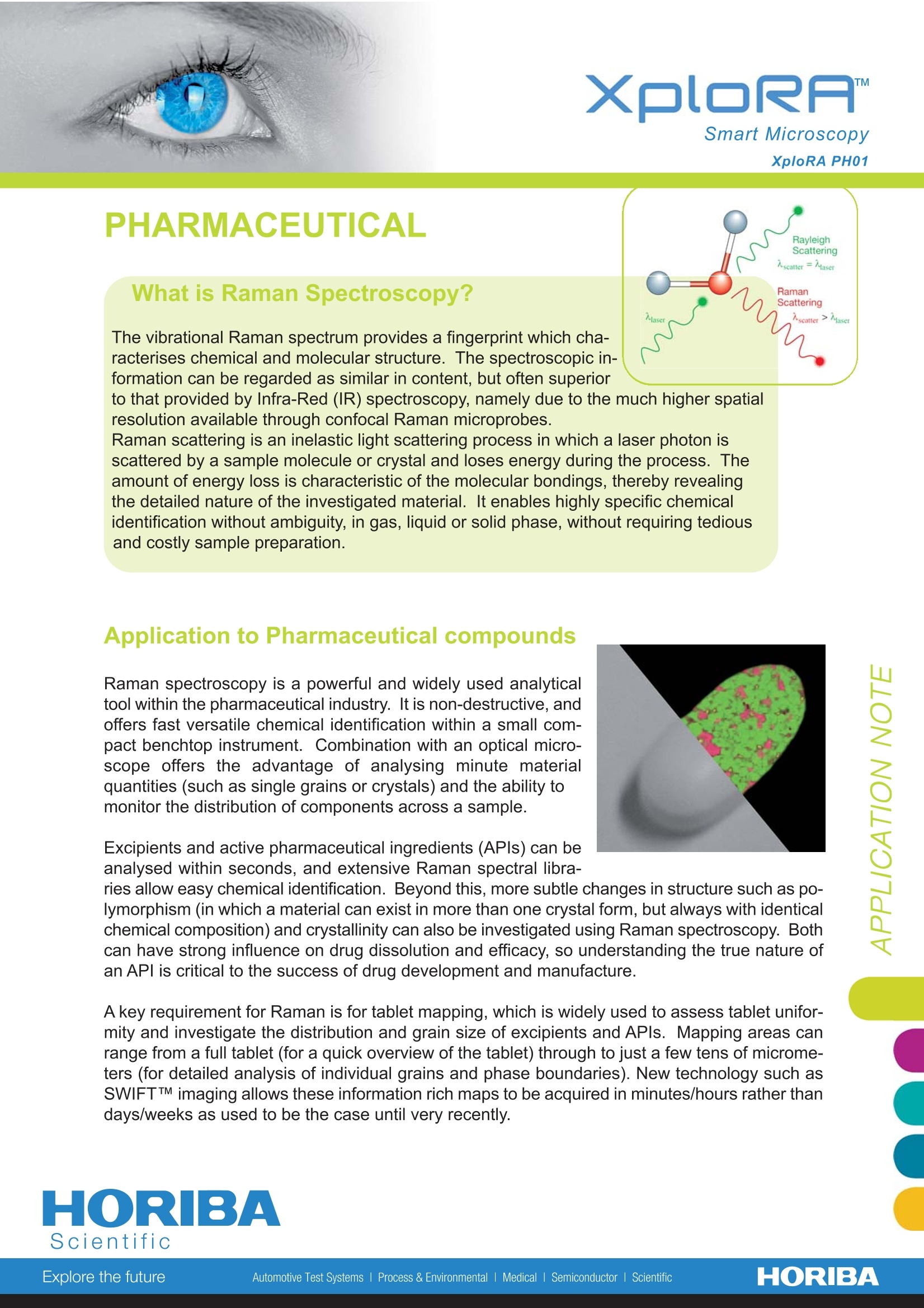
The vibrational Raman spectrum provides a fingerprint which characterises chemical and molecular structure. The spectroscopic information can be regarded as similar in content, but often superior to that provided by Infra-Red (IR) spectroscopy, namely due to the much higher spatial resolution available through confocal Raman microprobes. Raman scattering is an inelastic light scattering process in which a laser photon is scattered by a sample molecule or crystal and loses energy during the process. The amount of energy loss is characteristic of the molecular bondings, thereby revealing the detailed nature of the investigated material. It enables highly specific chemical identification without ambiguity, in gas, liquid or solid phase, without requiring tedious and costly sample preparation.
方案详情

XploRA PH01 Smart Microscopy XploRA PH01 PHARMACEUTICAL What is Raman Spectroscopy? The vibrational Raman spectrum provides a fingerprint which cha-racterises chemical and molecular structure. The spectroscopic in-formation can be regarded as similar in content, but often superiorto that provided by Infra-Red (IR) spectroscopy, namely due to the much higher spatialresolution available through confocal Raman microprobes. Raman scattering is an inelastic light scattering process in which a laser photon isscattered by a sample molecule or crystal and loses energy during the process. Theamount of energy loss is characteristic of the molecular bondings, thereby revealingthe detailed nature of the investigated material. It enables highly specific chemicalidentification without ambiguity, in gas, liquid or solid phase, without requiring tediousand costly sample preparation. Application to Pharmaceutical compounds Raman spectroscopy is a powerful and widely used analyticaltool within the pharmaceutical industry. It is non-destructive, andoffers fast versatile chemical identification within a small com-pact benchtop instrument. Combination with an optical micro-scope offers the advantage of analysing minute materialquantities (such as single grains or crystals) and the ability tomonitor the distribution of components across a sample. Excipients and active pharmaceutical ingredients (APIs) can beanalysed within seconds, and extensive Raman spectral libra- ries allow easy chemical identification. Beyond this, more subtle changes in structure such as po-lymorphism (in which a material can exist in more than one crystal form, but always with identicalchemical composition) and crystallinity can also be investigated using Raman spectroscopy. Bothcan have strong influence on drug dissolution and efficacy, so understanding the true nature ofan APl is critical to the success of drug development and manufacture. A key requirement for Raman is for tablet mapping,which is widely used to assess tablet unifor-mity and investigate the distribution and grain size of excipients and APls. Mapping areas canrange from a full tablet (for a quick overview of the tablet) through to just a few tens of microme-ters (for detailed analysis of individual grains and phase boundaries). New technology such asSWIFTTM imaging allows these information rich maps to be acquired in minutes/hours rather thandays/weeks as used to be the case until very recently. Raman Shift (cm') Colour-coded Raman images of a pharmaceutical tablet highlighting the spatialdistribution of the various components at different scales,allowing to explore thetablet uniformity as well the grain size and boundaries. The spectral signaturesunderneath are linked to the different chemical constituents The XploRA The XploRA is a new concept in Raman microscopy bringing Ramanchemical identification directly to your microscope. Combining micro-scopy and chemical analysis the system retains the full functionality ofyour microscope coupled with high performance Raman spectro-scopy. Compact and rugged in design, the XploRA is easy to use andtransport due to its minimal footprint, making it the ideal smart micro-scope for every R&D, QA/QC and forensic lab. Now you can explorethe true nature of your sample with rapid compound identification andchemical imaging, with no sample preparation and at atmosphericconditions. This non-destructive technique of analysis will boost youinto the new dimension of microscopy. Intuitive operation through newfully compliant software modules including GO!IM, Guided Operationwizard ensures complete ease of use and gets you up to full speedimmediatelv. In this application note Raman fast mapping data froman aspirin containing painkiller are shown. Pharmaceutical tablets contain a number of compo-nents in addition to the APl which is chosen for itstherapeutic effects. These components are used to bulkout the tablet, provide lubrication during mixing andcompaction, and aid digestion. Three Raman maps have been acquired, moving froma large area low resolution whole tablet image, throughto a high resolution small area of interest to analyseindividual grains/particles in detail. In the whole tablet map (1), which comprises 50,901pixels over a 7 x 18 mm² area, the major constituents(aspirin, paracetamol and caffeine) are visible, in ad-dition to the tablet coating. A higher resolution image(2) highlights a fourth component (cellulose) widelyspread across the tablet, but present only in small, dis-crete areas. The final image (3) was acquired with 2 umstep (90,601 data points), and allows the size and shapeof individual cellulose grains to be observed.Spectrafrom these four main components are also shown,illustrating the ease with which they can be distingui- shed using Raman. www.smartmicroscopy.com Find us at www.horiba.com or telephone: Email: mma-info@jobinyvon.comUSA: France:Scientific Germany: UK: +1-732-494-8660+33 (0)320 5918 00+49 (0)625184750+44(0)20 82048142China:+86(0)10 8567 9966Italy:+39 02 57603050 HORIBAExplore the futureAutomotive Test Systems Process & Environmental Medical Semiconductor l Scientific The vibrational Raman spectrum provides a fingerprint which characterises chemical and molecular structure. The spectroscopic information can be regarded as similar in content, but often superior to that provided by Infra-Red (IR) spectroscopy, namely due to the much higher spatial resolution available through confocal Raman microprobes. Raman scattering is an inelastic light scattering process in which a laser photon is scattered by a sample molecule or crystal and loses energy during the process. The amount of energy loss is characteristic of the molecular bondings, thereby revealing the detailed nature of the investigated material. It enables highly specific chemical identification without ambiguity, in gas, liquid or solid phase, without requiring tedious and costly sample preparation.
确定

还剩1页未读,是否继续阅读?

产品配置单

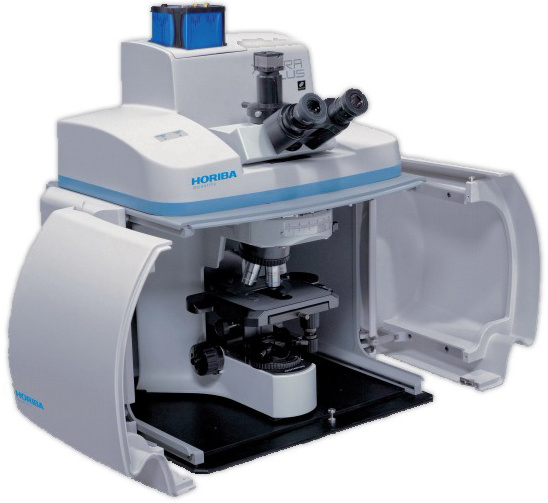
HORIBA(中国)为您提供《药品中表面形貌检测方案(激光拉曼光谱)》,该方案主要用于原料药中限度检查检测,参考标准--,《药品中表面形貌检测方案(激光拉曼光谱)》用到的仪器有HORIBA XploRA PLUS超快速拉曼成像光谱仪
推荐专场
相关方案
更多
该厂商其他方案
更多