


方案详情
文
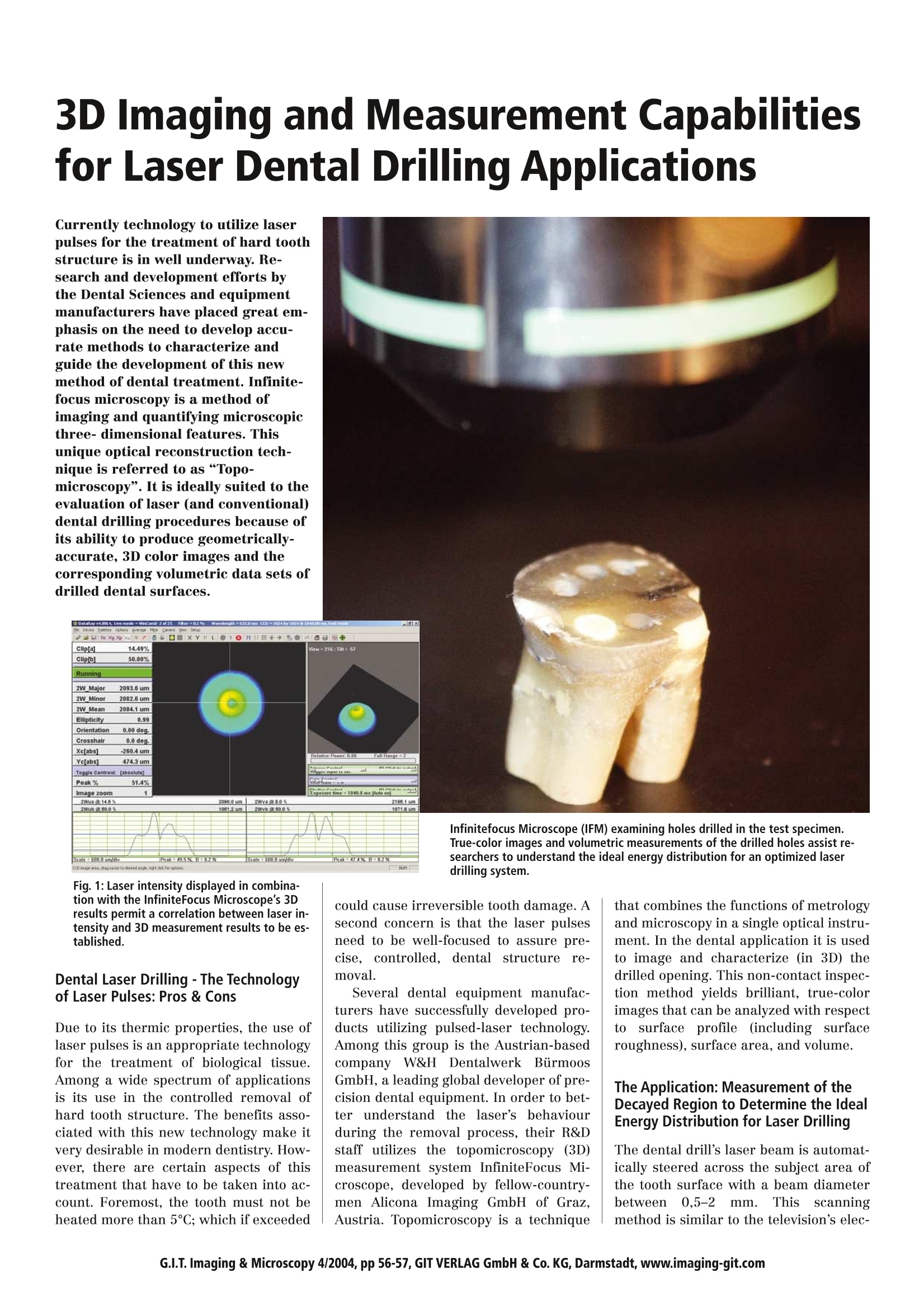
Currently technology to utilize laser pulses for the treatment of hard tooth structure is in well underway. Research and development efforts by the Dental Sciences and equipment manufacturers have placed great emphasis on the need to develop accurate methods to characterize and guide the development of this new method of dental treatment. Infinitefocus microscopy is a method of imaging and quantifying microscopic three- dimensional features. This unique optical reconstruction technique is referred to as “Topomicroscopy”. It is ideally suited to the evaluation of laser (and conventional) dental drilling procedures because of its ability to produce geometricallyaccurate, 3D color images and the corresponding volumetric data sets of drilled dental surfaces.
方案详情

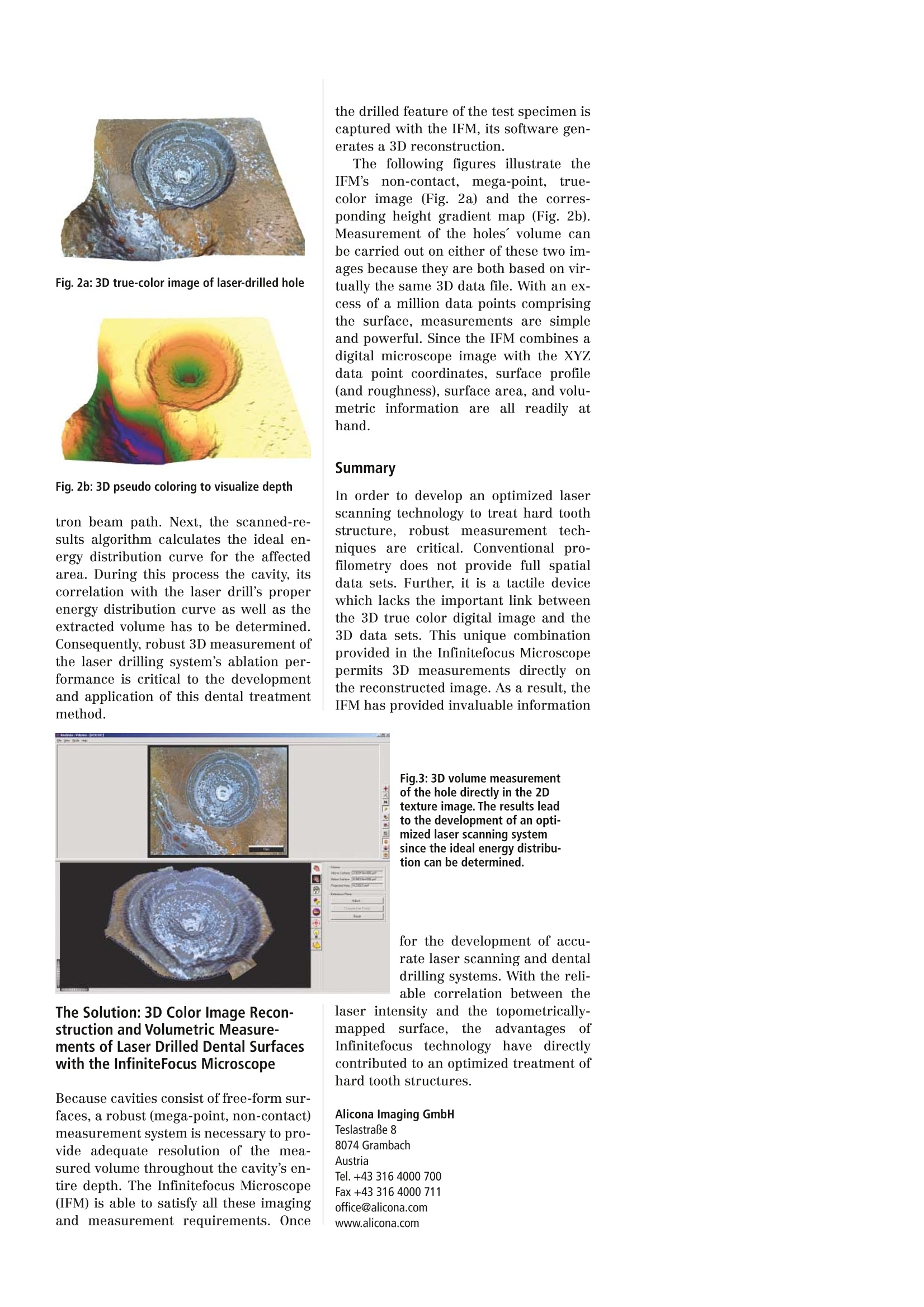
G.I.T. Imaging & Microscopy 4/2004, pp 56-57, GIT VERLAG GmbH & Co. KG, Darmstadt, www.imaging-git.com 3D Imaging and Measurement Capabilitiesfor Laser Dental Drilling Applications Currently technology to utilize laserpulses for the treatment of hard toothstructure is in well underway. Re-search and development efforts bythe Dental Sciences and equipmentmanufacturers have placed great em-phasis on the need to develop accu-rate methods to characterize andguide the development of this newmethod of dental treatment. Infinite-focus microscopy is a method ofimaging and quantifying microscopicthree-dimensional features. Thisunique optical reconstruction tech-nique is referred to as“Topo-microscopy”. It is ideally suited to theevaluation of laser (and conventional)dental drilling procedures because ofits ability to produce geometrically-accurate, 3D color images and thecorresponding volumetric data sets of drilled dental surfaces. Infinitefocus Microscope (IFM) examining holes drilled in the test specimen.True-color images and volumetric measurements of the drilled holes assist re-searchers to understand the ideal energy distribution for an optimized laserdrilling system. Fig. 1: Laser intensity displayed in combina-tion with the InfiniteFocus Microscope’s 3Dresults permit a correlation between laser in-tensity and 3D measurement results to be es-tablished. Dental Laser Drilling-The Technologyof Laser Pulses: Pros & Cons Due to its thermic properties, the use oflaser pulses is an appropriate technologyfor the treatment of biological tissue.Among a wide spectrum of applicationsis its use in the controlled removal ofhard tooth structure. The benefits asso-ciated with this new technology make itvery desirable in modern dentistry. How-ever, there are certain aspects of thistreatment that have to be taken into ac-count. Foremost, the tooth must not beheated more than 5C; which if exceeded could cause irreversible tooth damage. Asecond concern is that the laser pulsesneed to be well-focused to assure pre-cise, controlled, dental structure removal. Several dental equipment manufac-turers have successfully developed pro-ducts utilizing pulsed-laser technology.Among this group is the Austrian-basedcompany W&H DentalwerkkBirmoosGmbH, a leading global developer of pre-cision dental equipment. In order to bet-ter understand the laser's behaviourduring the removal process, their R&Dstaff utilizes the topomicroscopy (3D)measurement system InfiniteFocus Mi-croscope, developed by fellow-country-men Alicona Imaging GmbH of Graz,Austria. Topomicroscopy is a technique that combines the functions ofmetrologyand microscopy in a single optical instru-ment. In the dental application it is usedto image and characterize (in 3D) thedrilled opening. This non-contact inspec-tion method yields brilliant, true-colorimages that can be analyzed with respecttosurfacee profile(including surfaceroughness), surface area, and volume.The dental drill's laser beam is automat-ically steered across the subject area ofthe tooth surface with a beam diameterbetweenn0,5-22mm. Thisscanningmethod is similar to the television's elec- The Application: Measurement of theDecayed Region to Determine the IdealEnergy Distribution for Laser Drilling Fig. 2a: 3D true-color image of laser-drilled hole Fig. 2b: 3D pseudo coloring to visualize depth tron beam path. Next, the scanned-re-sults algorithm calculates the ideal en-ergy distribution curve for the affectedarea. During this process the cavity, itscorrelation with the laser drill's properenergy distribution curve as well as theextracted volume has to be determined.Consequently, robust 3D measurement ofthe laser drilling system's ablation per-formance is critical to the developmentand application of this dental treatmentmethod. the drilled feature of the test specimen iscaptured with the IFM, its software gen-erates a 3D reconstruction. The following figures illustrate theIFM'ss non-contact, mega-point, true-color image (Fig. 2a) and the corres-ponding height gradient map (Fig. 2b).Measurement of the holes’ volume canbe carried out on either of these two im-ages because they are both based on vir-tually the same 3D data file. With an ex-cess of a million data points comprisingthe surface, measurements are simpleand powerful. Since the IFM combines adigital microscope image with the XYZdata point coordinates, surface profile(and roughness), surface area, and volu-metric information are all readily athand. Summary In order to develop an optimized laserscanning technology to treat hard toothstructure, robust measurementttiech-niques are critical. Conventional pro-filometry does not provide full spatialdata sets. Further, it is a tactile devicewhich lacks the important link betweenthe 3D true color digital image and the3D data sets. This unique combinationprovided in the Infinitefocus Microscopepermits 3D measurements directly onthe reconstructed image. As a result, theIFM has provided invaluable information 的一一 The Solution: 3D Color Image Recon-struction and Volumetric Measure-ments of Laser Drilled Dental Surfaceswith the InfiniteFocus Microscope Because cavities consist of free-form sur-faces, a robust (mega-point, non-contact)measurement system is necessary to pro-vide adequate resolution of the mea-sured volume throughout the cavity’s en-tire depth. The Infinitefocus Microscope(IFM) is able to satisfy all these imagingand measurement requirements. Once
确定
还剩1页未读,是否继续阅读?

北京东方德菲仪器有限公司为您提供《牙齿中激光钻孔检测方案(轮廓仪)》,该方案主要用于牙齿中激光钻孔检测,参考标准--,《牙齿中激光钻孔检测方案(轮廓仪)》用到的仪器有
相关方案
更多