


方案详情
文
As these examples show microspectroscopic techniques are fast becoming invaluable tools for the life sciences, allowing researchers to move closer to a complete understanding of living processes. The images resulting from techniques such as Raman or XRF are more than just pretty pictures—they provide the scientist with a new dimension of information, based upon real biochemistry and composition. The possibilities are unlimited.
方案详情

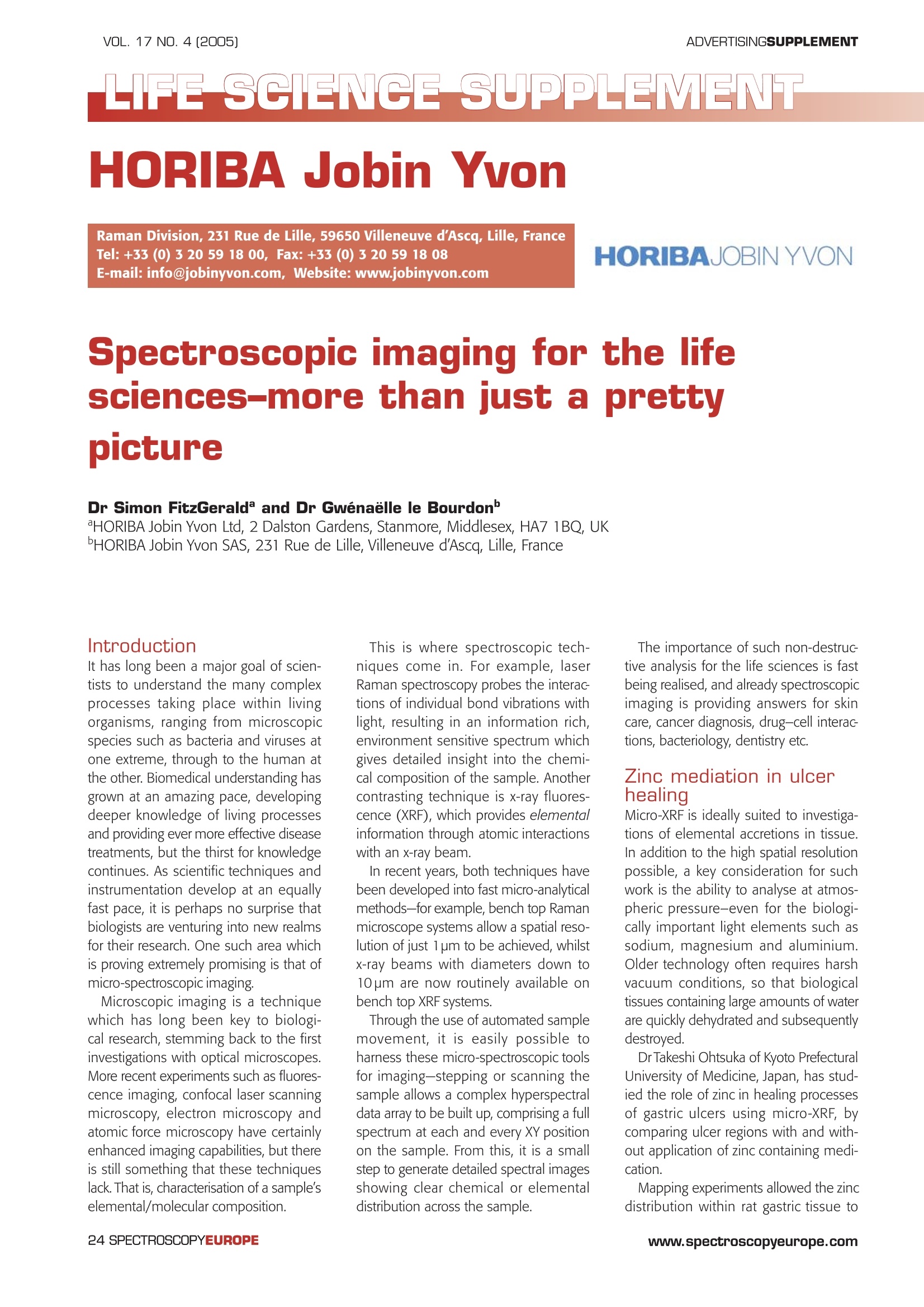
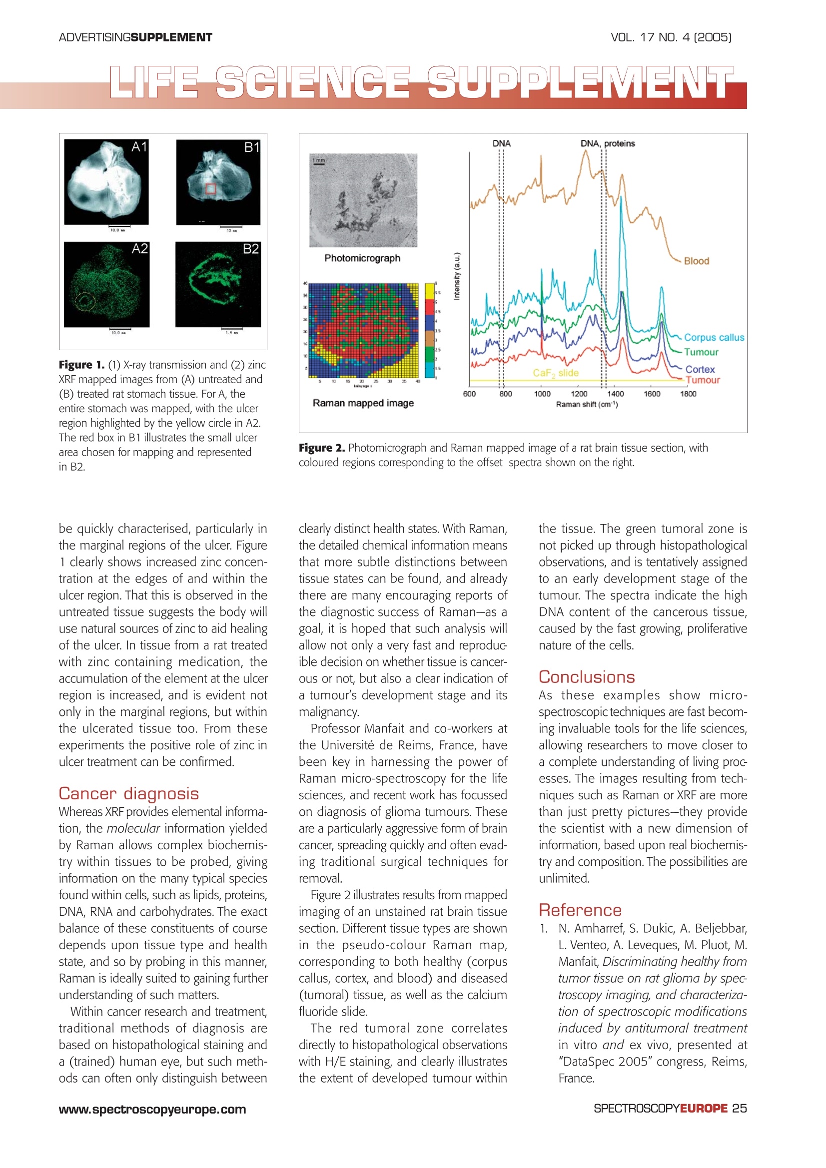
VOL. 17 NO. 4 [2005]ADVERTISINGSUPPLEMENT ADVERTISINGSUPPLEMENTVOL. 17 NO. 4 [2005] LIFE SCIENCE SUPPLEMENT HORIBA Jobin Yvon Raman Division, 231 Rue de Lille, 59650 Villeneuve d'Ascq, Lille, FranceTel: +33 (0) 3 20 59 18 00, Fax: +33 (0) 3 20 59 18 08E-mail: info@jobinyvon.com, Website: www.jobinyvon.com HORIBAJOBIN YVON Spectroscopic imaging for the lifesciences-more than just a pretty picture Dr Simon FitzGerald and Dr Gwenaelle le Bourdon' HORIBA Jobin Yvon Ltd, 2 Dalston Gardens, Stanmore, Middlesex, HA7 1BQ, UKHORIBA Jobin Yvon SAS, 231 Rue de Lille, Villeneuve d'Ascq, Lille, France Introduction It has long been a major goal of scien-tists to understand the many complexprocesses taking place within livingorganisms, ranging from microscopicspecies such as bacteria and viruses atone extreme, through to the human atthe other. Biomedical understanding hasgrown at an amazing pace, developingdeeper knowledge of living processesand providing ever more effective diseasetreatments, but the thirst for knowledgecontinues. As scientific techniques andinstrumentation develop at an equallyfast pace, it is perhaps no surprise thatbiologists are venturing into new realmsfor their research. One such area whichis proving extremely promising is that ofmicro-spectroscopic imaging. Microscopic imaging is a techniquewhich has long been key to biologi-cal research, stemming back to the firstinvestigations with optical microscopes.More recent experiments such as fluores-cence imaging, confocal laser scanningmicroscopy, electron microscopy andatomic force microscopy have certainlyenhanced imaging capabilities, but thereis still something that these techniqueslack. That is, characterisation of a sample'selemental/molecular composition. This is where spectroscopic tech-niques come in. For example, laserRaman spectroscopy probes the interac-tions of individual bond vibrations withlight, resulting in an information rich,environment sensitive spectrum whichgives detailed insight into the chemi-cal composition of the sample. Anothercontrasting technique is x-ray fluores-cence (XRF), which provides elementalinformation through atomic interactionswith an x-ray beam. In recent years, both techniques havebeen developed into fast micro-analyticalmethods-for example, bench top Ramanmicroscope systems allow a spatial reso-lution of just 1 pm to be achieved, whilstx-ray beams with diameters down to10 pm are now routinely available onbench top XRFsystems. Through the use of automated samplemovement, it is easily possible toharness these micro-spectroscopic toolsfor imaging-stepping or scanning thesample allows a complex hyperspectraldata array to be built up, comprising a fullspectrum at each and every XY positionon the sample. From this, it is a smallstep to generate detailed spectral imagesshowing clear chemical or elementaldistribution across the sample. The importance of such non-destruc-tive analysis for the life sciences is fastbeing realised, and already spectroscopicimaging is providing answers for skincare, cancer diagnosis, drug-cell interac-tions, bacteriology, dentistry etc. Zinc mediation in ulcerhealing Micro-XRF is ideally suited to investiga-tions of elemental accretions in tissue.In addition to the high spatial resolutionpossible, a key consideration for suchwork is the ability to analyse at atmos-pheric pressure-even for the biologi-callyimportant light elements such assodium, magnesium and aluminium.Older technology often requires harshvacuum conditions, so that biologicaltissues containing large amounts of waterare quickly dehydrated and subsequentlydestroyed. DrTakeshi Ohtsuka of Kyoto PrefecturalUniversity of Medicine, Japan, has stud-ied the role of zinc in healing processesof gastric ulcers using micro-XRF, bycomparing ulcer regions with and with-out application of zinc containing medi-cation. Mapping experiments allowed the zincdistribution within rat gastric tissue to Figure 1. (1) X-ray transmission and (2) zinc XRF mapped images from (A) untreated and(B) treated rat stomach tissue. ForA, theentire stomach was mapped, with the ulcerregion highlighted by the yellow circle in A2.The red box in B1 illustrates the small ulcerarea chosen for mapping and representedin B2. be quickly characterised, particularly inthe marginal regions of the ulcer. Figure1clearly shows increased zinc concen-tration at the edges of and within theulcer region. That this is observed in theuntreated tissue suggests the body willuse natural sources of zinc to aidhealingof the ulcer. In tissue from a rat treatedwith zinc containing medication, theaccumulation of the element at the ulcerregion is increased, and is evident notonly in the marginal regions, but withinthe ulcerated tissue too. From theseexperiments the positive role of zinc inulcer treatment can be confirmed. Cancer diagnosis Whereas XRF provides elemental informa-tion, the molecular information yieldedby Raman allows complex biochemis-try within tissues to be probed, givinginformation on the many typical speciesfound within cells, such as lipids, proteins,DNA, RNA and carbohydrates. The exactbalance of these constituents of coursedepends upon tissue type and healthstate, and so by probing in this manner,Raman is ideally suited to gaining furtherunderstanding of such matters. Within cancer research and treatment,traditional methods of diagnosis arebased on histopathological staining anda (trained) human eye, but such meth-ods can often only distinguish between Figure 2. Photomicrograph and Raman mapped image ofa rat brain tissue section, withcoloured regions corresponding to the offset spectra shown on the right. clearly distinct health states. With Raman,the detailed chemical information meansthat more subtle distinctions betweentissue states can be found, and alreadythere are many encouraging reports ofthe diagnostic success of Raman-as agoal, it is hoped that such analysis willallow not only a very fast and reproduc-ible decision on whether tissue is cancer-ous or not, but also a clear indication ofa tumour's development stage and itsmalignancy. Professor Manfait and co-workers atthe Universite de Reims, France, havebeen key in harnessing the power ofRaman micro-spectroscopy for the lifesciences, and recent work has focussedon diagnosis of glioma tumours. Theseare a particularly aggressive form of braincancer, spreading quickly and often evad-ing traditional surgical techniques forremovaall. Figure 2 illustrates results from mappedimaging of an unstained rat brain tissuesection. Different tissue types are shownin the pseudo-colour Raman map,corresponding to both healthy (corpuscallus, cortex, and blood) and diseased(tumoral) tissue,as well as the calciumfluoride slide. The red tumoral zone correlatesdirectly to histopathological observationswith H/E staining, and clearly illustratesthe extent of developed tumour within the tissue. The green tumoral zone isnot picked up through histopathologicalobservations, and is tentatively assignedto an early development stage of thetumour. The spectra indicate the highDNA content of the cancerous tissue,caused by the fast growing, proliferativenature of the cells. Conclusions As these examples show micro-spectroscopic techniques are fast becom-ing invaluable tools for the life sciences,allowing researchers to move closer toa complete understanding of living processes. The images resulting from tech-niques such as Raman or XRF are morethan just pretty pictures-they providethe scientist with a new dimension ofinformation, based upon real biochemis-try and composition. The possibilities areunlimited. Reference 1.IN. Amharref, S. Dukic, A. Beljebbar,L. Venteo, A. Leveques, M. Pluot, M.Manfait, Discriminating healthy fromtumor tissue on rat glioma by spec-troscopy imaging, and characteriza-tion of spectroscopic modificationsinduced by antitumoral treatmentin vitro and ex vivo, presented at"DataSpec 2005" congress, Reims,France. SPECTROSCOPYEUROPEwww.spectroscopyeurope.com www.spectroscopyeurope.comSPECTROSCOPYEUROPE As these examples show microspectroscopic techniques are fast becoming invaluable tools for the life sciences, allowing researchers to move closer to a complete understanding of living processes. The images resulting from techniques such as Raman or XRF are more than just pretty pictures—they provide the scientist with a new dimension of information, based upon real biochemistry and composition. The possibilities are unlimited.
确定
还剩1页未读,是否继续阅读?

HORIBA(中国)为您提供《大鼠中化学组成及分布检测方案(激光拉曼光谱)》,该方案主要用于其他中化学组成及分布检测,参考标准--,《大鼠中化学组成及分布检测方案(激光拉曼光谱)》用到的仪器有
相关方案
更多
该厂商其他方案
更多