


采用立陶宛Ekspla公司的由脉冲皮秒激光器PL2251-50和皮秒参量激光器PG401构成的振动和频光谱测量系统,对纤维素样品进行的测量分析揭示了纤维素表面层结构。
方案详情

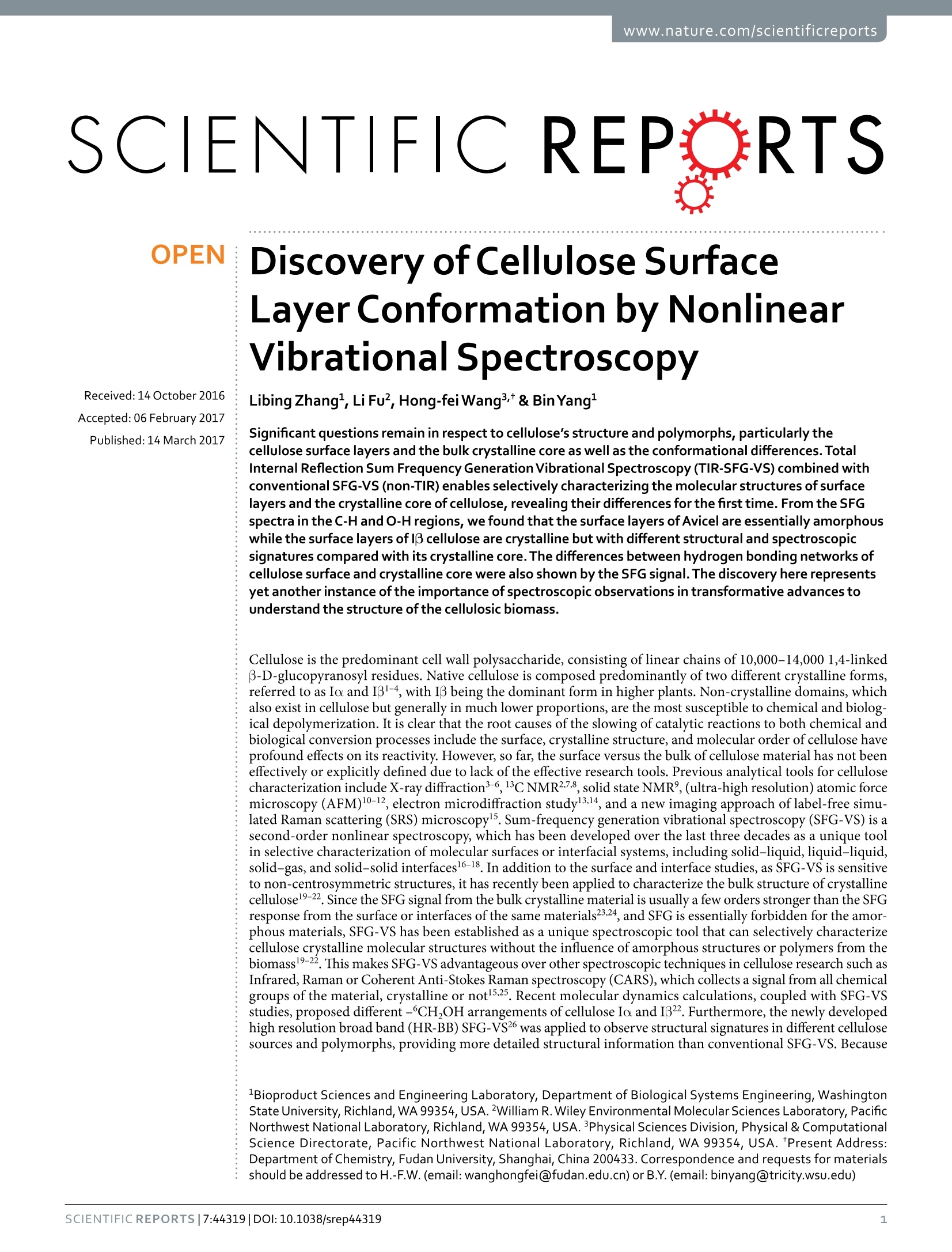
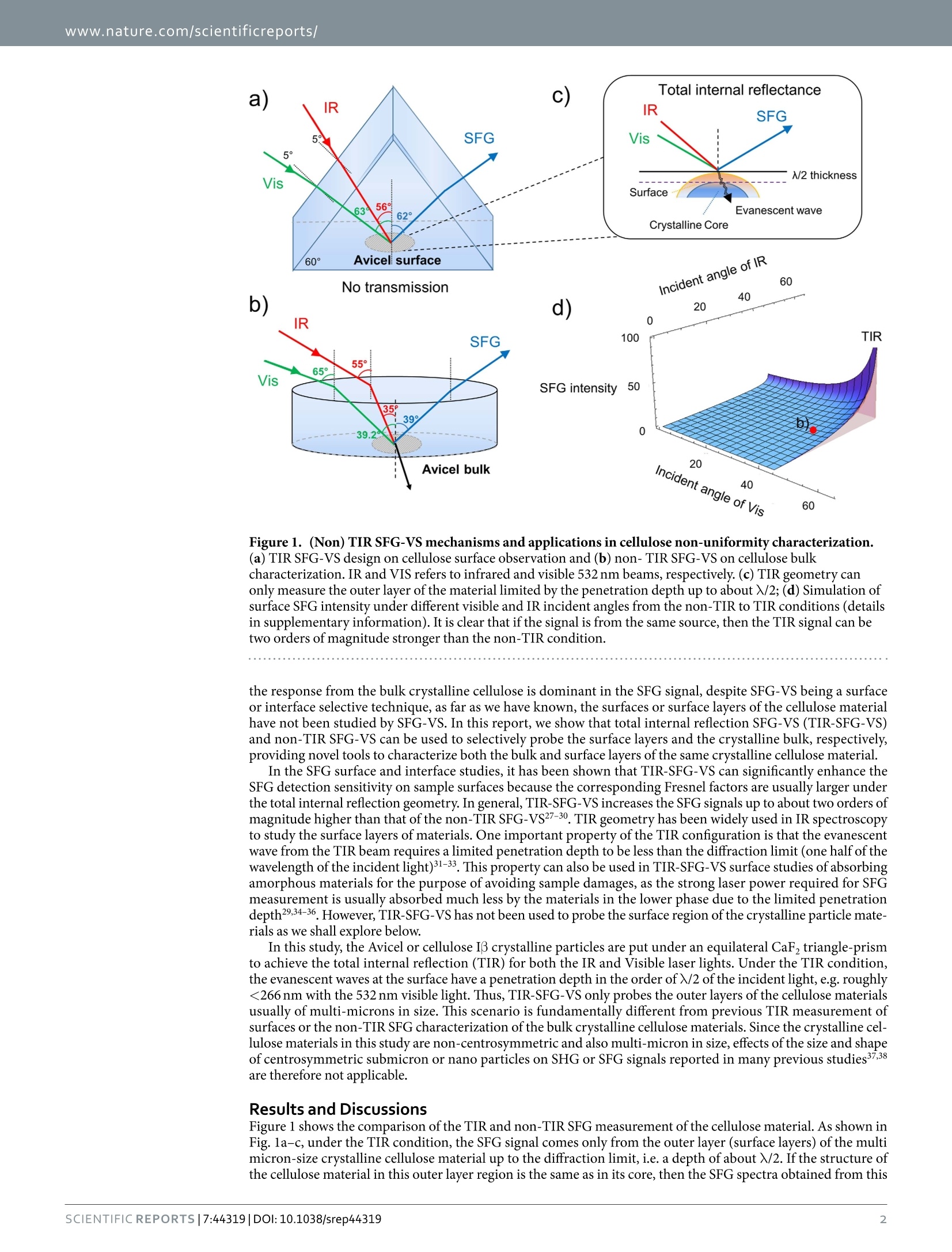
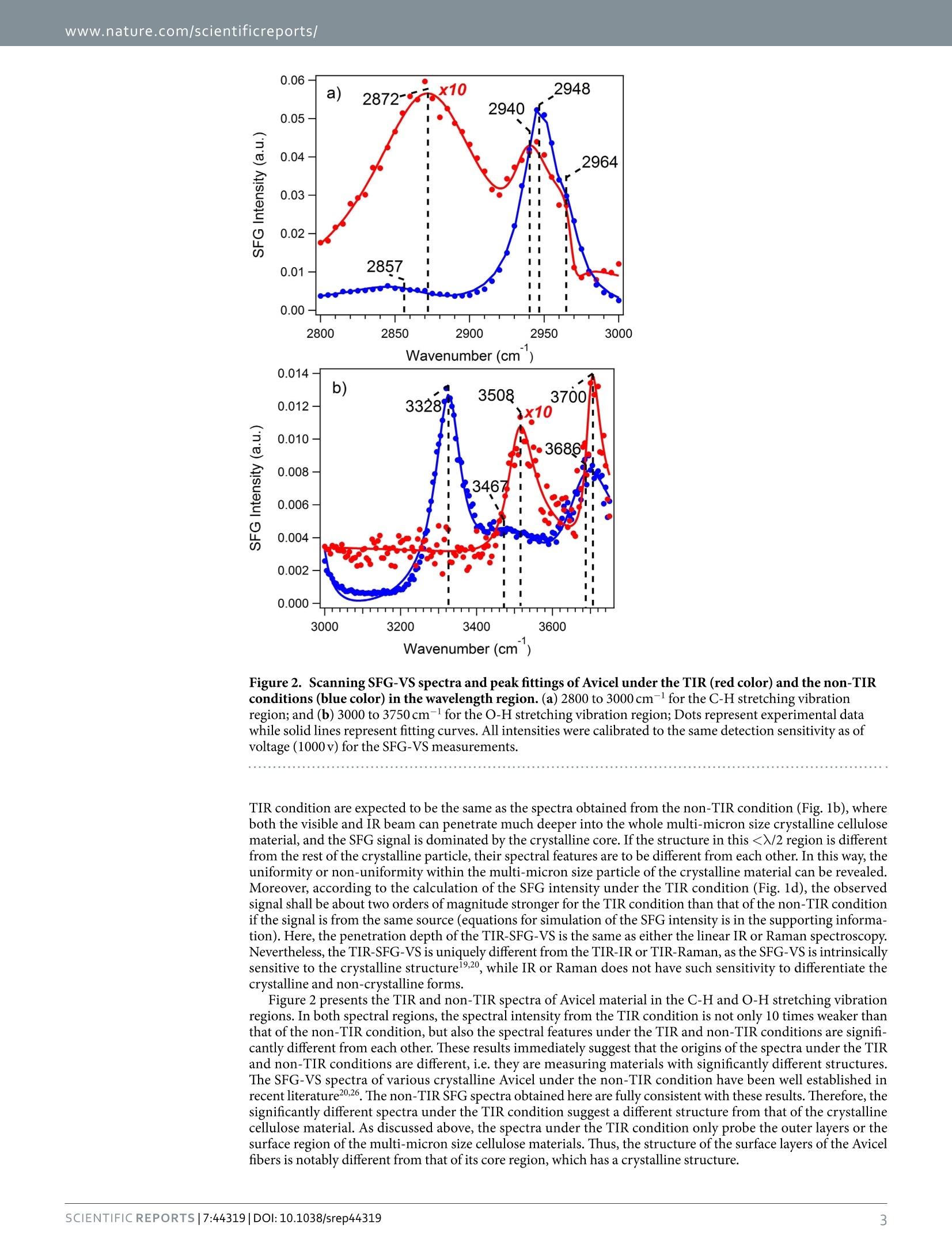
www.nature.com/scientificreports SCIENTIFIC REP RTS Discovery of Cellulose SurfaceLayerConformation by NonlinearVibrational Spectroscopy Received:14October2016 Libing Zhang1, Li Fu2, Hong-fei Wang3& Bin Yang1 Accepted:06 February 2017 Published:14 March 2017 Significant questions remain in respect to cellulose's structure and polymorphs, particularly thecellulose surface layers and the bulk crystalline core as well as the conformational differences.TotalInternal Reflection Sum Frequency Generation Vibrational Spectroscopy (TIR-SFG-VS) combined withconventional SFG-VS (non-TIR) enables selectively characterizing the molecular structures of surfacelayers and the crystalline core of cellulose, revealing their differences for the first time. From the SFGspectra in the C-H and O-H regions, we found that the surface layers of Avicel are essentially amorphouswhile the surface layers of l3 cellulose are crystalline but with different structural and spectroscopicsignatures compared with its crystalline core. The differences between hydrogen bonding networks ofcellulose surface and crystalline core were also shown by the SFG signal.The discovery here representsyet another instance of the importance of spectroscopic observations in transformative advances tounderstand the structure of the cellulosic biomass. Cellulose is the predominant cell wall polysaccharide, consisting of linear chains of 10,000-14,000 1,4-linkedB-D-glucopyranosyl residues. Native cellulose is composed predominantly of two different crystalline forms,referred to as Io and I31-4, with I3 being the dominant form in higher plants. Non-crystalline domains, whichalso exist in cellulose but generally in much lower proportions, are the most susceptible to chemical and biologg-ical depolymerization. It is clear that the root causes of the slowing of catalytic reactions to both chemical andbiological conversion processes include the surface, crystalline structure, and molecular order of cellulose haveprofound effects on its reactivity. However,so far, the surface versus the bulk of cellulose material has not beeneffectively or explicitly defined due to lack of the effective research tools. Previous analytical tools for cellulosecharacterization include X-ray diffraction3-6,13CNMR27,8, solid state NMR, (ultra-high resolution) atomic forcemicroscopy (AFM)10-12, electron microdiffraction studyl3,14, and a new imaging approach of label-free simu-lated Raman scattering (SRS) microscopy. Sum-frequency generation vibrational spectroscopy (SFG-VS) is asecond-order nonlinear spectroscopy, which has been developed over the last three decades as a unique toolin selective characterization of molecular surfaces or interfacial systems, including solid-liquid,liquid-liquid,solid-gas, and solid-solid interfaces16-18. In addition to the surface and interface studies, as SFG-VS is sensitiveto non-centrosymmetric structures, it has recently been applied to characterize the bulk structure of crystallinecellulose19-22. Since the SFG signal from the bulk crystalline material is usually a few orders stronger than the SFGresponse from the surface or interfaces of the same materials23,24, and SFG is essentially forbidden for the amor-phous materials, SFG-VS has been established as a unique spectroscopic tool that can selectively characterizecellulose crystalline molecular structures without the influence of amorphous structures or polymers from thebiomass19-22. This makes SFG-VS advantageous over other spectroscopic techniques in cellulose research such asInfrared, Raman or Coherent Anti-Stokes Raman spectroscopy (CARS), which collects a signal from all chemicalgroups of the material, crystalline or not15.25. Recent molecular dynamics calculations, coupled with SFG-VSstudies, proposed different -6CH,OH arrangements of cellulose Io and I322. Furthermore, the newly developedhigh resolution broad band (HR-BB) SFG-VS26 was applied to observe structural signatures in different cellulosesources and polymorphs, providing more detailed structural information than conventional SFG-VS. Because Bioproduct Sciences and Engineering Laboratory, Department of Biological Systems Engineering, WashingtonState University, Richland, WA 99354, USA.William R. Wiley Environmental Molecular Sciences Laboratory, PacificNorthwest National Laboratory, Richland, WA 99354, USA. Physical Sciences Division, Physical & ComputationalScience Directorate, Pacific Northwest National Laboratory, Richland, WA 99354, USA. *Present Address:Department of Chemistry, Fudan University, Shanghai,China 200433. Correspondence and requests for materialsshould be addressed to H.-F.W. (email:wanghongfei@fudan.edu.cn) or B.Y. (email: binyang@tricity.wsu.edu) No transmission Incident angle of IR 60 Figure 1. (Non) TIR SFG-VS mechanisms and applications in cellulose non-uniformity characterization.(a) TIR SFG-VS design on cellulose surface observation and (b) non- TIR SFG-VS on cellulose bulkcharacterization. IR and VIS refers to infrared and visible 532 nm beams, respectively. (c) TIR geometry canonly measure the outer layer of the material limited by the penetration depth up to about X/2; (d) Simulation ofsurface SFG intensity under different visible and IR incident angles from the non-TIR to TIR conditions (detailsin supplementary information). It is clear that if the signal is from the same source, then the TIR signal can betwo orders of magnitude stronger than the non-TIR condition. the response from the bulk crystalline cellulose is dominant in the SFG signal, despite SFG-VS being a surfaceor interface selective technique, as far as we have known, the surfaces or surface layers of the cellulose materialhave not been studied by SFG-VS. In this report, we show that total internal reflection SFG-VS (TIR-SFG-VS)and non-TIR SFG-VS can be used to selectively probe the surface layers and the crystalline bulk, respectively,providing novel tools to characterize both the bulk and surface layers of the same crystalline cellulose material. In the SFG surface and interface studies, it has been shown that TIR-SFG-VS can significantly enhance theSFG detection sensitivity on sample surfaces because the corresponding Fresnel factors are usually larger underthe total internal reflection geometry. In general, TIR-SFG-VS increases the SFG signals up to about two orders ofmagnitude higher than that ofthe non-TIR SFG-VS27-30. TIR geometry has been widely used in IR spectroscopyto study the surface layers of materials. One important property of the TIR configuration is that the evanescentwave from the TIR beam requires a limited penetration depth to be less than the diffraction limit (one half of thewavelength of the incident light)31-33. This property can also be used in TIR-SFG-VS surface studies of absorbingamorphous materials for the purpose of avoiding sample damages, as the strong laser power required for SFGmeasurement is usually absorbed much less by the materials in the lower phase due to the limited penetrationdepth29,34-36. However, TIR-SFG-VS has not been used to probe the surface region of the crystalline particle mate-rials as we shall explore below. In this study, the Avicel or cellulose I3 crystalline particles are put under an equilateral CaFztriangle-prismto achieve the total internal reflection (TIR) for both the IR and Visible laser lights. Under the TIR condition,the evanescent waves at the surface have a penetration depth in the order of /2 of the incident light, e.g. roughly<266 nm with the 532 nm visible light. Thus, TIR-SFG-VS only probes the outer layers of the cellulose materialsusually of multi-microns in size. This scenario is fundamentally different from previous TIR measurement ofsurfaces or the non-TIR SFG characterization of the bulk crystalline cellulose materials. Since the crystalline cel-lulose materials in this study are non-centrosymmetric and also multi-micron in size, effects of the size and shapeof centrosymmetric submicron or nano particles on SHG or SFG signals reported in many previous studies37.38are therefore not applicable. Results and Discussions Figure 1 shows the comparison of the TIR and non-TIR SFG measurement of the cellulose material. As shown inFig. la-c, under the TIR condition, the SFG signal comes only from the outer layer (surface layers) of the multimicron-size crystalline cellulose material up to the diffraction limit, i.e. a depth of about X/2. If the structure ofthe cellulose material in this outer layer region is the same as in its core, then the SFG spectra obtained from this Figure2. Scanning SFG-VS spectra and peak fittings of Avicel under the TIR (red color) and the non-TIRconditions (blue color) in the wavelength region. (a) 2800 to 3000 cm- for the C-H stretching vibrationregion;and (b) 3000 to 3750 cm-l for the O-H stretching vibration region; Dots represent experimental datawhile solid lines represent fitting curves. All intensities were calibrated to the same detection sensitivity as ofvoltage (1000v) for the SFG-VS measurements. TIR condition are expected to be the same as the spectra obtained from the non-TIR condition (Fig.1b), whereboth the visible and IR beam can penetrate much deeper into the whole multi-micron size crystalline cellulosematerial, and the SFG signal is dominated by the crystalline core. If the structure in this
确定
还剩5页未读,是否继续阅读?

产品配置单

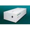
北京欧兰科技发展有限公司为您提供《纤维素中振动和频光谱检测方案(其它光谱仪)》,该方案主要用于其他中理化分析检测,参考标准--,《纤维素中振动和频光谱检测方案(其它光谱仪)》用到的仪器有Ekspla SFG 表面和频光谱分析系统、PL2250 闪光灯泵浦皮秒Nd:YAG激光器
推荐专场


该厂商其他方案
更多