


方案详情
文
利用激光剥蚀-电感耦合等离子体质谱(LA-ICP-MS)研究了水培环境中水稻根和茎对于金纳米粒子的吸收及其空间分布。水稻植株分别在含有直径为2 nm的带正、中、负电荷的的金纳米粒子(AuNP1(+), AuNP2(0), AuNP3(-))中水培。植株在含有1.6 mg Au/L的AuNPs暴露5天或者在0.14 mg Au/L的AuNPs暴露3个月用以研究纳米粒子的表面电荷如何影响活体植株组织吸收。结果表明末端的功能基团很大程度上影响了AuNP吸收进去植物组织。利用LA-ICP-MS测定了暴露5天下的水稻根,含量分布为:AuNP1(+)> AuNP2(0)>AuNP3(-),但在茎中的分布却是相反的,证明了AuNP3(-)的优先转移。生物成像揭示了叶肉和叶脉的AuNP分布情况依赖于器官或AuNP浓度。
方案详情

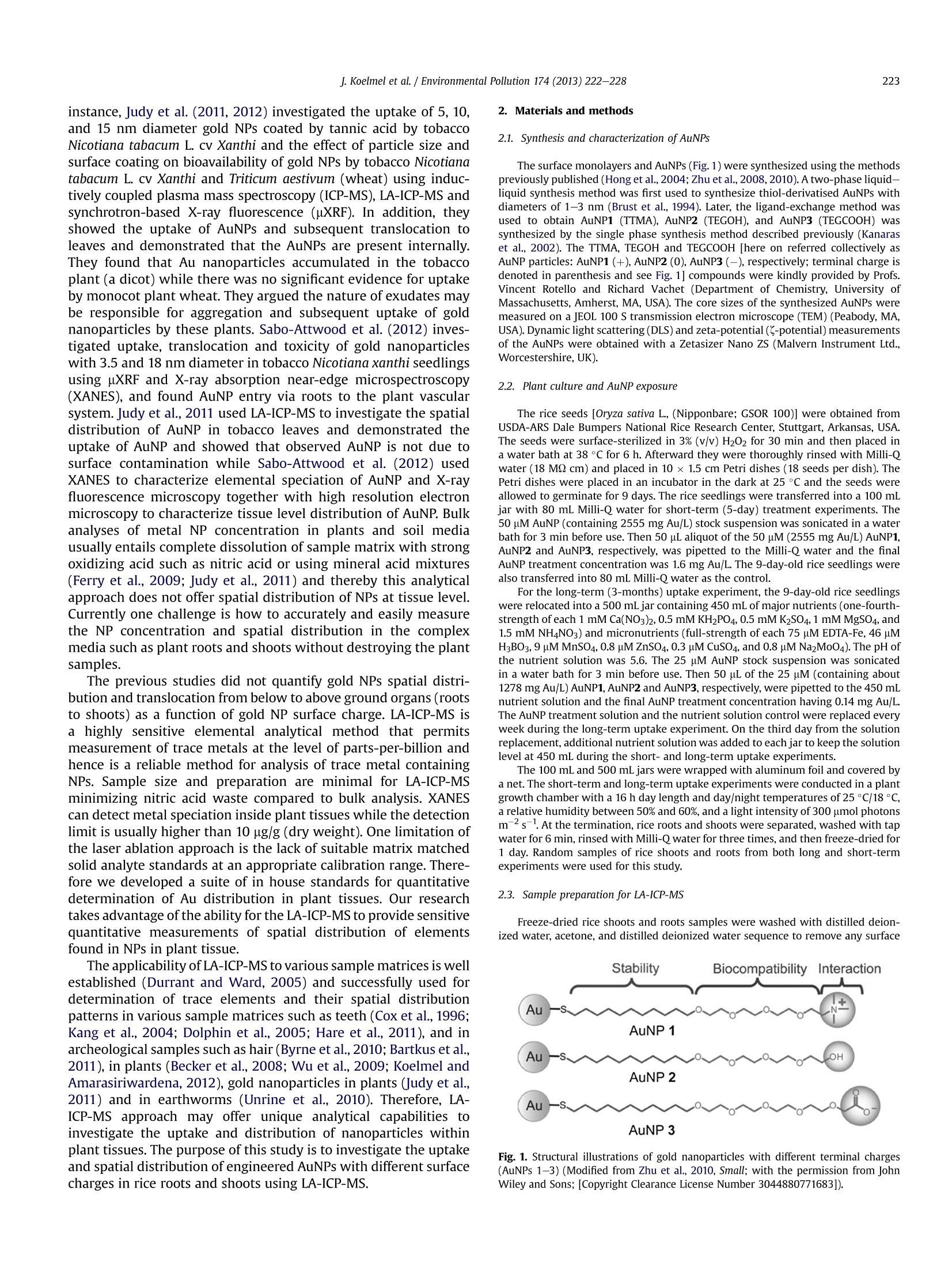
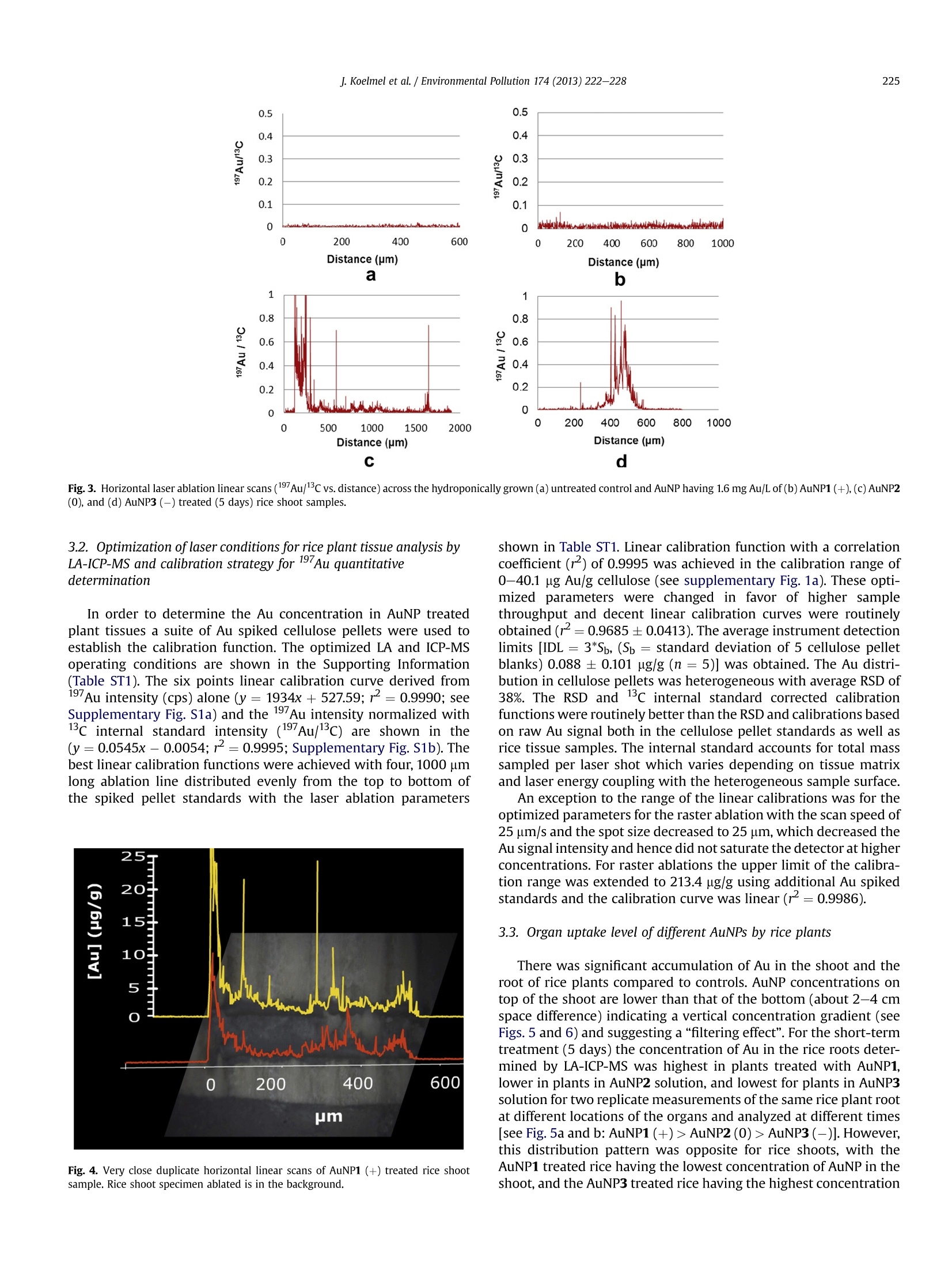
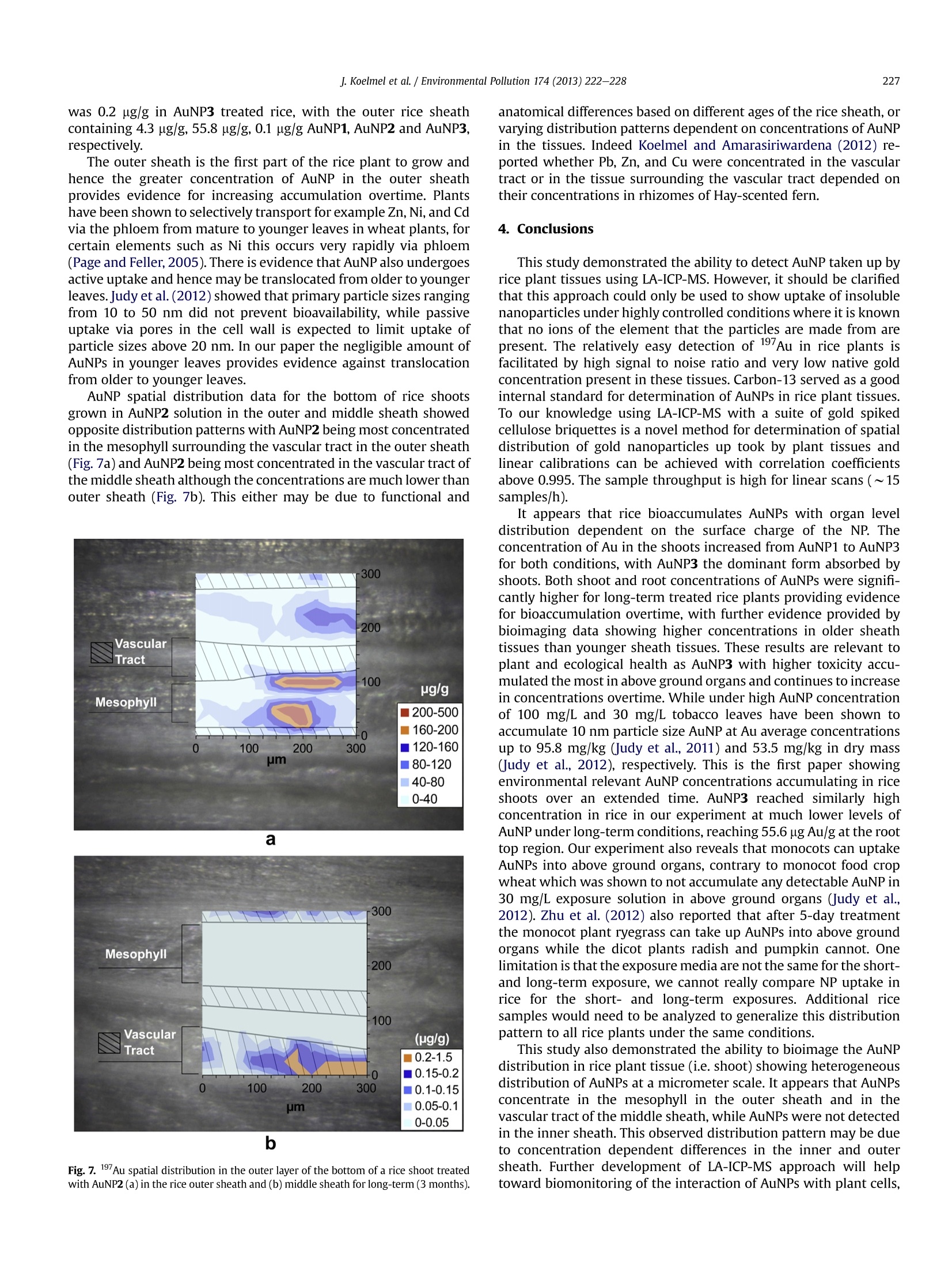
Environmental Pollution 174 (2013)222-228Contents lists available at SciVerse ScienceDirect 223J. Koelmel et al./Environmental Pollution 174(2013)222-228 0269-7491/$- see front matter @ 2012 Elsevier Ltd. All rights reserved.http://dx.doi.org/10.1016/j.envpol.2012.11.026 Environmental Pollution ELSEVIER journal homepage: www.elsevier.com/locate/envpol Investigation of gold nanoparticles uptake and their tissue level distribution inrice plants by laser ablation-inductively coupled-mass spectrometry Jeremy Koelmel, Thomas Leland, Huanhua Wang, Dulasiri Amarasiriwardena*, Baoshan Xingb School of Natural Science, Hampshire College, Amherst, MA 01002, USA ”Stockbridge School of Agriculture, University of Massachusetts, Amherst, MA 01003,USA ARTICLEINFO ABSTRACT Article history:Received 15 July 2012Received in revised form16 November 2012Accepted 20 November 2012 Keywords:Gold nanoparticleUptakeLA-ICP-MSRice (Oryza sativa L.)Bioimaging The tissue level uptake and spatial distribution of gold nanoparticles (AuNPs) in rice (Oryza sativa L.)roots and shoots under hydroponic conditions was investigated using laser ablation-inductivelycoupled plasma-mass spectrometry (LA-ICP-MS). Rice plants were hydroponically exposed to posi-tively, neutrally, and negatively charged AuNPs [AuNP1(+), AuNP2(0), AuNP3(-)] with a core diameterof 2 nm. Plants were exposed to AuNPs having 1.6 mg Au/L for 5 days or 0.14 mg Au/L for 3 months toelucidate how the surface charges of the nanoparticles affects their uptake into living plant tissues. Theresults demonstrate that terminal functional groups greatly affected the AuNP uptake into plant tissues.Au concentration determined by LA-ICP-MS in 5 day treated rice roots followed this order:AuNP1(+)> AuNP2(0)> AuNP3(-) but this order was reversed for rice shoots, indicating preferentialtranslocation of AuNP3(-). Bioimages revealed distributions of mesophyll and vascular AuNP depen-dent on organ or AuNP concentration. ◎ 2012 Elsevier Ltd. All rights reserved. 1. Introduction The use of nanostructures as a mode for bioimaging and sensingin biomedicine is one of the most intensely studied areas innanotechnology (Dreaden et al., 2012; Loo et al., 2005; Jain et al.,2008). However, the application of nanotechnology to plant andenvironmental systems has lagged behind. With the increasedprevalence of nano products it is vital to monitor the transport, fate,and toxicity of engineered nanoparticles once they are released intothe environment and how they behave in biological systems. Goldnanoparticles (NPs) are well suited for examining the bioavail-ability and toxicity of manufactured nanomaterials in environ-mentally complex systems, because of their resistance to oxidativedissolution and negligible release of dissolved Au ions as well as thelow natural background concentrations of gold that facilitateseasily discernible signals.They also have the ability to remain intactin soil and aqueous media, and under the physiological conditionsin plants and invertebrates (Unrine et al., 2012). In addition, goldNPs can be functionalized with different surface charge, e.g. posi-tive, neutral, and negative charges. In recent years, a tremendousincrease in the applications of gold NPs in biomedical imaging anddetection (Copland et al., 2004; Sreenivasan, 2010; Jiang et al., ( * Corresponding author. ) ( E-mail address: dans@hamp s hire.edu (D . Amarasiriwardena). ) 2006), cancer diagnostics (El-Sayed et al., 2005) and therapy, andbiological and sensing of heavy metals (Kim et al., 2001) have beenobserved. However, there are only a few studies about the detectionand fate of gold NPs in plant systems. Research is progressing on the fate and transport of goldnanoparticles in the environment, their accumulation in the foodchain has been documented via amphibian consumption of wormson AuNP treated soil (Unrine et al., 2012), and from producers toprimary consumers (e.g. Judy et al., 2011). Biomagnification hasbeen observed for example by up to a factor of 11.6 with 10 nmdiameter AuNP in hornworms feeding off tobacco leaves (Judy et al.,2011) raising additional means for concern. Evidence shows thatAuNP can be cytotoxic depending on nanoparticle characteristicsand can be up taken into various human organs, with positivelycharged terminal amine groups more cytotoxic than negativelycharged carboxyl groups (Lipka et al.,2010), showing potentialcause for concern for human health if AuNPs particles enter foodcrops and livestock. Therefore understanding plant uptake andaccumulation of gold nanoparticles is important for developingcrop species that can prevent accumulation in above ground organsas well as accumulators for the removal of AuNP from the soil andwater. Unrine et al., 2010 first used laser ablation-inductively coupledplasma-mass spectrometry (LA-ICP-MS) to image gold NP distri-bution in earthworms (Eisenia fetida). Recently, the application ofgold NPs in plant systems has attracted considerable interest. For instance, Judy et al.(2011, 2012) investigated the uptake of 5, 10,and 15 nm diameter gold NPs coated by tannic acid by tobaccoNicotiana tabacum L. cv Xanthi and the effect of particle size andsurface coating on bioavailability of gold NPs by tobacco Nicotianatabacum L. cv Xanthi and Triticum aestivum (wheat) using induc-tively coupled plasma mass spectroscopy (ICP-MS), LA-ICP-MS andsynchrotron-based X-ray fluorescence (uXRF). In addition, theyshowed the uptake of AuNPs and subsequent translocation toleaves and demonstrated that the AuNPs are present internally.They found that Au nanoparticles accumulated in the tobaccoplant (a dicot) while there was no significant evidence for uptakeby monocot plant wheat. They argued the nature of exudates maybe responsible for aggregation and subsequent uptake of goldnanoparticles by these plants. Sabo-Attwood et al. (2012) inves-tigated uptake, translocation and toxicity of gold nanoparticleswith 3.5 and 18 nm diameter in tobacco Nicotiana xanthi seedlingsusing uXRF and X-ray absorption near-edge microspectroscopy(XANES), and found AuNP entry via roots to the plant vascularsystem. Judy et al., 2011 used LA-ICP-MS to investigate the spatialdistribution of AuNP in tobacco leaves and demonstrated theuptake of AuNP and showed that observed AuNP is not due tosurface contamination while Sabo-Attwood et al. (2012) usedXANES to characterize elemental speciation of AuNP and X-rayfluorescence microscopy together with high resolution electronmicroscopy to characterize tissue level distribution of AuNP. Bulkanalyses of metal NP concentration in plants and soil mediausually entails complete dissolution of sample matrix with strongoxidizing acid such as nitric acid or using mineral acid mixtures(Ferry et al., 2009; Judy et al., 2011) and thereby this analyticalapproach does not offer spatial distribution of NPs at tissue level.Currently one challenge is how to accurately and easily measurethe NP concentration and spatial distribution in the complexmedia such as plant roots and shoots without destroying the plantsamples. The previous studies did not quantify gold NPs spatial distri-bution and translocation from below to above ground organs (rootsto shoots) as a function of gold NP surface charge. LA-ICP-MS isa highly sensitive elemental analytical method that permitsmeasurement of trace metals at the level of parts-per-billion andhence is a reliable method for analysis of trace metal containingNPs. Sample size and preparation are minimal for LA-ICP-MSminimizing nitric acid waste compared to bulk analysis. XANEScan detect metal speciation inside plant tissues while the detectionlimit is usually higher than 10 ug/g (dry weight). One limitation ofthe laser ablation approach is the lack of suitable matrix matchedsolid analyte standards at an appropriate calibration range.There-fore we developed a suite of in house standards for quantitativedetermination of Au distribution in plant tissues. Our researchtakes advantage of the ability for the LA-ICP-MS to provide sensitivequantitative measurements of spatial distribution of elementsfound in NPs in plant tissue. The applicability of LA-ICP-MS to various sample matrices is wellestablished (Durrant and Ward, 2005) and successfully used fordetermination of trace elements and their spatial distributionpatterns in various sample matrices such as teeth (Cox et al., 1996;Kang et al., 2004; Dolphin et al., 2005; Hare et al., 2011), and inarcheological samples such as hair (Byrne et al., 2010; Bartkus et al.,2011), in plants (Becker et al., 2008; Wu et al., 2009; Koelmel andAmarasiriwardena, 2012), gold nanoparticles in plants (Judy et al.,2011) and in earthworms (Unrine et al., 2010). Therefore, LA-ICP-MS approach may offer unique analytical capabilities toinvestigate the uptake and distribution of nanoparticles withinplant tissues. The purpose of this study is to investigate the uptakeand spatial distribution of engineered AuNPs with different surfacecharges in rice roots and shoots using LA-ICP-MS. 2.1. Synthesis and characterization of AuNPs The surface monolayers and AuNPs (Fig. 1) were synthesized using the methodspreviously published (Hong et al., 2004; Zhu et al., 2008, 2010). A two-phase liquid-liquid synthesis method was first used to synthesize thiol-derivatised AuNPs withdiameters of 1-3 nm (Brust et al.,1994). Later, the ligand-exchange method wasused to obtain AuNP1 (TTMA), AuNP2 (TEGOH), and AuNP3 (TEGCOOH) wassynthesized by the single phase synthesis method described previously (Kanaraset al., 2002). The TTMA, TEGOH and TEGCOOH [here on referred collectively asAuNP particles: AuNP1 (+), AuNP2 (0), AuNP3 (-), respectively; terminal charge isdenoted in parenthesis and see Fig. 1] compounds were kindly provided by Profs.Vincent Rotello and Richard Vachet (Department of Chemistry, University ofMassachusetts, Amherst, MA, USA). The core sizes of the synthesized AuNPs weremeasured on a JEOL 100 S transmission electron microscope (TEM) (Peabody, MA,USA). Dynamic light scattering (DLS) and zeta-potential (<-potential) measurementsof the AuNPs were obtained with a Zetasizer Nano ZS (Malvern Instrument Ltd.,Worcestershire,UK). 2.2. Plant culture and AuNP exposure The rice seeds [Oryza sativa L., (Nipponbare; GSOR 100)] were obtained fromUSDA-ARS Dale Bumpers National Rice Research Center, Stuttgart, Arkansas, USA.The seeds were surface-sterilized in 3% (v/v) H202 for 30 min and then placed ina water bath at 38 °C for 6 h. Afterward they were thoroughly rinsed with Milli-Qwater (18 MQ cm) and placed in 10 x 1.5 cm Petri dishes (18 seeds per dish). ThePetri dishes were placed in an incubator in the dark at 25 C and the seeds wereallowed to germinate for 9 days. The rice seedlings were transferred into a 100 mLjar with 80 mL Milli-Q water for short-term (5-day) treatment experiments. The50 uM AuNP (containing 2555 mg Au/L) stock suspension was sonicated in a waterbath for 3 min before use. Then 50 pL aliquot of the 50 uM (2555 mg Au/L) AuNP1,AuNP2 and AuNP3, respectively, was pipetted to the Milli-Q water and the finalAuNP treatment concentration was 1.6 mg Au/L. The 9-day-old rice seedlings werealso transferred into 80 mL Milli-Q water as the control. For the long-term (3-months) uptake experiment, the 9-day-old rice seedlingswere relocated into a 500 mL jar containing 450 mL of major nutrients (one-fourth-strength of each 1 mM Ca(NO3)2,0.5 mM KH2PO4,0.5 mM K2SO4,1 mM MgSO4, and1.5 mM NHNO3) and micronutrients (full-strength of each 75 pM EDTA-Fe, 46 uMH3BO3,9 pM MnSO4,0.8 pM ZnSO4,0.3 pM CuSO4,and 0.8 uM Na2Mo04). The pH ofthe nutrient solution was 5.6. The 25 uM AuNP stock suspension was sonicatedin a water bath for 3 min before use. Then 50 pL of the 25 uM (containing about1278 mg Au/L)AuNP1, AuNP2 and AuNP3, respectively, were pipetted to the 450 mLnutrient solution and the final AuNP treatment concentration having 0.14 mg Au/L.The AuNP treatment solution and the nutrient solution control were replaced everyweek during the long-term uptake experiment. On the third day from the solutionreplacement, additional nutrient solution was added to each jar to keep the solutionlevel at 450 mL during the short- and long-term uptake experiments. The 100 mL and 500 mL jars were wrapped with aluminum foil and covered bya net. The short-term and long-term uptake experiments were conducted in a plantgrowth chamber with a 16 h day length and day/night temperatures of 25 C/18°C,a relative humidity between 50% and 60%, and a light intensity of 300 umol photonsm-2s-. At the termination, rice roots and shoots were separated, washed with tapwater for 6 min, rinsed with Milli-Q water for three times, and then freeze-dried for1 day. Random samples of rice shoots and roots from both long and short-termexperiments were used for this study. 2.3. Sample preparation for LA-ICP-MS Freeze-dried rice shoots and roots samples were washed with distilled deion-ized water, acetone, and distilled deionized water sequence to remove any surface Fig.1. Structural illustrations of gold nanoparticles with different terminal charges(AuNPs 1-3)(Modified from Zhu et al., 2010, Small; with the permission from JohnWiley and Sons; [Copyright Clearance License Number 3044880771683]). contaminants (IAEA, 1978). Cleaned samples were dried and mounted on a 3Mmounting tape (3M, Minneapolis, MN, USA) and each sample was fixed on a petro-graphic slide (Buehler, Lake Bluff, IL,USA).(See Fig. 2a and b). 2.4. Standard gold pellet preparation Suitable solid standards for gold determination in plant tissue are not readilyavailable. In order to assess Au concentration distribution in the rice tissue by LA-ICP-MS the instrument was calibrated using a series of prepared gold spiked solidcellulose standards. Gold standard solutions in the range of 0.1-50 pg/L wereprepared by diluting 1000 pg/L Au standard solution (Certiprep, Spex Metuchen,NJ, USA). A 1 mL aliquot of liquid gold standard solution was added to ~250 mgof cellulose XRF binder (Spex Cellulose Prep Aid, particle size <20 um, No 3642,Metuchen, NJ) and mixed thoroughly in scintillation vials. Fifteen drops of liquidbinder (Chemplex, Palm City, FL, USA) were added and dried at 60 °C for 3 h toevaporate, methylene chloride binder solvent and later each Au/cellulose mixturewas further ground in a Wig-L-Bug mixer mill (Spex, Metuchen, NJ). Dried gold-cellulose-binder mixture powder standards were stored in a desiccator. Later150 mg gold-cellulose binder mixtures were pressed with a pneumatic pressapplying 12-ton pressure for 30 s. A suite of 1.2 cm diameter pressed pellets withknown quantities of gold including a blank pellet was prepared and the final mass ofeach pellet was measured. The Au concentration ranged from 0.5 to 213.4 pg Au/g ofcellulose. 2.5. LA-ICP-MS Nd:YAG deep UV (213 nm) laser ablation system (NWR 213; New WaveResearch, Fremont CA, USA) coupled to an ICP-MS spectrometer PE-Elan 6000a(Perkin Elmer Instruments, Shelton, CT, USA) was used for Au determination in riceshoots and roots specimens. Mounted specimens were placed in the laser ablationchamber, focused,and the chamber air was evacuated and the chamber purged withcarrier gas. A mixture of helium and argon was used as a carrier gas with a mass flowrate of 0.25 L/min and 0.75 L/min, respectively. ICP-MS operating parameters firstwere optimized under liquid nebulizer mode and later with LA. A suite of Au spiked cellulose standard pellets discussed previously were firstloaded into the ablation chamber before the analysis of rice tissue samples. Four orthree 1 mm long replicate lines were ablated to measure the 197Au and 1’c signals foreach standard. Laser conditions for both standards and sample scans were kept thesame during the analysis session. The LA-ICP-MS operating parameters were listedunder the supplementary information Table ST1. The rice shoot or root samples treated with AuNP1, AuNP2, and AuNP3 alongwith untreated control samples were focused using x,y, and z stage settings at eachlaser ablation session, and later plant tissue regions were identified and photo-graphed. Linear scans were made in continuous firing mode across the bottom andtop part of roots and shoots for each experiment (long-term and short-term) andeach treatment (AuNP1(+),AuNP2(0), and AuNP3(-)). Only one plant sample wasanalyzed for each experiment and treatment. For the short-term experiment abla-tion lines were repeated below the original ablation scans to verify quantification at Shoot Root a b Fig. 2. Linear ablation lines across (approximately 2-4 cm apart; lines denote abla-tions locations) the shoot (left) and root (right) at their top and bottom positions (a),and a young shoot showing curled inner, middle, and outer sheath (top); uncurledtissue was laser ablated on a raster area of 300 by 300 um dimension. two separate times (Fig. 2a). For raster ablations the bottom of the shoot was cut atabout 0.5 inch intervals and the inner, middle, and outer leaf sheath were separatedwith cleaned tweezers working under a low magnification microscope (Fig. 2b).Each separated layer was uncurled and placed on 3M mounting tape for laserablation. Raster areas with 300 by 300 um dimension were tested in select areas ofthese tissues using single pass laser ablation mode. The relevant laser ablationoperating conditions such as laser energy, repetition rate, spot size, and scan speedare shown in the supplementary information in Table ST1. 3. Results and discussion 3.1. Evidence for AuNP uptake The AuNP1, AuNP2 and AuNP3 used in this study have similarcore sizes (~2 nm) but different surface monolayers. The intro-duction of surface monolayers onto the AuNP surfaces stabilizesthem against aggregation. Depending on the structures of surfacemonolayer, at day 0 hydrodynamic sizes of these AuNPs rangedfrom 6 to 10 nm in deionized water and hydroponic nutrientsolution. But at day 1 and 3 the hydrodynamic sizes of the nega-tively charged AuNPs in hydroponic nutrient solution were largerthan 10 nm (Zhu et al., 2012). AuNPs used in this study were stablefor up to 5 days in hydroponic nutrient solutions. The terminalfunctional groups on each AuNP control the surface charge as evi-denced by their <-potentials that remain essentially constant over5 days: AuNP1, +24± 5 mV; AuNP2, -2±1mV; AuNP3,-17±6 mV. Overall, the polarities of <-potentials are consistentwith the chemical charges on the terminal functional groups.In a separate study by Zhu et al.(2012) we also measured the dis-solved Au from AuNPs in the nutrient solution, and demonstratedthat more than 98% of the added AuNPs were found as intactparticles. And no significant amount of dissolved gold was found inthe nutrient solutions with less than 5 pg/L of dissolved elementalgold found in the nutrient solution in each case. Horizontal linear laser ablation scans across the untreatedcontrol and 1.6 mg Au/L AuNP1, AuNP2,and AuNP3 treated riceshoot samples in the short-term uptake experiment are shown inFig.2. Laser energy of 35%, spot size of 35 um, repetition rate of5 Hz, and a scan speed of 5 pm/s were used for these initial laserablation experiments of rice plant tissues. Fig. 3a shows the back-ground 197Au/c signal across the control rice shoot blade. Asexpected it demonstrates a very low Au background signal akin tocontrol samples. However,AuNP2 and AuNP3 treated rice (Fig. 3cand d, respectively) showed highly elevated 197Au/13c signalsagainst the control (see Fig. 3a) while AuNP1 treated rice samplesslightly elevated in this shoot sample (Fig. 3b). These LA-ICP-MSresults show clear visual evidence for uptake of Au by rice plantsand they are heterogeneously distributed in the tissue. Forexample, AuNP2 (see Fig. 3c) treated rice shoot shows elevated Auaccumulation particularly at the left edge (100-300 um region)while AuNP3 (Fig.3d) treated sample indicates accretion of Au atthe mid zone of the blade (300-600 um). Similar linear laser scanswere acquired (not shown here) for both shoots and roots of severalindividual rice plant samples indicating clear uptake of AuNPsdepending on their terminal charge. Fig. 4 shows an example of very close duplicate horizontal linearscan of AuNP2 treated rice shoot indicating comparable Au hori-zontal distribution pattern. Duplicate ablation scans in close prox-imity indicated that linear ablations are representative of a largerportion of the rice tissue than that just ablated, but it is importantto note that quantification of [Au] is only for very specific portionsof the rice plant and do not represent bulk analysis. The ability toeasily detect mono isotopic 197Au in AuNP treated rice tissues isfacilitated in part by very low native concentration of Au in riceplants. Consequently a high signal to background ratio of 197Ausignal was attained with LA-ICP-MS. a 3.2. Optimization of laser conditions for rice plant tissue analysis byLA-ICP-MS and calibration strategy for 197Au quantitativedetermination In order to determine the Au concentration in AuNP treatedplant tissues a suite of Au spiked cellulose pellets were used toestablish the calibration function. The optimized LA and ICP-MSoperating conditions are shown in the Supporting Information(Table ST1). The six points linear calibration curve derived from197Au intensity (cps) alone (y=1934x+ 527.59; r2=0.9990; seeSupplementary Fig. S1a) and the 197Au intensity normalized with1’c internal standard intensity (197Au/lc) are shown in the(y=0.0545x- 0.0054; r²=0.9995; Supplementary Fig. S1b). Thebest linear calibration functions were achieved with four, 1000 umlong ablation line distributed evenly from the top to bottom ofthe spiked pellet standards with the laser ablation parameters Fig. 4. Very close duplicate horizontal linear scans of AuNP1 (+) treated rice shootsample. Rice shoot specimen ablated is in the background. d Fig. 3. Horizontal laser ablation linear scans (197Au/13Cvs. distance) across the hydroponically grown (a) untreated control and AuNP having 1.6 mg Au/L of (b)AuNP1(+), (c)AuNP2(0), and (d) AuNP3 (-) treated (5 days) rice shoot samples. shown in Table ST1. Linear calibration function with a correlationcoefficient (r-) of 0.9995 was achieved in the calibration range of0-40.1 pg Au/g cellulose (see supplementary Fig. 1a). These opti-mized parameters were changed in favor of higher samplethroughput and decent linear calibration curves were routinelyobtained (r²=0.9685±0.0413). The average instrument detectionlimits [IDL 3*Sb, (Sb standard deviation of 5 cellulose pelletblanks) 0.088±0.101 ug/g(n = 5)] was obtained. The Au distri-bution in cellulose pellets was heterogeneous with average RSD of38%. The RSD and 13c internal standard corrected calibrationfunctions were routinely better than the RSD and calibrations basedon raw Au signal both in the cellulose pellet standards as well asrice tissue samples. The internal standard accounts for total masssampled per laser shot which varies depending on tissue matrixand laser energy coupling with the heterogeneous sample surface. An exception to the range of the linear calibrations was for the optimized parameters for the raster ablation with the scan speed of 25 pm/s and the spot size decreased to 25 um, which decreased the Au signal intensity and hence did not saturate the detector at higher concentrations. For raster ablations the upper limit of the calibra- tion range was extended to 213.4 ug/gusing additional Au spiked standards and the calibration curve was linear (r²=0.9986). 3.3. Organ uptake level of different AuNPs by rice plants There was significant accumulation of Au in the shoot and theroot of rice plants compared to controls. AuNP concentrations ontop of the shoot are lower than that of the bottom (about 2-4 cmspace difference) indicating a vertical concentration gradient (seeFigs. 5 and 6) and suggesting a "filtering effect". For the short-termtreatment (5 days) the concentration of Au in the rice roots deter-mined by LA-ICP-MS was highest in plants treated with AuNP1,lower in plants in AuNP2 solution, and lowest for plants in AuNP3solution for two replicate measurements of the same rice plant rootat different locations of the organs and analyzed at different times[see Fig. 5a and b:AuNP1(+)>AuNP2(0)> AuNP3 (-)]. However,this distribution pattern was opposite for rice shoots, with theAuNP1 treated rice having the lowest concentration of AuNP in theshoot, and the AuNP3 treated rice having the highestconcentration [Au] Versus Terminal Charge andOrgan Fig.5. Organ level distribution of AuNP1(+), AuNP2 (0), and AuNP3(-) and control:[197Au] distribution across bottom and top of rice roots and rice shoots with short-termtreatment for two replicate measurements (Fig. 5a and b) at different locations of thesame rice specimen. [AuNP3 (-)> AuNP2 (0)> AuNP1 (+)] (Fig. 5a and b). Fig. 5a andb show the results of analysis using different locations on thebottom and top of the same plant organs (root and shoot, respec-tively). It showed the same uptake pattern of 3 types of AuNP havingdifferent terminal charged groups verifying the AuNP uptake orderdiscussed above although their Au concentrations varied signifi-cantly in both replicates. This could be attributed to heterogeneousspatial distribution of AuNPs at micrometer distance tissue levels inindividual plant organ (i.e.,shoot or root) as discussed earlier. [Au] Versus Terminal Charge and Fig.6. Organ level distribution of AuNP1 (+),AuNP2(0), and AuNP3(-) and control atdifferent locations of the same rice plant:[1Au] distribution across bottom and top ofroots and shoots with long-term treatment. Based on this evidence it appears that AuNP1 is bound up by theroots and hence is not easily transported to the shoots, while lessAuNP3 is bound to the roots. In contrast more AuNP3 concentratedin the shoots with neutral AuNP2 accumulates somewhat moder-ately in both organs. One hypothesis for the observed distributionpatterns is that the positively charged AuNP1 active site binds tothe carboxyl group of pectins (-COOH group at the C6 position in d-galacturonic acid monomer) found surrounding the cell wall (Mohrand Schopfer, 1995), and other membrane and cell wall derivedbiopolymers with ligand sites such as glycoproteins and lignin. Itappears that the negatively charged AuNP3 would not bind to thesesites in the root cells and xylem and would hence be more easilytransferred to aerial organs where AuNP3 bioconcentrates byunknown mechanism(s). In the long-term (3 months) treatment of AuNP with riceseedlings the concentrations of Au in the different organs wassignificantly higher than short-term (5 days) experiment (compareFig. 6 vs. 5). Concentrations for the long-term treatment (Fig. 6) inthe bottom of rice roots were 44.3, 21.1, and 654.4 ug/g for AuNP1,AuNP2, and AuNP3, respectively, and in the root top were 204.8,402.4, and 55.6 pg/g, respectively. Average concentrations for tworeplicates of the short-term treatment (Fig. 5a and b) in the bottomof rice roots were 8.4±3.1,3.5±1.0, and 0.1±0.1ug g for AuNP1,AuNP2, and AuNP3, respectively, and in the root top were 3.9±1.2,1.7±0.1, and 0.1 ±0.1 pg/g, respectively. Concentrations for thelong-term treatment in the bottom of rice shoots were 3.6,12.6, and72.9 ug/g for AuNP1, AuNP2, and AuNP3, respectively, and in theshoot top were all below detection limit (DL) for this experiment(<0.053 pg/g). Concentrations for the short-term treatment in thebottom of rice shoots were 0.1±0.2,1.5±2.1, and 3.1±1.7 pg/gfor AuNP1, AuNP2, and AuNP3, respectively, and in the top were0.02±0.0.01 (below the detection limit), 0.8±0.5, and 1.7±1.3 pg/g,respectively. It appears that the amount of accumulation of AuNPsand their organ level distribution depend on the AuNP concentra-tion in the solution, the growth time and on the charge of theAuNPs. One difference was that no AuNPs were detected in the shoottops for the long-term treatment. This may be due to the lowerconcentration of AuNP in the solution for the long-term treatment(AuNP with 0.14 mg Au/L) if the mechanism for AuNP translocatefrom the vascular transport stream in the shoot cannot completelyachieve removal of AuNP once AuNP concentration exceedsa perhaps presently unknown threshold in vascular tissues or both.This shows that bioaccumulation of AuNP continues overtime andthe rate of accumulation may depend on the age of rice plant. Thedistribution of Au in the roots depending on the charge of theAuNPs differed for the two treatments. While in the short-termtreatment AuNP concentration in rice roots decreased fromAuNP1 to AuNP3, in the long-term treatment (see Fig. 6) AuNPconcentration order reversed in rice roots (i.e., increased fromAuNP1 to AuNP3; see Fig. 6 versus Fig.5a and b). Hence the accu-mulation and organ level distribution of AuNPs may be dependenton the rate of uptake, AuNP surface charge, exposure time, andoverall AuNP dose concentration and translocation mechanismfrom rice roots to shoots. 3.4. Bioimaging of AuNP in rice shoots (culm) The raster ablations were used to bioimage AuNP distribution inthe inner, middle, and outer sheath for the long-term treatment(Fig. 2b). The experiment using long-term treated rice plantsshowed that concentrations of Au were all below the detectionlimit (DL) for raster ablation (0.065 ug/g for this elemental bio-imaging experiment) in the inner sheath for AuNP1,AuNP2 andAuNP3 treated rice. The only detectable AuNP in the middle sheath was 0.2 pg/g in AuNP3 treated rice, with the outer rice sheathcontaining 4.3 pg/g,55.8 ug/g, 0.1 pg/g AuNP1, AuNP2 and AuNP3,respectively. The outer sheath is the first part of the rice plant to grow andhence the greater concentration of AuNP in the outer sheathprovides evidence for increasing accumulation overtime. Plantshave been shown to selectively transport for example Zn,Ni,and Cdvia the phloem from mature to younger leaves in wheat plants, forcertain elements such as Ni this occurs very rapidly via phloem(Page and Feller,2005). There is evidence that AuNP also undergoesactive uptake and hence may be translocated from older to youngerleaves. Judy et al. (2012) showed that primary particle sizes rangingfrom 10 to 50 nm did not prevent bioavailability, while passiveuptake via pores in the cell wall is expected to limit uptake ofparticle sizes above 20 nm. In our paper the negligible amount ofAuNPs in younger leaves provides evidence against translocationfrom older to younger leaves. AuNP spatial distribution data for the bottom of rice shootsgrown in AuNP2 solution in the outer and middle sheath showedopposite distribution patterns with AuNP2 being most concentratedin the mesophyll surrounding the vascular tract in the outer sheath(Fig.7a) and AuNP2 being most concentrated in the vascular tract ofthe middle sheath although the concentrations are much lower thanouter sheath (Fig. 7b). This either may be due to functional and a b Fig. 7. 197Au spatial distribution in the outer layer of the bottom of a rice shoot treatedwith AuNP2 (a) in the rice outer sheath and (b) middle sheath for long-term (3 months). anatomical differences based on different ages of the rice sheath, orvarying distribution patterns dependent on concentrations of AuNPin the tissues. Indeed Koelmel and Amarasiriwardena (2012) re-ported whether Pb, Zn, and Cu were concentrated in the vasculartract or in the tissue surrounding the vascular tract depended ontheir concentrations in rhizomes of Hay-scented fern. 4. Conclusions This study demonstrated the ability to detect AuNP taken up byrice plant tissues using LA-ICP-MS. However, it should be clarifiedthat this approach could only be used to show uptake of insolublenanoparticles under highly controlled conditions where it is knownthat no ions of the element that the particles are made from arepresent. The relatively easy detection of 197Au in rice plants isfacilitated by high signal to noise ratio and very low native goldconcentration present in these tissues. Carbon-13 served as a goodinternal standard for determination of AuNPs in rice plant tissues.To our knowledge using LA-ICP-MS with a suite of gold spikedcellulose briquettes is a novel method for determination of spatialdistribution of gold nanoparticles up took by plant tissues andlinear calibrations can be achieved with correlation coefficientsabove 0.995. The sample throughput is high for linear scans (~15samples/h). It appears that rice bioaccumulates AuNPs with organ leveldistribution dependent on the surface charge of the NP. Theconcentration of Au in the shoots increased from AuNP1 to AuNP3for both conditions, with AuNP3 the dominant form absorbed byshoots. Both shoot and root concentrations of AuNPs were signifi-cantly higher for long-term treated rice plants providing evidencefor bioaccumulation overtime, with further evidence provided bybioimaging data showing higher concentrations in older sheathtissues than younger sheath tissues. These results are relevant toplant and ecological health as AuNP3 with higher toxicity accu-mulated the most in above ground organs and continues to increasein concentrations overtime. While under high AuNP concentrationof 100 mg/L and 30 mg/L tobacco leaves have been shown toaccumulate 10 nm particle size AuNP at Au average concentrationsup to 95.8 mg/kg (Judy et al., 2011) and 53.5 mg/kg in dry mass(Judy et al., 2012), respectively. This is the first paper showingenvironmental relevant AuNP concentrations accumulating in riceshoots over an extended time. AuNP3 reached similarly highconcentration in rice in our experiment at much lower levels ofAuNP under long-term conditions, reaching 55.6 pg Au/g at the roottop region. Our experiment also reveals that monocots can uptakeAuNPs into above ground organs, contrary to monocot food cropwheat which was shown to not accumulate any detectable AuNP in30 mg/L exposure solution in above ground organs (Judy et al.,2012). Zhu et al. (2012) also reported that after 5-day treatmentthe monocot plant ryegrass can take up AuNPs into above groundorgans while the dicot plants radish and pumpkin cannot. Onelimitation is that the exposure media are not the same for the short-and long-term exposure, we cannot really compare NP uptake inrice for the short- and long-term exposures. Additional ricesamples would need to be analyzed to generalize this distributionpattern to all rice plants under the same conditions. This study also demonstrated the ability to bioimage the AuNPdistribution in rice plant tissue (i.e. shoot) showing heterogeneousdistribution of AuNPs at a micrometer scale. It appears that AuNPsconcentrate in the mesophyll in the outer sheath and in thevascular tract of the middle sheath, while AuNPs were not detectedin the inner sheath. This observed distribution pattern may be dueto concentration dependent differences in the inner and outersheath. Further development of LA-ICP-MS approach will helptoward biomonitoring of the interaction of AuNPs with plant cells, and thus allow designing strategies, such as for nanosensors andnano carriers in agriculture. This study reveals the uptake of AuNPby rice under hydroponic conditions, a staple food source in manyparts of the world, and that LA-ICP-MS can be used as versatileanalytical approach to elucidate the fate and transport of engi-neered nanoparticles in biological systems where no soluble ionicspecies from the test nanoparticle is present under controlledexperimental conditions. Acknowledgments The authors wish to thank USDA-ARS Dale Bumpers NationalRice Research Center, Stuttgart, Arkansas for providing rice seedsOryza sativa (Nipponbare). Special thanks also extended toProfessors Vincent Rotello and Richard Vachet of Department ofChemistry, University of Massachusetts at Amherst for providing usAuNPs for this work. We would also like to acknowledge theNational Science Foundation (NSF) MRI-R2 (Grant: BRI 0959028)for funding the LA-ICP-MS system, USDA-AFRI (2011-67006-30181)and USDA-AFRI Hatch program (MAS 00978) for financial support.JK and TL would like to thank the Society for Analytical Chemists inPittsburgh for funding their summer undergraduate researchinternships and making this research possible. Appendix A. Supplementary data References Bartkus, L., Amarasiriwardena, D., Arriaza, B., Bellis, D., Yanez, J., 2011. Exploringlead exposure in ancient Chilean mummies usinga single strand of hair by laserablation-inductively coupled plasma-mass spectrometry (LA-ICP-MS).Micro-chem. J. 98,267-274. Becker, J.S., Dietrich, R.C., Matush,A., Pozebon, D., Dressler, V.L., 2008. Quantitativeimages of metals in plant tissues measured by laser ablation inductivelycoupled plasma mass spectrometry. Spectrochim. Acta B 63, 1248-1252. Brust, M., Walker, M., Bethell, D., Schiffrin, D.J., Whyman,R.,1994. Synthesis of thiol-derivatised gold nanoparticles in a two-phase liquid-liquid system. J. Chem.Soc. Chem. Commun. 7, 801-802. Byrne, S., Amarasiriwardena, D., Bandak, B., Bartkus, L., Kane, J., Jones, J., Yaniez, J.,Arriaza, B., Cornejo, L., 2010. Were Chinchorros exposed to arsenic? Arsenicdetermination in Chinchorro mummies’ hair by laser ablation inductivelycoupled plasma-mass spectrometry (LA-ICP-MS). Microchem.J. 94,28-35. Copland, J.A., Eghtedari, M., Popov, V.L., Kotov, N., Mamedova, N., Motamedi, M.,Oraevsky, A.A., 2004. Bioconjugated gold nanoparticles as a molecular basedcontrast agent: implications for imaging of deep tumors using optoacoustictomography. Mol. Imaging Biol. 6 (5),341-349. Cox, A., Keenan, F., Cooke, M., Appleton, J., 1996. Trace element profiling of dentaltissues using laser ablation-inductively coupled plasma-mass spectrometry.Fresenius. J. Anal. Chem. 354, 254-258. Dolphin, A.E., Goodman, A.H., Amarasiriwardena, D.D., 2005. Variation in elementalintensities among teeth and between pre-and postnatal regions of enamel. Am.J. Phys. Anthropol. 128 (4), 878-888. Dreaden, E.C., Alkilany, A.M., Huang,X., 2012. The golden age: gold nanoparticles forbiomedicine. Chem. Soc. Rev. 41.2740-2779. Durrant, S.F., Ward,N.I., 2005. Recent biological and environmental applications oflaser ablation inductively coupled plasma mass spectrometry (LA-ICP-MS).J. Anal. At. Spectrom. 20,821-829. El-Sayed, I.H., Huang, X.H., El-Sayed, M.A., 2005. Surface plasmon resonance scat-tering and absorption of anti-EGFR antibody conjugated gold nanoparticles incancer diagnostics: Applications in oral cancer. Nano Lett. 5(5), 829-834. Ferry, J.L., Craig, P., Hexel, C., Sisco, P., Frey, R., Pennington, P.L., Fulton, M.H.,Geoff, S.I., Decho, A.W.,Kashiwada, S., Murphy, C.J., Shaw, T.J.,2009. Transfer ofgold nanoparticles from the water column to the estuarine food web. Nat.Nanotechnol.4,441-444. Hare, D., Austin, C., Doble, P., Arora, M., 2011. Elemental bio-imaging of traceelements in teeth using laser ablation-inductively coupled plasma-mass spec-trometry.J. Dentistry 39, 397-403. Hong, R., Emrick, T., Rotello, V.M., 2004. Monolayer-controlled substrate selectivityusing noncovalent enzyme-nanoparticle conjugates. J. Am. Chem. Soc. 126,13572-13573. International Atomic Energy Agency (IAEA), 1978.Activation Analysis of Hair as anIndicator of Contamination on Man by Environmental Trace Element Pollutants.IAEA/RL/50, Vienna. Jain, P.K., Huang, X., El-Sayed, I.H., El-Sayed, M.A., 2008. Noble metals on thenanoscale: optical and photothermal properties and some applications inimaging, sensing, biology, and medicine. Acc. Chem.Res. 41,1578-1586. Jiang, Z., Sun, S.,Liang, A., Huang, W.,Qin, A., 2006. Gold-labeled nanoparticle-basedimmune resonance scattering spectral assay for trace apolipoprotein AI andapolipoprotein B. Clin. Chem. 52(7),1389-1394. Judy, J.D., Unrine,J.M., Bertsch, P.M., 2011. Evidence for biomagnification of goldnanoparticles within a terrestrial food chain. Environ. Sci. Technol. 45, 776-781. Judy, J.D., Unrine,J.M., Rao, W., Wirick, S., Bertsch, P.M., 2012. Bioavailability of goldnanomaterials to plants: importance of particle size and surface coating.Environ. Sci. Technol. 46,8467-8474. Kanaras, A.G., Kamounah, F.S., Schaumburg, K., Kiely, C.J, Brust, M., 2002.Thioalkylated tetraethylene glycol: a new ligand for water soluble monolayerprotected gold clusters. Chem. Commun. 20, 2294-2295. Kang, D.,Amarasiriwardena, D., Goodman,A.H., 2004. Application of laser ablation-inductively coupled plasma-mass spectrometry (LA-ICP-MS) to investigatetrace metal spatial distributions in human tooth enamel and dentine growthlayers and pulp. Anal. Bioanal. Chem. 378, 1608-1615. Kim, Y.,Johnson, R.C., Hupp, J.T., 2001. Gold nanoparticle-based sensing of “spec-troscopically silent" heavy metal ions. Nano Lett. 1 (4), 165-167. Koelmel, J.,Amarasiriwardena, D.,2012. Imaging of metal bioaccumulation in Hay-scented fern (Dennstaedtia punctilobula) rhizomes growing on contaminatedsoils by laser ablation ICP-MS. Environ. Pollut. 168, 62-70. Lipka, J., Semmler-Behnke, M., Sperling, R.A., Wenk, A., Takenaka, S., Schleh, C.,Kissel, T., Parak, W.J., Kreyling, W.G., 2010. Biodistribution of PEG-modified goldnanoparticles following intratracheal instillation and intravenous injection.Biomaterials 31, 6574-6581. Loo, C., Lowery, A., Halas, N., West, J.,Drezek,R., 2005. Immunotargeted nanoshellsfor integrated cancer imaging and therapy. Nano Lett. 5 (4), 709-711. Mohr, H., Schopfer, P., 1995. Plant Physiology (G., Lawl27or, D.W., Lawlor, Trans.).Springer-Verlag, Berlin, pp. 21-37. Page, V., Feller, U., 2005. Selective transport of zinc, manganese, nickel, cobalt andcadmium in the root system and transfer to the leaves in young wheat plants.Ann. Bot. 96, 425-434. Sabo-Attwood, T., Unrine, J.M., Stone, J.W., Murphy, C.J., Ghoshroy, S., Blom, D.,Bertsch, P.M., Newman, L.A., 2012. Uptake, distribution and toxicity of goldnanoparticles in tobacco (Nicotiana xanthi) seedlings. Nanotoxicology 6 (4),353-360. Sreenivasan, V.R.K., 2010. Selective detection and estimation of C-reactive protein inserum using surface-functionalized gold nano-particles gold nano-particles.Anal. Chim. Acta 662,186-192. Unrine, J.M., Hunyadi, S.E., Tsyusko, O.V., Rao, W., Aaron Shoults-Wilson, W.,Bertsch, P.M., 2010. Evidence for bioavailability of Au nanoparticles from soiland biodistribution within earthworms (Eisenia fetida). Environ. Sci. Technol.44, 8308-8318. Unrine, J.M., Shoults-Wilson, W.A., Zhurbich,O., Bertsch, P.M., Tsyusko, O.V., 2012.Trophic transfer of Au nanoparticles from soil along a simulated terrestrial foodchain. Environ.Sci. Technol. 46,9753-9760. Wu, B., Zoriy,M., Chen, Y., Becker, J.S., 2009. Imaging of nutrient elements in theleaves of Elsholtzia splendens by laser ablation inductively coupled plasma massspectrometry. Talanta 78, 132-137. Zhu, ZJ., Ghosh, P.S., Miranda, O.R., Vachet, R.W., Rotello, V.M., 2008. Multiplexedscreening of cellular uptake of gold nanoparticles using laser desorption/ioni-zation mass spectrometry. J. Am. Chem. Soc. 130 (43),14139-14143. ( Zhu, Z J., Carboni, R., Quercio J r . , M.J, Y a n, B . , Miranda, O. R ., Anderton, D.L . , Arcaro, K .F., Rotello,V.M., Vachet, R .W., 2 010. Surface p r operties dictate uptake,distribution, excretion, and t oxicity of nanoparticles i n fish. Small 6, 2261 -2 265. ) ( Zhu, ZJ., Wang, H., Ya n , B., Zh e ng, H., Jiang, Y., Mi r anda, O.R. , Rote l lo, V.M., Xing, X.,Vachet, R.W., 2012. Effect ofSurface Charge on the Uptake and Distribution of Gold Nanoparticles i n four Plants Species. Environ. Sc i . Technol. 46(22),12391 - 12398. ) 利用激光剥蚀-电感耦合等离子体质谱(LA-ICP-MS)研究了水培环境中水稻根和茎对于金纳米粒子的吸收及其空间分布。水稻植株分别在含有直径为2 nm的带正、中、负电荷的的金纳米粒子(AuNP1(+), AuNP2(0), AuNP3(-))中水培。植株在含有1.6 mg Au/L的AuNPs暴露5天或者在0.14 mg Au/L的AuNPs暴露3个月用以研究纳米粒子的表面电荷如何影响活体植株组织吸收。结果表明末端的功能基团很大程度上影响了AuNP吸收进去植物组织。利用LA-ICP-MS测定了暴露5天下的水稻根,含量分布为:AuNP1(+)> AuNP2(0)>AuNP3(-),但在茎中的分布却是相反的,证明了AuNP3(-)的优先转移。生物成像揭示了叶肉和叶脉的AuNP分布情况依赖于器官或AuNP浓度。
确定


还剩5页未读,是否继续阅读?

产品配置单
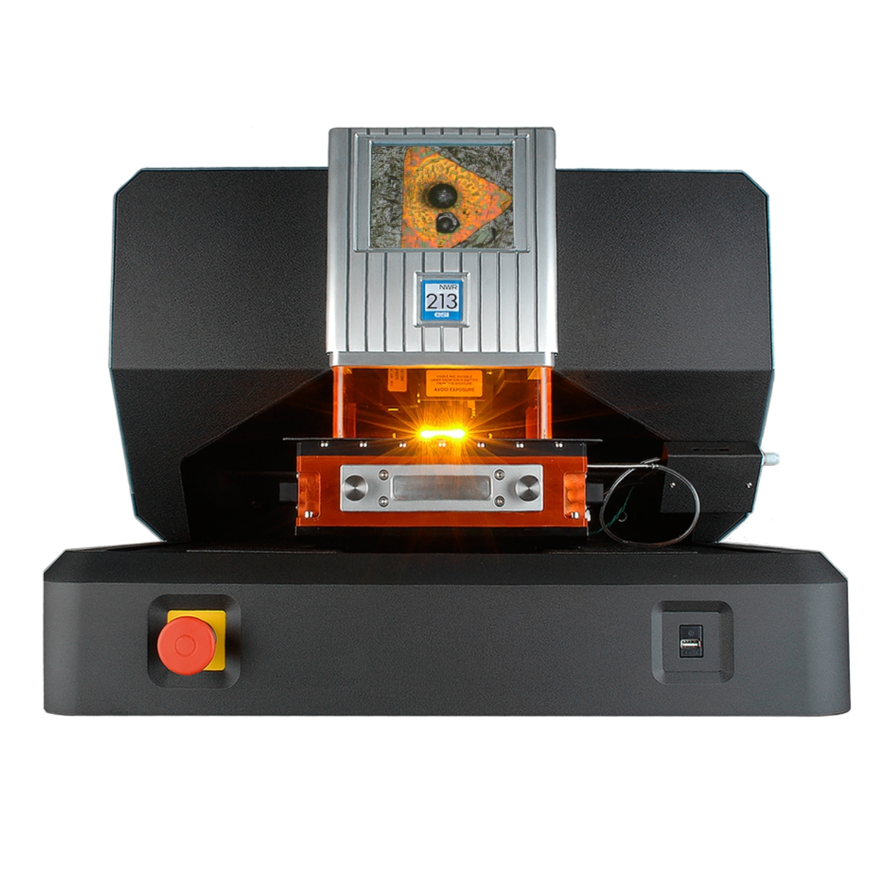
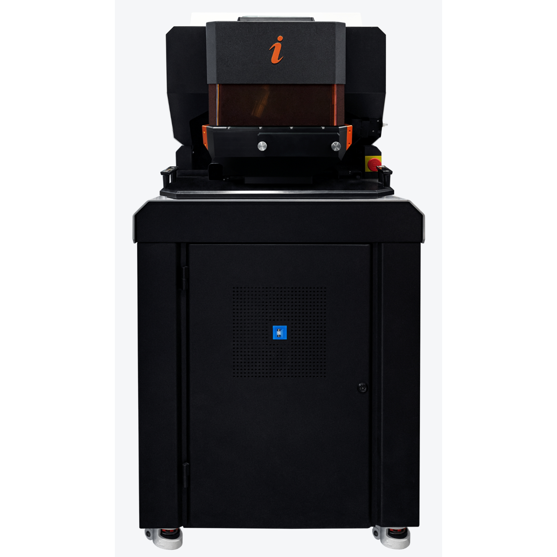
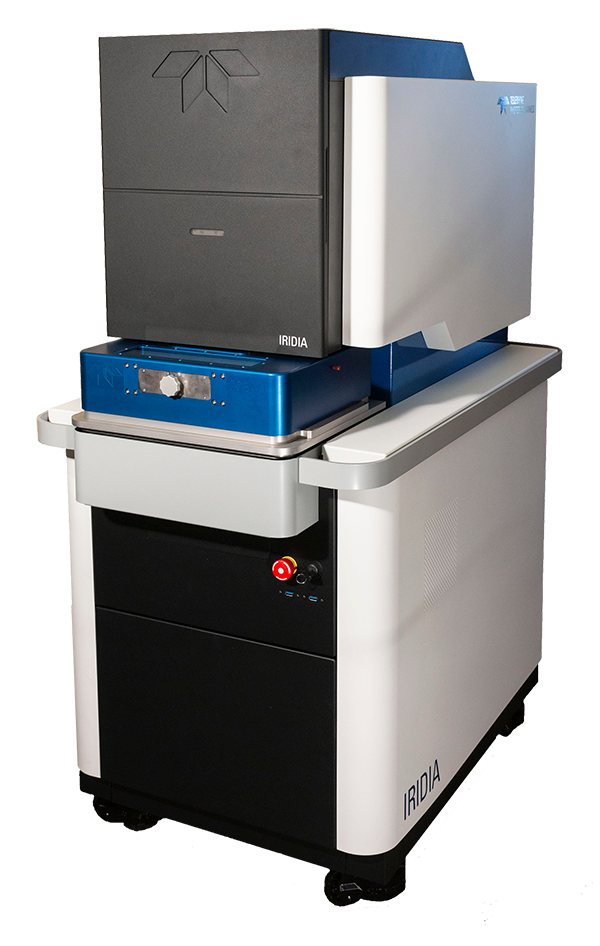
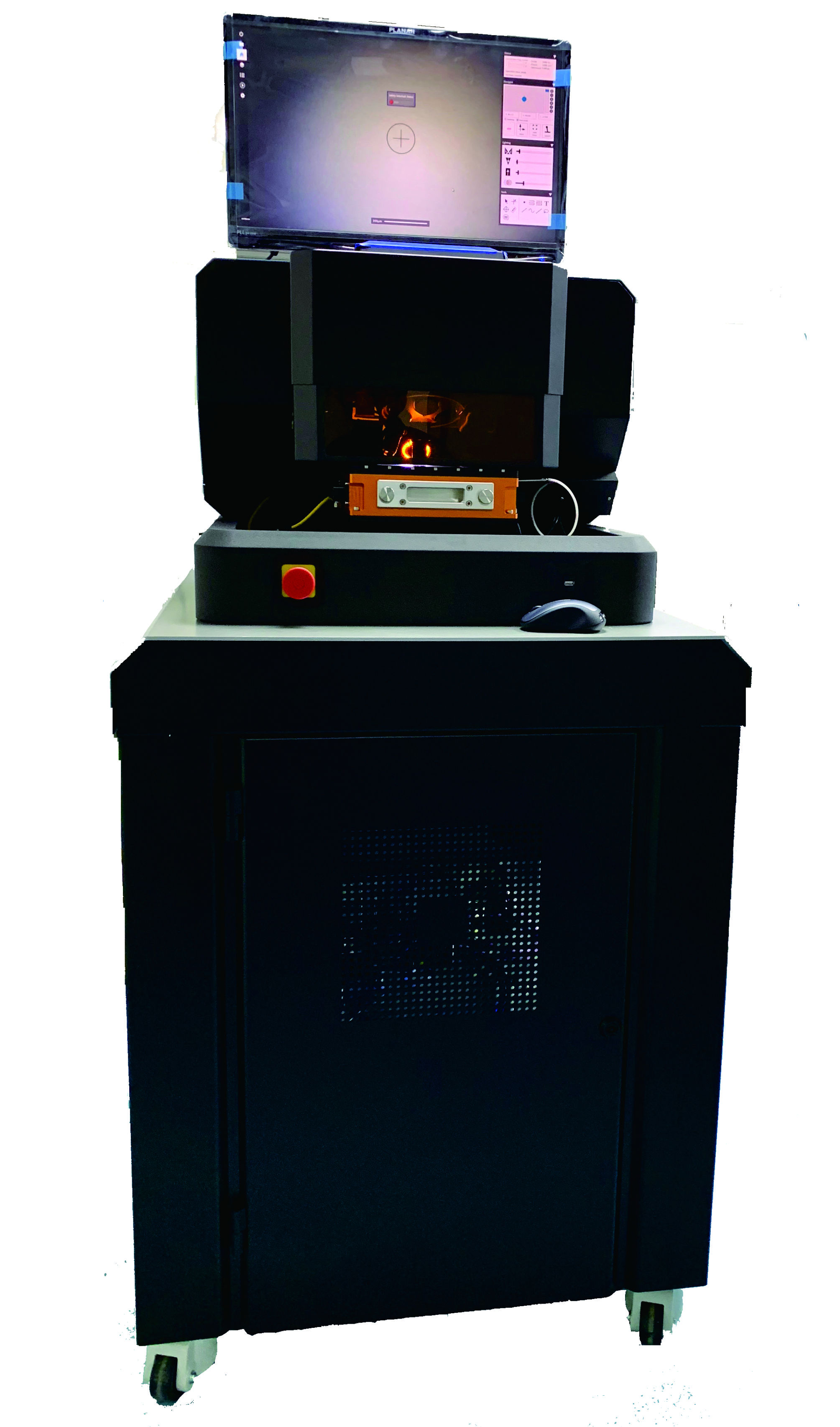
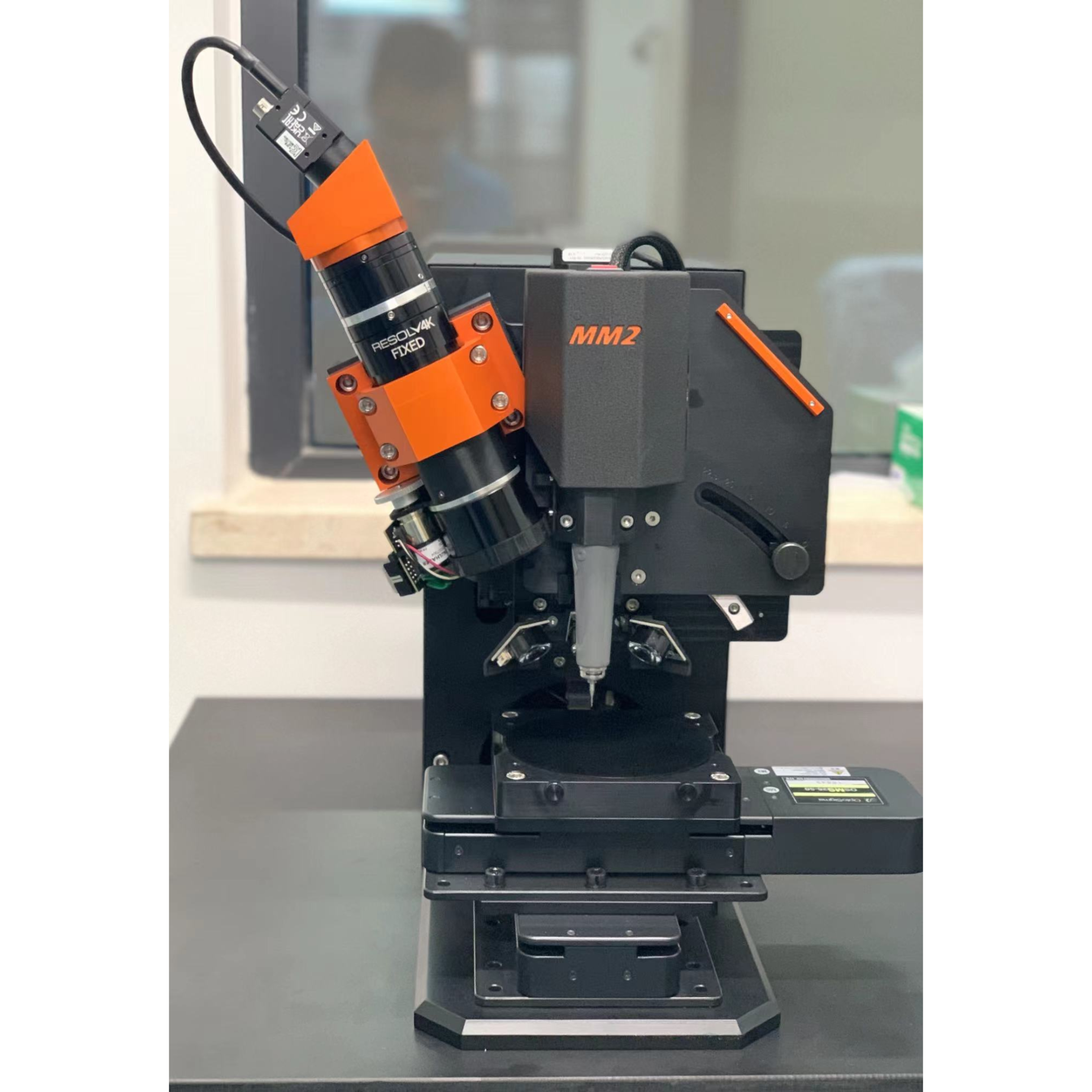
上海凯来仪器有限公司为您提供《水稻植株中金纳米粒子的吸收及其组织水平上的分布情况检测方案(激光剥蚀进样)》,该方案主要用于其他中金纳米粒子的吸收及其组织水平上的分布情况检测,参考标准--,《水稻植株中金纳米粒子的吸收及其组织水平上的分布情况检测方案(激光剥蚀进样)》用到的仪器有ESL213 灵活的激光剥蚀系统
推荐专场
相关方案
更多