


方案详情
文
Fluorescence spectroscopy has demonstrated a way to characterize and quantify skin aging and photoaging. The Fluorolog® allows direct in-vivo monitoring of endogenous skin fluorescence, which makes it possible to follow markers (tryptophan and collagen) in skin, and their changes in both healthy and diseased tissue. It also provides the possibility of evaluating the effect on skin from the modifications of these native proteins or the induction of The Fluorolog® will find a wide range of applications in skin research and photo-dynamic therapy.
方案详情

Endogenous Skin Fluorescence In Vivo on Human Skin Introduction Thefluorescencespectraotintrinsic protein fluorophores have beenstudiedextensively and used iininvestigating biological events. Nativefluorophores in human tissue includeNADH, collagen, elastin, tryptophan,tyrosine, porphyrins and FAD. Recentstudies haveproven that skinfluorescence originating from tryptophanand collagen can serve as a quantitativemarker for normal. diseased.chronically-aged, and sun-damagedskin. Characterizing fluorescencespectra of these native fluorophoresalso allows the study of changes inthese markersirin bothhealthy anddiseased skin. The HORIBA Jobin YvonFluorolog spectrofluorometer canmeasure endogenous fluorescence invivo on human skin. When configuredwith double-grating monochromators atbothexcitationand emission,theinstrumentvides extreme pr highrejection of stray light. A fiber-opticbundle accessory transmits light to theskini and returnsthefluorescenceemission to the detector. The Fluorologis capable of in-vivo monitoring ofendogenous skin fluorescence from ahighly scattering background such ashuman skin, with high sensitivity andprecision. Experimental method Fluorescence spectra weremeasured in vivo on human skin. Asshown in Fig. 1, light from a Xe lampwas passed through a double-gratingmonochromator.where the selectedwavelengths were focused into one arm of a randomized, bifurcated fiber-opticbundle. This light traveled to the com-mon end of the bundle irradiating skin incontact with the bundle. The emittedlight was then collected by the emissionbundle, passed through another double-grating monochromator, and finally di-rected to the emission photomultiplier-tube detector. The probe was held by anadapter to ensure good contact andeven pressure between the bundle andthe skin. Fig. 1. Diagram of skin layers and light path dur-ing a fluorescence excitation or emission scan. Three excitation spectr3awerecollected from human skin. The first ex-citation spectrum1 wasscanned from260-320 nm with the emission mono-chromator parked at 340 nm, the sec-ond spectrum from 260-380 nm with theemission parked at 400 nm, and thethird spectrum from 260-420 nm withthe emission set to 440 nm. The band- pass of the instrument was set to 5 nmfor both excitation and emission, and theintegration time was 0.2 s. Results and discussion Fig. 2 shows three excitation fluo-rescent spectra from normal healthyskin. These spectra are differentiated bythree peak maxima: 295 nm, 340 nm,and 360 nm. The excitation spectrumwith a peak maximum at 295 nm origi-nated from tryptophan fluorescence inthe epidermis. The excitation spectrumat 340 nm corresponds to the pepsin-digestible collagen cross-links’fluores-cence, and the 360-nm band is the col-lagenase-digestible collagen cross-links'tluorescence. Fig.2. Fluorescence-excitation spectra of en-dogenous human skin in vivo. Fluorescence spectra of trypto-phan and collagen can serve as quanti-tative markers for photoaging and natu-ral aging. Exposure to UV light causessignificant qualitative and quantitativechanges iinn cutaneousfluorescencespectra. In photoaged skin, the trypto-phan signal (Aexc = 295 nm) increases ( K o llias, N .; G illies, R.; Moran, M.; Kochevar, I .E.;Anderson, R .R. J. I nvestig. Dermatol., 1998, 111(5), 776-780. ) with photoaging and the two distinct col-lagen bands became one broad band,centered at 355 nm. In addition, twonew bands, i.e., excitation at 270 and350nm., were observed in thephotoaged skin. In contrast, the trypto-phan band decreases with natural agingwhile the 335-340-nm band correspond-ing to the pepsin-digestible collagencross-links increases. The 360-nm bandof the collagenase-digestible collagencross-links fluorescence rremains ttrhesame with natural aging. Conclusions Fluorescence spectroscopy hasdemonstrated a way to characterize andquantify skin aging and photoaging. TheFluorologe allows direct in-vivomonitoring of endogenous skinfluorescence, which makes it possible tofollow markers (tryptophan andcollagen) in skin, and their changes inboth healthy and diseased tissue. It alsoprovides the possibility of evaluating theeffect on skin from the modifications ofthese native proteins or the induction ofexogenous chromophores. TheFluorolog@ will find a wide range ofapplications in skin research and photo-dynamic therapy. ( USA: H ORIBA Jobin Yvon In c ., 3 8 80 Park Avenue, Edison, NJ 08 8 20-3012, Toll-Free:+1 - 866-jobinyvon Tel: +1-732-494-8660, F ax: + 1 - 732-549-5125, E - mail: info@jobinyvon.com,www.jobinyvon.com ) ( France: H ORIBA Jobin Yvon S . A . S., 1 6-18, rue du Canal , 9116 5 Longjumeau Cedex, T el: + 33 (0) 1 64 54 13 00,F a x: + 33 (0) 1 69 09 93 1 9 , w ww.jobinyvon.f r ) ( Japan: H ORIBA Ltd., J Y Optical Sales Dept, Hig a s h i-Kanda, Daij i Building, 1-7-8 Higashi-Kanda ) ( Chiyoda-ku, T okyo 1 0 1-0031, T e l: +81 (0)3 3861 8231, w ww.jyhoriba.jp Germany: +49(0)89 462317-0 I taly:+390 2 57603050 U K: +44 (0)20 82048142 ) Copyright C HORIBA Jobin Yvon; version .HORIBAExplore the future HORIBAJOBIN YVONChina: +() ORIBAExplore the future Fluorescence spectroscopy has demonstrated a way to characterize and quantify skin aging and photoaging. The Fluorolog® allows direct in-vivo monitoring of endogenous skin fluorescence, which makes it possible to follow markers (tryptophan and collagen) in skin, and their changes in both healthy and diseased tissue. It also provides the possibility of evaluating the effect on skin from the modifications of these native proteins or the induction of The Fluorolog® will find a wide range of applications in skin research and photo-dynamic therapy.
确定


还剩1页未读,是否继续阅读?

产品配置单
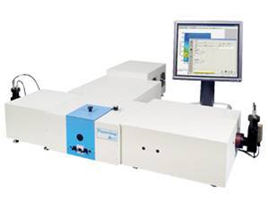
HORIBA(中国)为您提供《活体皮肤中老化、光老化检测方案(分子荧光光谱)》,该方案主要用于其他中老化、光老化检测,参考标准--,《活体皮肤中老化、光老化检测方案(分子荧光光谱)》用到的仪器有HORIBA Fluorolog®-3科研级荧光光谱仪
推荐专场


相关方案
更多
该厂商其他方案
更多