One of the major fields of investigation of L'OREAL is to improve the understanding of the chemical composition and structure of skin and hair. To enable a better design of cosmetic products, a thorough understanding of the mechanisms and the nature of the interactions of cosmetic ingredients with these substrates is necessary.Raman is a key analytical method allowing the needed understanding of such mechanisms. In this context,HORIBA Jobin Yvon & L''OREAL Research have established a close collaboration in applying the Raman technique under in vitro conditions or directly in vivo,for hair,skin,nail, eyelashes and model substrates.
方案详情

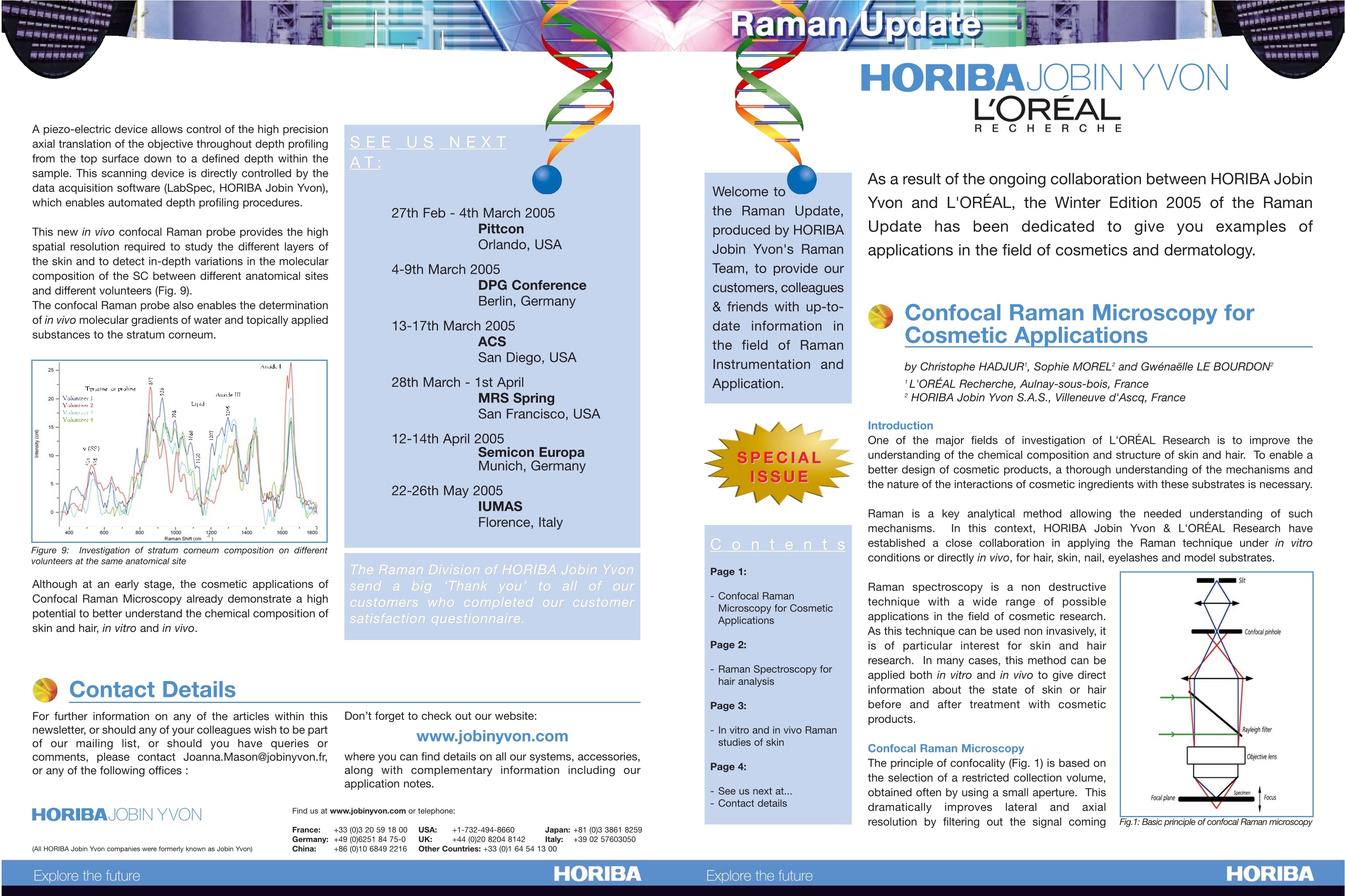
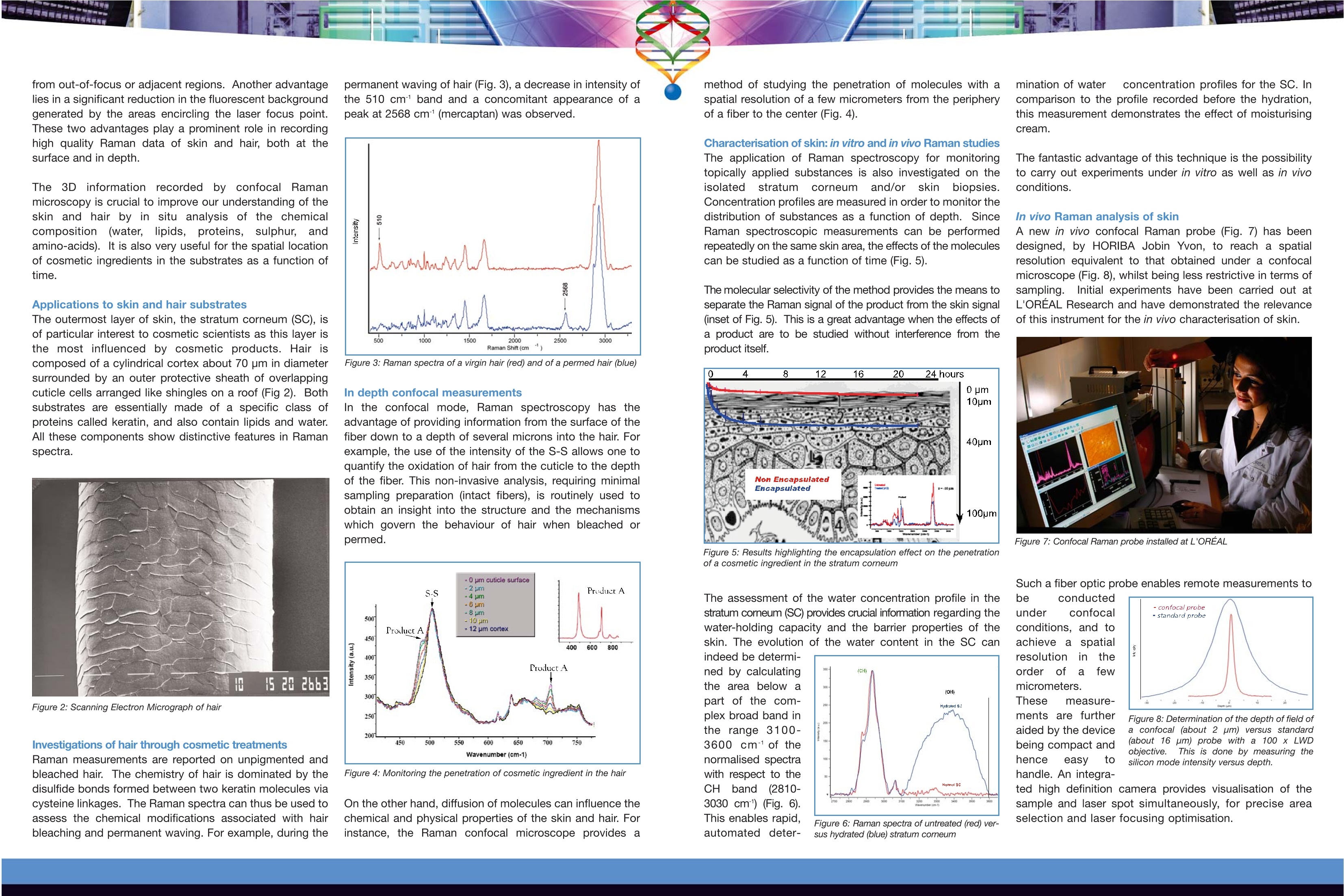
所一尚(All HORIBA Jobin Yvon companies were formerly known as Jobin Yvon)HORIBAHORIBAExplore the future A piezo-electric device allows control of the high precisionaxial translation of the objective throughout depth profilingfrom the top surface down to a defined depth within thesample. This scanning device is directly controlled by thedata acquisition software (LabSpec, HORIBA Jobin Yvon),which enables automated depth profiling procedures. This new in vivo confocal Raman probe provides the highspatial resolution required to study the different layers ofthe skin and to detect in-depth variations in the molecularcomposition of the SC between different anatomical sitesand different volunteers (Fig.9). The confocal Raman probe also enables the determinationof in vivo molecular gradients of water and topically appliedsubstances to the stratum corneum. Figure 9: Investigation of stratum corneum composition on differentvolunteers at the same anatomical site Although at an early stage, the cosmetic applications ofConfocal Raman Microscopy already demonstrate a highpotential to better understand the chemical composition ofskin and hair, in vitro and in vivo. SEEUSNEXT The Raman Division of HORIBA Jobin Yvonsend a bigThank you’ to all ofourcustomers who completed our customersatisfaction questionnaire. Don’t forget to check out our website: Contact Details For further information on any of the articles within thisnewsletter, or should any of your colleagues wish to be partof our mailing list, or should you have queries orcomments, please contact Joanna.Mason@jobinyvon.fr,or any of the following offices : Welcome tothe Raman Update,produced by HORIBAJobin Yvon's RamanTeam, to provide ourcustomers, colleagues& friends with up-to-date information inthe field of RamanInstrumentation andApplication. Conte nt s Page 1: 一. Confocal RamanMicroscopy for Cosmetic Applications Page 2: - Raman Spectroscopy forhair analysis Page 3: - In vitro and in vivo Ramanstudies of skin Page 4: - See us next at... -Contact details RECHERCH E As a result of the ongoing collaboration between HORIBA JobinYvon and L'OREAL, the Winter Edition 2005 of the RamanUpdate has been dedicated to give you examples ofapplications in the field of cosmetics and dermatology. Confocal Raman Microscopy forCosmetic Applications by Christophe HADJURI, Sophie MOREL’ and Gwenaelle LE BOURDON’ 'L'OREAL Recherche, Aulnay-sous-bois, France ?HORIBA Jobin Yvon S.A.S., Villeneuve d'Ascq, France Introduction one of the major fields of investigation of L'OREAL Research is to improve theunderstanding of the chemical composition and structure of skin and hair. To enableabetter design of cosmetic products, a thorough understanding of the mechanisms andthe nature of the interactions of cosmetic ingredients with these substrates is necessary. Raman is a key analytical method allowing the needed understanding of suchmechanisms. In this context, HORIBA Jobin Yvon & L'OREAL Research haveestablished a close collaboration in applying the Raman technique under in vitroconditions or directly in vivo, for hair, skin, nail, eyelashes and model substrates. Raman spectroscopy is a non destructivetechnique with a wide range of possibleapplications in the field of cosmetic research.As this technique can be used non invasively, itis of particular interest for skin and hairresearch. In many cases, this method can beapplied both in vitro and in vivo to give directinformation about the state of skin or hairbefore and after treatment with cosmeticproducts. Confocal Raman MicroscopyThe principle of confocality (Fig. 1) is based onthe selection of a restricted collection volume.obtained often by using a small aperture. Thisdramatically improves lateral and axialresolution by filtering out the signal coming Fig.1: Basic principle of confocal Raman microscopy from out-of-focus or adjacent regions. Another advantagelies in a significant reduction in the fluorescent backgroundgenerated by the areas encircling the laser focus point.These two advantages play a prominent role in recordinghigh quality Raman data of skin and hair, both at thesurface and in depth. The 3D information recorded by confocal Ramanmicroscopy is crucial to improve our understanding of theskin and hair by in situ analysis of the chemicalcomposition (water, lipids, proteins, sulphur, andamino-acids). It is also very useful for the spatial locationof cosmetic ingredients in the substrates as a function oftime. Applications to skin and hair substrates The outermost layer of skin, the stratum corneum (SC), isof particular interest to cosmetic scientists as this layer isthe most influenced by cosmetic products. Hair iscomposed of a cylindrical cortex about 70 pm in diametersurrounded by an outer protective sheath of overlappingcuticle cells arranged like shingles on a roof (Fig 2). Bothsubstrates are essentially made of a specific class ofproteins called keratin, and also contain lipids and water.All these components show distinctive features in Ramanspectra. Figure 2: Scanning Electron Micrograph of hair Investigations of hair through cosmetic treatments Raman measurements are reported on unpigmented andbleached hair. The chemistry of hair is dominated by thedisulfide bonds formed between two keratin molecules viacysteine linkages. The Raman spectra can thus be used toassess the chemical modifications associated with hairbleaching and permanent waving. For example, during the permanent waving of hair (Fig.3), a decrease in intensity ofthe 510 cm band and a concomitant appearance of apeak at 2568 cm"(mercaptan) was observed. Figure 3: Raman spectra of avirgin hair (red) and of a permed hair (blue) In depth confocal measurements ln the confocal mode, Raman spectroscopy has theadvantage of providing information from the surface of thefiber down to a depth of several microns into the hair. Forexample, the use of the intensity of the S-S allows one toquantify the oxidation of hair from the cuticle to the depthof the fiber. This non-invasive analysis, requiring minimalsampling preparation (intact fibers), is routinely used toobtain an insight into the structure and the mechanismswhich govern the behaviour of hair when bleached orpermed. Figure4: Monitoring the penetration of cosmetic ingredient in the hair On the other hand, diffusion of molecules can influence thechemical and physical properties of the skin and hair. Forinstance, the Raman confocal microscope provides a Characterisation of skin: in vitro and in vivo Raman studiesThe application of Raman spectroscopy for monitoringtopically applied substances is also investigated on theisolatedstratum corneum and/or)rskinbiopsies.Concentration profiles are measured in order to monitor thedistribution of substances as a function of depth. SinceRaman spectroscopic measurements can be performedrepeatedly on the same skin area, the effects of the moleculescan be studied as a function of time (Fig.5). The molecular selectivity of the method provides the means toseparate the Raman signal of the product from the skin signal(inset of Fig. 5). This is a great advantage when the effects ofa product are to be studied without interference from theproduct itself. Figure 5: Results highlighting the encapsulation effect on the penetrationof a cosmetic ingredient in the stratum corneum The assessment of the water concentration profile in thestratum corneum (SC) provides crucial information regarding thewater-holding capacity and the barrier properties of theskin. The evolution of the water content in the SC can indeed be determi-ned by calculatingthe area below apart of the com-plex broad band inthe range 3100-3600 cmofthenormalised spectrawith respect to theCHbband (2810-3030 cm) (Fig. 6).This enables rapid, Figure 6: Raman spectra ofuntreated (red) ver-sus hydrated (blue) stratum corneum mination of water concentration profiles for the SC. Incomparison to the profile recorded before the hydration,this measurement demonstrates the effect of moisturisingcream. The fantastic advantage of this technique is the possibilityto carry out experiments under in vitro as well as in vivoconditions. In vivo Raman analysis of skin A new in vivo confocal Raman probe (Fig. 7) has beendesigned, by HORIBA Jobin Yvon, to reach a spatialresolution equivalent to that obtained under a confocalmicroscope (Fig.8), whilst being less restrictive in terms ofsampling. Initial experiments have been carried out atL'OREAL Research and have demonstrated the relevanceof this instrument for the in vivo characterisation of skin. Figure 7: Confocal Raman probe installed at L'OREAL Such a fiber optic probe enables remote measurements to Figure 8:Determination of the depth of field ofa confocal (about 2 pm) versus standard(about 16 um) probe with a 100 x LWDobjective. This is done by measuring thesilicon mode intensity versus depth. be conductedunder confocalconditions, and toachieve a sspatialresolutionir theorder01 a fewmicrometers.These measure-ments are furtheraided by the devicebeing compact andhence easy tohandle. An integra- ted high definition camera provides visualisation of thesample and laser spot simultaneously, for precise areaselection and laser focusing optimisation.
确定
还剩1页未读,是否继续阅读?
HORIBA(中国)为您提供《化妆品中成分的相互作用检测方案 》,该方案主要用于美容/修饰类化妆品中成分的相互作用检测,参考标准--,《化妆品中成分的相互作用检测方案 》用到的仪器有HORIBA XploRA PLUS超快速拉曼成像光谱仪
推荐专场
相关方案
更多
