


方案详情
文
The IMV-4000 enables infrared imaging by a figure of two or more
faster than using a conventional single element detector. A
technique that quickly analyzes the results is also desirable. JASCO
has proposed an analysis of imaging data with a variety of
multivariate analysis techniques to accelerate the processing of
infrared imaging data. For example, we have made it possible to
easily create a visualization of the distribution map for the various
components of multilayer film by analyzing imaging data using
principle component analysis (PCA). We have also created a model
that performs secondary structure estimation (SSE) on proteins
based on IR spectra by principal components regression (PCR) and
方案详情

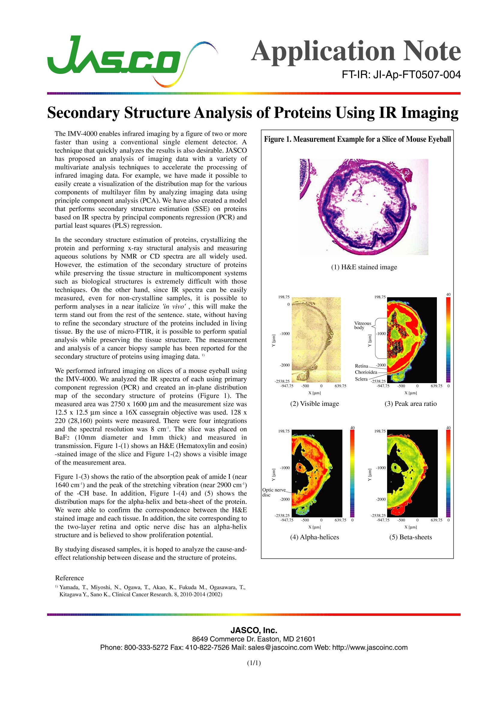
JASCO,Inc.8649 Commerce Dr. Easton, MD 21601Phone:800-333-5272 Fax: 410-822-7526 Mail: sales@jascoinc.com Web: http://www.jascoinc.com(1/1) s.coApplication NoteFT-IR:JI-Ap-FT0507-004 Secondary Structure Analysis of Proteins Using IR Imaging The IMV-4000 enables infrared imaging by a figure oftwo or morefaster than using a conventional single element detector. Atechnique that quickly analyzes the results is also desirable. JASCOhas proposed an analysis of imaging data with a variety ofmultivariate analysis techniques to accelerate the processing ofinfrared imaging data. For example, we have made it possible toeasily create a visualization of the distribution map for the variouscomponents of multilayer film by analyzing imaging data usingprinciple component analysis (PCA). We have also created a modelthat performs secondary structure estimation (SSE) on proteinsbased on IR spectra by principal components regression (PCR) andpartial least squares (PLS) regression. In the secondary structure estimation of proteins, crystallizing theprotein and performing x-ray structural analysis and measuringaqueous solutions by NMR or CD spectra are all widely used.However, the estimation of the secondary structure of proteinswhile preserving the tissue structure in multicomponent systemssuch as biological structures is extremely difficult with thosetechniques. On the other hand, since IR spectra can be easilymeasured, even for non-crystalline samples, it is possible toperform analyses in a near italicize in vivo’, this will make theterm stand out from the rest of the sentence. state, without havingto refine the secondary structure of the proteins included in livingtissue. By the use of micro-FTIR, it is possible to perform spatialanalysis while preserving the tissue structure. The measurementand analysis of a cancer biopsy sample has been reported for thesecondary structure of proteins using imaging data." We performed infrared imaging on slices of a mouse eyeball usingthe IMV-4000. We analyzed the IR spectra of each using primarycomponent regression (PCR) and created an in-plane distributionmap of the secondary structure of proteins (Figure 1). Themeasured area was 2750 x 1600 um and the measurement size was12.5 x 12.5 um since a 16X cassegrain objective was used. 128x220 (28,160) points were measured. There were four integrationsand the spectral resolution was 8 cm. The slice was placed onBaF2 (10mm diameter anddj1mm thick) and1 measuredd intransmission. Figure 1-(1) shows an H&E (Hematoxylin and eosin)-stained image of the slice and Figure 1-(2) shows a visible imageof the measurement area. Figure 1-(3) shows the ratio of the absorption peak of amide I (near1640 cm') and the peak of the stretching vibration (near 2900 cm')of the -CH base. In addition, Figure 1-(4) and (5) shows thedistribution maps for the alpha-helix and beta-sheet of the protein.We were able to confirm the correspondence between the H&Estained image and each tissue. In addition, the site corresponding tothe two-layer retina and optic nerve disc has an alpha-helixstructure and is believed to show proliferation potential. By studying diseased samples, it is hoped to analyze the cause-and-effect relationship between disease and the structure of proteins. Reference Figure 1. Measurement Example for a Slice of Mouse Eyeball
确定
还剩1页未读,是否继续阅读?

产品配置单

佳士科商贸有限公司为您提供《FTIR IR红外成像分析蛋白质的二级结构》,该方案主要用于其他中检测,参考标准--,《FTIR IR红外成像分析蛋白质的二级结构》用到的仪器有JASCO傅立叶变换红外光谱仪FT/IR-6000、JASCO FTIR-4000傅立叶变换红外光谱仪、jascoRFT-6000傅立叶变换红外拉曼光谱仪
推荐专场

相关方案
更多
该厂商其他方案
更多