


方案详情
文
Introduction
CD spectra provide information on the secondary structure of proteins and the environment of
aromatic side chains. Therefore, CD measurement using a stopped-flow system is considered as one of
the best methods for analyzing the unfolding and refolding of proteins.
The existence of an intermediate between denaturated state and natural state during the refolding of
proteins has been reported. The CD stopped-flow method is used for examining this refolding process. In
this report the refolding process of cytochrome c (cyt c) measured using a SFS-492 stopped-flow system
will be explained.
Keywords: Stopped-flow, Circular Dichroism, Refolding
Sample Preparation
Aqueous solution of Cytochrome c denaturated by guanidine hydrochloride (GuHCl) was diluted with
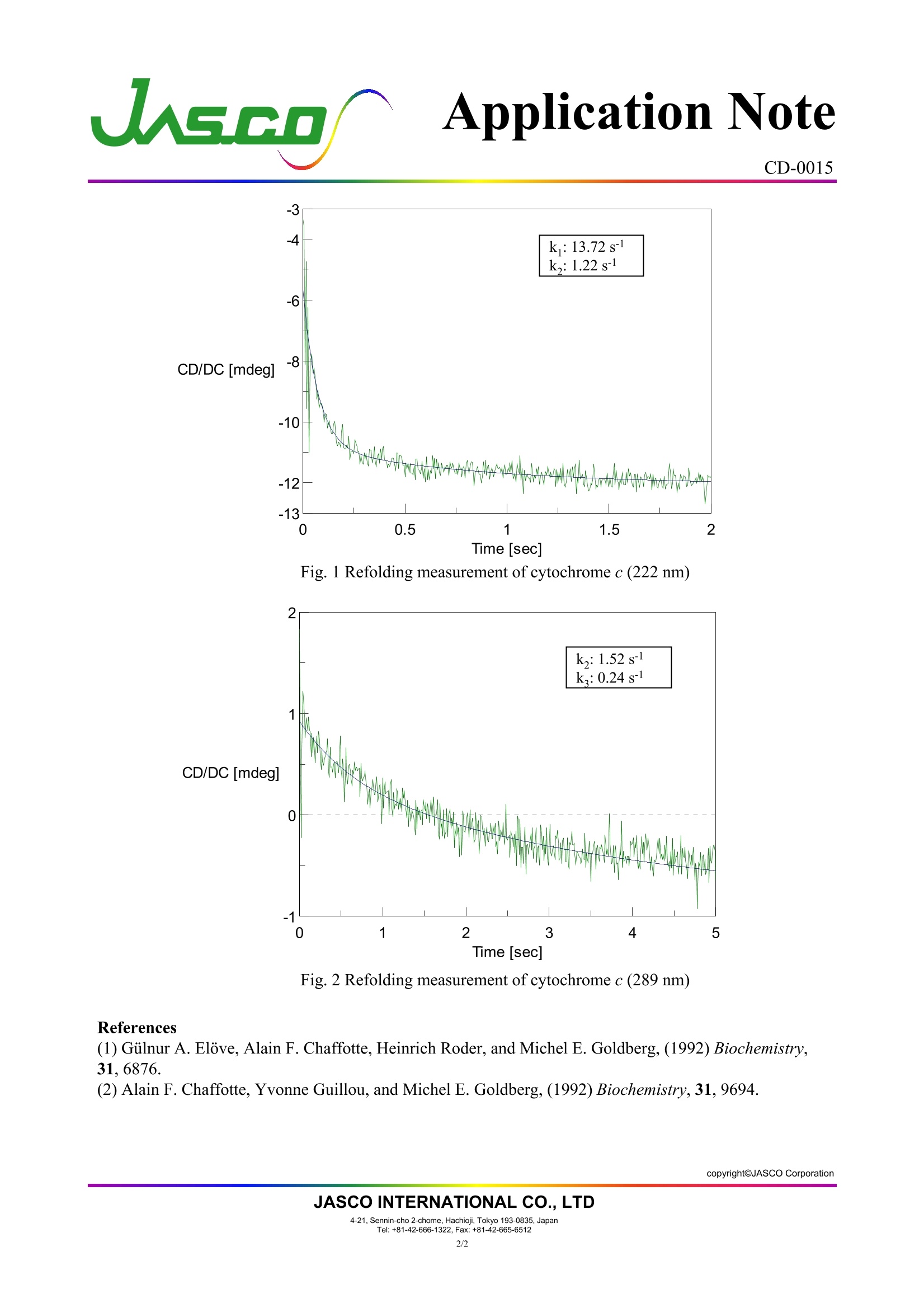
0.1 M acetic acid buffer solution (1:9). The refolding process was observed at 222 nm for the secondary
方案详情

Application Note CD-0015 Refolding of Cytochrome c Introduction CD spectra provide information on the secondary structure of proteins and the environment ofaromatic side chains. Therefore, CD measurement using a stopped-flow system is considered as one ofthe best methods for analyzing the unfolding and refolding of proteins.The existence of an intermediate between denaturated state and natural state during the refolding ofproteins has been reported. The CD stopped-flow method is used for examining this refolding process. Inthis report the refolding process of cytochrome c (cyt c) measured using a SFS-492 stopped-flow systemwill be explained. Keywords: Stopped-flow, Circular Dichroism,Refolding Sample Preparation Aqueous solution of Cytochrome c denaturated by guanidine hydrochloride (GuHCl) was diluted with0.1 M acetic acid buffer solution (1:9). The refolding process was observed at 222 nm for the secondarystructure and at 289 nm for the environment of the aromatic side chain. Measurement conditions Measurement system: J-815 + SFS-492 stopped-flow system Optical pathlength: 2mm Temperature: Room temperature Flow rate: 1.5 mL/sec Each sample was prepared at an optimal concentration because CD intensities at 222 nm for thesecondary structure and at 289 nm for aromatic side chains are quite different. Wavelength 222 nm 289 nm Data interval 5 msec 10 msec Response 4 msec 8 msec Bandwidth 4 nm 2nm Syringe 1 2 mg/mL cytc/4.3 M GuHC1 10 mg/mL cyt c/4.3M GuHCl Loading volume 30 uL 30 uL Syringe 2 0.1 M acetic acid buffersolution (pH 6.3) 0.1 M acetic acid buffersolution (pH 6.3) Loading volume 270 uL 270 uL Accumulation 36 times 24 times Results The change in the CD value at 222 nm reflects fast refolding of the secondary structure within 200msec (Fig. 1), but the change at 289 nm reflecting the environment of the aromatic side chain was slower(Fig. 2). This slower change appears in the latter step of the refolding process. These results indicate thebrief existence of an intermediate state with a refolded secondary structure but with aromatic side chainsremained unfolded. s.c.o, CD-0015 -3 Fig. 1 Refolding measurement of cytochrome c (222 nm) Fig. 2 Refolding measurement of cytochrome c (289 nm) ( References ) (1) Gulnur A. Elove, Alain F. Chaffotte, Heinrich Roder, and Michel E. Goldberg, (1992) Biochemistry, ( 31,6876. (2) Alain F . Chaffotte, Yvonne Guillou, and Michel E. Goldberg, (1992) Biochem i stry, 31,9694. ) copyrightJASCO Corporation JASCO INTERNATIONAL CO., LTD copyrightOJASCO CorporationJASCO INTERNATIONAL CO., LTD-Sennin-cho -chome, Hachioji, Tokyo JapanTel:+ Fax: + IntroductionCD spectra provide information on the secondary structure of proteins and the environment ofaromatic side chains. Therefore, CD measurement using a stopped-flow system is considered as one of
确定
还剩1页未读,是否继续阅读?

产品配置单

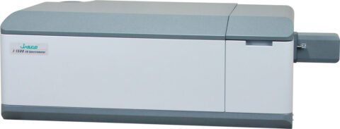
佳士科商贸有限公司为您提供《细胞色素中折叠过程检测方案 》,该方案主要用于其他中折叠过程检测,参考标准--,《细胞色素中折叠过程检测方案 》用到的仪器有JASCO圆二色光谱仪CD J-1500
推荐专场

相关方案
更多
该厂商其他方案
更多