白皮书《采用先进的细胞分析方案加速病毒生物学和宿主免疫的发现》重点介绍了如何使用Incucyte®活细胞分析系统和iQue® 3先进的高通量流式细胞术解决这些挑战,从而更加深入地了解病毒感染过程中宿主病原体的相互作用。
方案详情

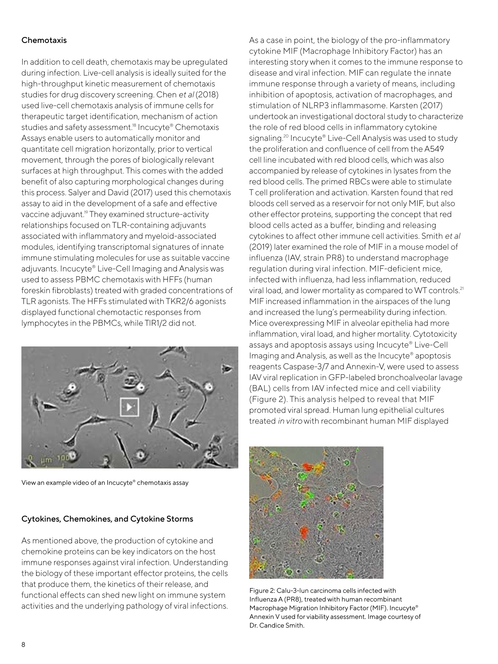
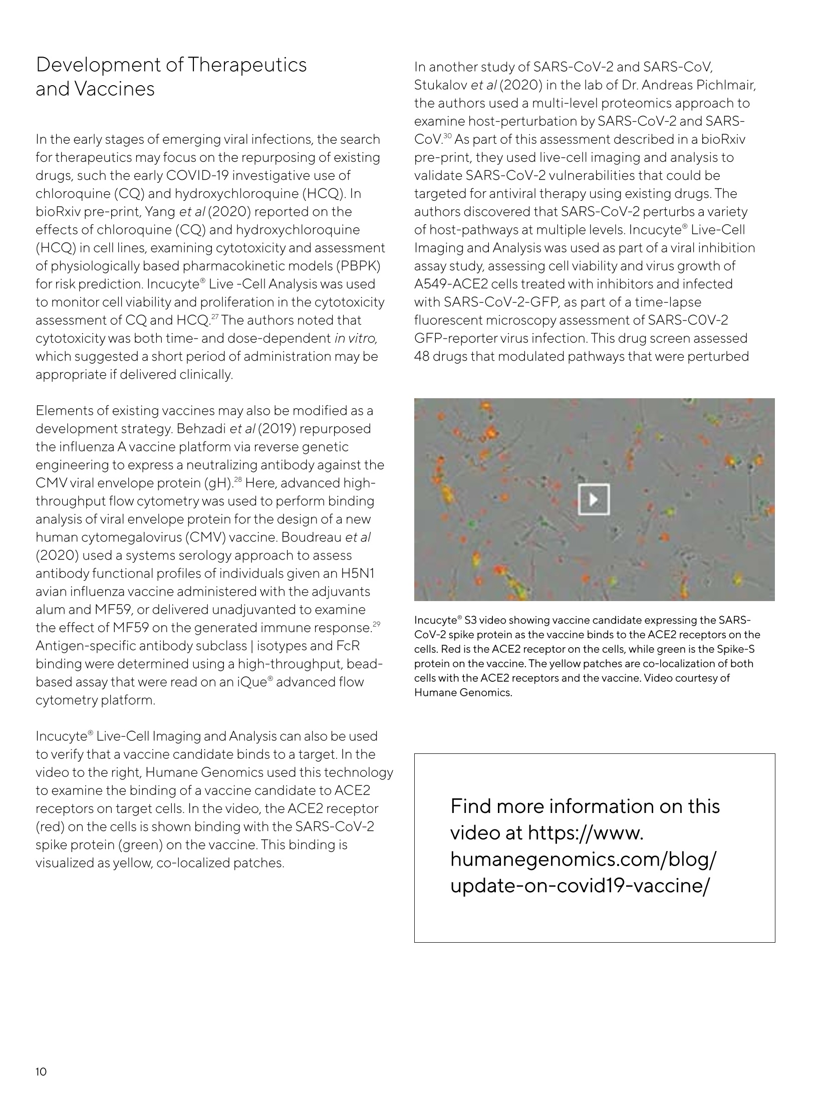
White Paper Cell Death June24,2020 Keywords or phrases:ViralInfection, Viral Replication,Antibody ResponseEpitope Mapping,Immune Cell Activation, Chemotaxis,Cytokine Storm, Cell Death,Immune Cell Killing,NETosis Accelerating Discoveries for Viral Biology andHost Immunity with Advanced Cell AnalysisSolutions Jill Granger,Product Management, Essen BioScience, Inc.-a Sartorius Company,300 West Morgan Road, Ann Arbor, Michigan, 48108, USA Introduction In the wake of global pandemics such as COVID-19, MERS-CoV, and Ebola, virology studies take center stage, capturing theattention of both scientists and the public alike. But within this "pathogen cloud"lies a silver lining-increased investment inviral biology research, the establishment of new scientific collaborations for rapid discovery, and renewed curiosity among thegeneral public on the long-term health impact of emergent pathogens. This increased awareness is accompanied by an urgent need to characterize viral biology and the host immune response forthe identification of therapeutics and development of vaccines. Successfully executing a rapid,effective response toemergent pathogens requires the use of experimental approaches that are high throughput, yet properly balanced withthorough analysis for scientific rigor, leading to reproducible results. Traditional endpoint analysis can be limited, utilizingtime-consuming, tedious"hands-on"workflows with multiple instruments, which also presents safety concerns throughincreased exposure to pathogens.The experimental manipulations of these assays introduce environmental fluctuations andlack of control that are not truly representative of host physiology. Traditional end-point assays lack kinetic insight on viralbiology and immune responses, quickly consume limited sampleand reagents, and require multiple instruments and softwarethat could introduce variability. Taken together, traditional analysis assays are hampered by a slow time-to-result, lack kineticinformation, and present workflow challenges that are problematic for fast-paced therapeutic screening and vaccinedevelopment. This is especially important during emerging infections when time-to-result is critical, but may impede othertranslational virology research activities, such as the development of therapeutics for regenerative medicine and cancervaccines using oncolytic viruses. The IncucyteLive-Cell Analysis System and iQue° 3Advanced High-Throughput Flow Cytometry platformsaddress many of these challenges,providing rapid, yetcomprehensive and insightful analysis of viral biology andhost immune response studies.These instruments providethe automation, flexible applications, and throughputneeded to quickly analyze the dynamics of viral infectionand the immune response with reproducibility to providenewinsights. The Incucyte@ incorporates automated image acquisitionand analysis while maintaining physiological conditionsin the user's incubator for better physiological control,quantifying dynamic changes in real time for hours, days,or weeks. The system's flexible applications,multiplexingcapabilities, non-perturbing reagents, and integrated,user-friendly software permit rapid evaluation of cell health,function, movement, and morphology in a physiologicallyrelevant environment while minimizing operator exposureto pathogens via remote access. The iQue@ 3 is an automated,high-throughput,advancedflow cytometry platform that allows users to rapidlymultiplex protein analysis, immunophenotyping andperform functional assessments. Users can assess immunecell activation, perform functional profiling and antibodyscreening. This comes with the convenience of ready-to-use kits including the iQueT Cell Phenotyping kit,Human Qbeads@ Inflammation Panel for cytokine stormassessment,and kits for T cell activation, memory, killing,and exhaustion along with cytokine profiling for greaterindepth characterization of the host immune response ineach sample. Miniaturization is possible, as the platformrequires only small sample volumes and is reagent sparing.It accommodates a variety of microplates, including 384- Advanced cell analysis providesnew insights on viral biologyand host immune responsesthroughout the host|pathogenlifecycle. well for high-throughput analysis in minutes for profilingand pharmacological screening. Researchers can generatereproducible, high throughput tracking and analysis withintegrated,user-friendly Forecyt@ software. This timesaving,streamlined software offers plate-level annotation,analytics, and results visualization tools ideally suited forscreening large data sets. Users may also access linearrange data for deeper understanding on assay performance.Data from wells may be linked together and multiple assayoutcomes combined to facilitate "hit"identification. The research studies that follow provide real-worldexamples of how Incucyte° Live-Cell analysis andiQue° 3 Advanced High-Throughput Flow Cytometrywere incorporated into virology workflows to provide rapidanalysis solutions, simplification, and enhanced throughput,aiding the discovery of new insights on the host-pathogenlife cycle throughout the course of infection. Additionally,post-infection, these platforms were used to help monitorexposure, assess immune protection, and accelerate thedevelopment of anti-viral small molecules,neutralizingantibodies, and vaccines. Host Infection and Viral Replication The host| pathogen lifecycle involves a series ofdynamic events, including viral fusion, entry, replication,and spreading. Understanding the kinetic biology ofthese early events, including rates of infection andreplication, can provide important clues for theidentification of therapeutic targets, neutralizingantibodies, and vaccines Selecting and Developing a Viral Infection Model A critical, first step for virology studies is the selectionof an appropriate model that is both biologically andclinically relevant. Roberts et al(2017) evaluated celllines to identify the most physiologically relevant as an invivo model for the infectious cycle of Chikungunya virus(CHIKV) Incucyte@ Live-Cell Imaging and Analysis wasused to capture Brightfield images of cell morphologyduring differentiation, better recapitulating in vivoconditions within the physiologically relevantenvironment of the incubator. Morphological changesduring C2C12 (myoblast) and Huh7 (hepatocyte)differentiation were visualized. C2C12 cells were serum-starved in vitro, differentiated, and formed myotubes,similar to in vitro muscle tissue. This was also associatedwith enhanced CHIKV RNA replication.Huh7 cellsdifferentiated with DMSO underwent arrested growthand up-regulation of liver proteins, showing reducedlevels of CHIKV RNA replication compared to that ofundifferentiated cells. This illustrated how importantmorphological information from live-cell analysis canadd to biological understanding. The development of new infection models may berequired to enable the safe handling of viruses. There islimited availability of BSL-4facilitates that can safelyaccommodate particularly virulent viruses, which mayimpede research progress.Xiao et al(2018) developed anEbola virus pseudotype (E-S-FLU), a single-cycle viruscapable of mimicking Ebola cell entry, which could besafely handled in BSL1/2 containment facilities. TheE-S-FLU virus was created using an influenza virus thathad been coated the glycoprotein from Ebolavirus(EBOV-GP). E-S-Flu encoded a green fluorescentreporter that replaced the hemagglutinin (HA) genefrom influenza.Alentiviralvector was used to transduceMDCK-SIAT1 cells that stably expressed high levelsof EBOV-GP (E-SIAT). As part of the E-S-Flu viral titercharacterization, the Incucyte° was used to obtain imagesof plaques at 3 h intervals, revealing key morphologicalclues during infection kinetics: E-SIAT cells formed small diffuse plaques, while H5-SIAT cells produced denserplaques. A single production batch of the pseudotyped,E-S-FLU virus was leveraged to screen a library of 1280compounds as BSL1/2 for inhibition of viral entry. Viral Entry and Infection Characterizing viral entry and infection can provideinformation that may be exploited for therapeutic targetidentification or vaccine development. Stein et a(2019)examined Human cytomegalovirus (CMV) entry into hostcells for therapeutic target identification using a library ofmonoclonal antibodies directed against human retinalpigmented epithelial cell surface proteins (ARPE). Micewere injected with an ARPE-19-derived membrane fractionto produce immunized sera, which was analyzed forantibodies against ARPE-19 cell surface proteins usingiQue@Advanced High-Throughput Flow Cytometry. Aviralinhibition assay identified two clones that caused inhibitionyet failed to block entry into fibroblasts. Subsequentexperimentation led the authors to propose that CD46 viralentry is dependent on a post-binding event, wherein CD46interacts with CMV PC. Studies using CD-46KO epithelialcells showed a reduction in CMV dissemination,indicatingthat a CD46-dependent process may be involved in viralspreading. Theauthors further related the advantages ofusing this mAB library approach over genetic screening,including the lack of manipulation of transcriptional profiles,elimination of transfection and transduction toxic effects, aswell as flexibility for assay customization. Viral infection and entry can also be studied using live-cellimaging and analysis to monitor signals from fluorescentreporters in treated cells. Qui et al (2018) studied theinteractions between the Ebola virus (EBOV)glycoproteinwith Niemann-Pick C1 (NCP1) to gain insight on traffickingand delivery of EBOV to NCP1.4Incucyte@ Live-cell Imagingand Analysis was used to capture the entry of GFP-expressing MLV pseudotypes harboring EBOVAM GPor VSV G in ArPIKfyve and Sac3 KO cells through thevisualization of GFP+ foci. Additional analysis showed thatall subunits of the PAS complex were required for EBOVentry, and that cellular production of PtdIns (3, 5) P isneeded to promote efficient delivery to NPC1.Takentogether,these results shed new light on the infectionand entry of this virulent pathogen. Viral Replication In addition to viral entry, viral replication represents anotherarea where these platforms may provide new insights fortherapeutic exploitation.Rothan et al(2019) studied the useof host cell components as an alternative approach fortargeting viral pathogens in dengue virus (DENV) and Zikavirus (ZIKV) infections.Live-cell analysis was used toquantitate real-time proliferation of ZIKA-infected andDENV-infected Vero cells treated with Bardoxolne methyl(CDDO-me) or neutralization with ZIKV-neutralizingantiserum. CDDO-me is an inhibitor of the Hrd1 ubiquitinligase-mediated ERAD pathway that possibly targets hostfactor grp94 and was found to inhibit dislocation.ZIKV-infected Vero cells were treated with CDDO-me or ZIKV- neutralizing serum, and the Incucyte° was used toautomatically generate kinetic processing metrics fromthe imaging.The percentage confluence over time wascalculated to determine the cytopathic effects of infection.CCDO-me (0.13 uM) protected the Zika-infected Vero cellsas well as 5%ZIKV-neutralizing antiserum. CDDO-me andPU-WS13 (a grp94 inhibitor) also had anti-flaviviruses activity,and inhibition of grp94 may play a role in CCCO-meinhibition of both DENV and ZIKV. Herod et al (2019) usedlive-cell imaging and analysis to detect GFP reporter geneexpression to study the effects of the anti-influenza drugArbidol (ARB) against the foot-and-mouth disease (FMDV)and equine rhinitis virus (ERAV).Using replicons in a FMDVreplication assay, they were able to study viral replication atthe BSL2 level of containment. BHK-21 cells were treatedwith DMSO,GuHCl,or ARB and transfected with a wild-typeGFPFMDVreplicon. Expression of the fluorescent reporterprotein was monitored for 10 h using the Incucyte, revealingthat ARB treatment produced dose-dependent suppressionof FMDVreplication.Subsequent analysis showed that ARBinhibited the replication of FMDVRNAsub-genomicreplicons, suggesting a possible inhibition of picornavirusgenome replication. BHK-21 cells transfected with a foot-and-mouth disease virus (FMDV)replicon containing a GFP (green) reporter. Incucyte° Live-CellImaging and Analysis was used to visualize, in real-time, the cellsexpressing the GFP (green) reporter as a measure of virus replicationover time.A typical well was imaged for GFP (green) expression hourlyand used to compile a movie showing FMDV replication.Data courtesyof Dr. Morgan Herod (University of Leeds). Foradditional information onthis topic, watch webinar byDr. Fiona Tulloch“FurtheringInsight and Productivity inCell Biology with Real-TimeQuantitative Live-Cell Analysis." In addition to viral replication, live-cell analysis can providenew insights into viral transmission and spread. In the caseof Measles Virus (MeV), the virus can form syncytia whenuninfected cells fuse with infected cells expressing F-andH-glycoproteins from the viral envelope. MeV, part of theMorbilliviruses, may also spread by viral budding. Kelly et al(2019) used live-cell analysis to study the cell-to-cell spreadof Measles Virus (MeV) using a recombinant MeV thatexpressed a GFP fluorescent protein as a transcription unit.'The Incucyte° was used to quantitateviral replication,syncytia formation, and expansion, revealing that a proteininduced by interferon, bone marrow stromal antigen 2(BST2), inhibited the replication of MEV-GFP. Thesefindings were also concordant with a reduction in MeVnucelocapsid (N), Fand H proteins as detected by westernblot. Additional analysis with Incucyte, immunolabeling,and biochemistry were utilized to determine that BST2inhibited cell fusion (visualized as reduced syncytiaformation) and cell-cell spread. Anotherimportant point of study for viral transmission isthe element of viral dispersal. Viruses may be dispersedthrough multi-viron assemblies that infect the host cells.Anderu-Moreno and Sajuan (2018) studied the viralfitness offree vesicular stomatitis virus (VSV) particlesverses those that were aggregated in saliva. This studyleveraged the kinetic capabilities of real-time,live-cellimaging and analysis with Incucyte° to compare fitnesseffects of viral particles dispersed in aggregated form tothose in monodisperse. Real-time, live cell kinetic studieswere used to study mouse embryonic fibroblasts (MEFs)inoculated with either monodispersedVSV-mCherryparticles or aggregated VSV-GFP using human saliva.The growth curves, generated from the Incucyte@analysis, revealed leftward shift for viral populationsformed by aggregates, as compared to those which weremonodispersed.Aggregation of virus particles imparteda short-term fitness advantage and increased the releaseof viral progeny, which was dependent on cellularpermissively to infection and was correlated with thelevel of cellular innate immunity. Viral biology doesn't occur in isolation but is subject tothe forces of the host immune response,which is delicateinterplay between the innate and adaptive immunesystems that ebbs and flows as the pathogen is detected,combatted, and eliminated.The dynamics and extent ofthis response is complicated by individual genetics andinflammatory status, as well as viral mutational shifts.Kinetic, comprehensive analysis may uncover keyinformation on the timing and intensity of this dynamic,often complex story. Changes in cellular health, function,phenotype, and activity, as well as cyclic production ofpro-and anti-inflammatory mediators throughout thecourse of infection can hold critical informationabouthow the virus immune system interactions and responseto therapeutic candidates. Immune Cell Activation Viral exposure initiates immune cell activation andsustained responses for pathogen elimination. Zander etal(2019) studied how CD4+T cells can help sustainexhausted CD8+T cells during chronic infection andcancer. The authors performed a functional assessmentof CD8+ T cells that were specific for the GP33-41 peptideof lymphocytic choriomeningitis virus (LCMV). Subsets ofsorted GP33-41CD8+T cells were co-cultured with targetEL4 ells treated with GP22 peptide or a control peptide.The authors used Incucyte° S3 and Incucyte@Annexin VRed Reagent to quantitate cytotoxicity of the CD8+Tcells.Using additional scRNA-seq analysis and transcriptionalprofiling, they discovered that the CX3CR1+-expressingCD8+T cell subset had a cytolytic function that was key inviral control. This was also dependent on IL-21 producedfrom the CD4+Tcells. In another CD4+ T cell study, Kwon et al (2020) examinedlatent HIV-1 proviruses in human resting memory CD4+Tcell subsets to better understand latency and escape ofHIV-1 from immune clearance. A multiple stimulation viraloutgrowth assay was used to test naive and memory Tcells(central, transitional, and effector) from HIV-1 patients onantiretroviral therapy. The iQue Screener Plus was used toassess cellular activation levels of resting CD+4 T cellsubsets, finding no correlation between the subsets androunds of stimulation needed to produce viral outgrowth.Additional analysis revealed a lack of enrichment for intactor inducible proviruses in the Tcell subsets, highlighting thecomplexity of pursuing T cell memory subsets for HIV-1therapy. Antibody Responses and Epitope Mapping In addition to activation of innate immune responses, theadaptive immune system is called into action to produceantibodies for viral defense. Epitope mapping of viralantigens is an important characterization for study of earlyresponses, and advanced high-throughput flow cytometryis ideally suited to supplement such investigations. In aninsightful study by Williamson etal(2019), the authorsanalyzed early convalescent memory B cells from survivorsof Ebola infection." Monoclonal antibodies (mAbs) fromthe memory B cells were isolated from survivors 1-3months post infection to examine the frequency of Ebola-specific B cells. The iQue Advanced Flow CytometryPlatform was used as part of the alanine-scanning shotgunmutagenesis for epitope mapping. An alanine scanningmutagenesis library was created for Ebola glycoprotein(EBOVGP)to generate alanine-scanning mutagenesislibrary clones. HEK-293T cells were transfected with thelibrary, mutant GPs expressed, and the cells fixed andstained for flow cytometry with a Fab fragment ofEBVOV237 and a secondary Alexa Fluor 488 conjugated-antibody. The iQue° was used for analysis of cellularfluorescence. The reactivity of EBOV237 Fab fragments tothe mutants was compared to the wild-type EBOVGPAmucin. Clones that did not bind the EBOV237 Fabfragment, but bound to other control EBOV mAbs, weredeemed clones of interest. Subsequent analysis identifieda neutralizing epitope in the glycan cap of the surfaceglycoprotein,recognized by a mAB designated asEBOV237.This epitope may play an important role in theearlyresponse to Ebola virus and be a potential vaccinecandidate. In another study using alanine mutagenesis, Collins et al(2019)studied the human B cell antibody response to Zikavirus infection."Two type-specific mAbs were isolated froma SIKV patient with protective antibodies and were used tomap viral epitopes. Alanine scanning mutagenesis wascarried out to generate a ZIKV prM|Eslanin-scan librarywith mapping of cells expressing ZIKV E mutants. Thethroughput of the iQue@ Advanced Flow CytometryPlatform enabled rapid assessment of mean cellularfluorescence for the epitope mapping. The authorsconcluded that both the plasma antibody and memoryB cell responses are ZIKV-type specific. The neutralizingantibodies produced primarily target quaternary structureepitopes on the viral envelope, which the authors notedhad been found in DENV and other flaviviruses. Theyemphasized that the identification of targets that produce along-lived neutralizing Ab response was informative, andmay be used to guide antigen design and immunitygenerated by vaccines. Understanding the kinetics of cell death mechanismscan provide some valuable, and even previously unknown,insights for understanding immune cell killing and celldeath mechanisms of virally infected cells and identifynew therapeutic targets. One area of cell death that has received increasedattention in viral disease is NETosis. In a ground-breakingstudy by Barr and Roderiguez-Garcia et al, the authorsidentified NETosis formation by neutrophils in genitalmucosa as a protective defense against HIV infection, apreviously undescribed finding in the early stage of HIVinfection.Live-cell imaging and analysis with Incucyte°captured the in vitro dynamics of GFP-tagged HIV-VLP(HlVviral-like particles) entrapped by human genitalneutrophils (Figure 1). NETs were released within minutesof viral exposure. Figure 1:Neutrophils isolated from the female genital tract cultured inimpermeant DNA dye (red) and stimulated with GFP-labeled HIV viral-like particles (green) to induce NET formation. (A) shows the DNAstaining in NETs (red). (B) shows the GFP-labeled viral-like particlesentrapped in NETs. Bottom left panel (C) shows the phase contrast, inwhich NETs can also be visualized, and bottom right panels (D) showsthe merge with HIV trapped in the NETs appearing as yellow. Imagecourtesy of Dr. Marta Rodriquez-Garcia, Tufts University School ofMedicine. In another NETosis study, Drs. Chirivi,Raats, and colleagues(at ModiQuest B.V.) reported on NET release into theextracellular environment during inflammation.4Incucyte°Live-Cell Imaging and Analysis was used to capture NETrelease from neutrophils in the presence of an engineeredtherapeutic anti-citrullinated protein antibody (tACPA),which showed both therapeutic and prophylactic potentialfor inflammatory disease associated with NET pathology,being the first in kind described to interfere with NETexpulsion into the extracellular space. In addition to NETosis, cell death may also arise fromapoptosis. Brown et al (2018) examined the interaction ofcritical proteins involved in viral infections of poultry: theMeq from Marek’'s disease virus (MDV) and the apoptinprotein in chicken anemia virus (CAV)15Incucyte Live-CellImaging and Analysis and Incucyte° Caspase-3/7 ApoptosisReagent were used to study the functional, kinetic effectsof Meq-apoptin interactions on apoptosis on transfectedchicken fibroblast cells (DF-1).Meq protein inhibitedapoptin, which may have important implications for CAVvaccine production.Cubas-Gaona et al(2018) examinedthe loss of IgM B lymphocytes in Infectious Bursal Disease(IBDV) and the role of type 1 interferon (IFN) in IBDVinfection.Live-cell analysis was used to examine enhancedapoptosis of HeLa cells treated with IFN-a and infectedwith IBDVand DF-1 cells. This study demonstrated thatIBDV-infected cells undergo marked apoptosis whenexposed to IFN-a. A third mechanism of cell death is necroptosis, andviruses may avoid destruction by exploiting thismechanism to promote infection.Reddy et al(2020)studied the role of Nr-13, a Bcl-2 related protein, in thedeath of avian B cells." Herpesvirus of Turkey (HVT)VNR-13 is an alpha herpesvirus-encoded Bcl-2 homolog,and a lab-generated mutant (HVT-AvNr-13) was used tostudy the functional role of VNr-13. Live-cell analysis wasused to monitor apoptosis of DF-1 cells infected withHVTvNr-13 transfected cells, showing that HVT vNr-13 isinvolved in the inhibition of cellular apoptosis at laterstages of viral replication along with an increase of PFU.HVT was found to block apoptosis in cells that wereinfected, but also to activate apoptosis in bystander cellsthat were not infected. In addition to cell death, chemotaxis may be upregulatedduring infection. Live-cell analysis is ideally suited for thehigh-throughput kinetic measurement of chemotaxisstudies for drug discovery screening.Chen et al(2018)used live-cell chemotaxis analysis of immune cells fortherapeutic target identification,mechanism of actionstudies and safety assessment.Incucyte° ChemotaxisAssays enable users to automatically monitorandquantitate cell migration horizontally, prior to verticalmovement, through the pores of biologically relevantsurfaces at high throughput. This comes with the addedbenefit of also capturing morphological changes duringthis process. Salyer and David (2017) used this chemotaxisassay to aid in the development of a safe and effectivevaccine adjuvant.They examined structure-activityrelationships focused onTLR-containing adjuvantsassociated with inflammatory and myeloid-associatedmodules, identifying transcriptomal signatures of innateimmune stimulating molecules for use as suitable vaccineadjuvants. Incucyte@Live-Cell Imaging and Analysis wasused to assess PBMC chemotaxis with HFFs (humanforeskin fibroblasts) treated with graded concentrations ofTLRagonists. The HFFs stimulated with TKR2/6agonistsdisplayed functional chemotactic responses fromlymphocytes in the PBMCs, while TIR1/2 did not. View an example video of an Incucyte°chemotaxis assay Cytokines, Chemokines, and Cytokine Storms As mentioned above, the production of cytokine andchemokine proteins can be key indicators on the hostimmune responses against viral infection.Understandingthe biology of these important effector proteins, the cellsthat produce them, the kinetics of their release, andfunctional effects can shed new light on immune systemactivities and the underlying pathology of viral infections. As a case in point, the biology of the pro-inflammatorycytokine MIF (Macrophage Inhibitory Factor) has aninteresting story when it comes to the immune response todisease and viral infection. MIF can regulate the innateimmune response through a variety of means, includinginhibition of apoptosis,activation of macrophages, andstimulation of NLRP3 inflammasome. Karsten (2017)undertook an investigational doctoral study to characterizethe role of red blood cells in inflammatory cytokinesignaling.2Incucyte°Live-Cell Analysis was used to studythe proliferation and confluence of cell from the A549cell line incubated with red blood cells, which was alsoaccompanied by release of cytokines in lysates from thered blood cells. The primed RBCs were able to stimulateT cell proliferation and activation.Karsten found that redbloods cell served as a reservoir for not only MIF, but alsoother effector proteins, supporting the concept that redblood cells acted as a buffer, binding and releasingcytokines to affect other immune cell activities. Smith et al(2019) later examined the role of MIF in a mouse model ofinfluenza (IAV, strain PR8) to understand macrophageregulation during viral infection.MIF-deficient mice,infected with influenza, had less inflammation, reducedviral load, and lower mortality as compared to WT controls."MIF increased inflammation in the airspaces of the lungand increased the lung's permeability during infection.Mice overexpressing MIF in alveolar epithelia had moreinflammation, viral load, and higher mortality. Cytotoxicityassays and apoptosis assays using Incucyte@Live-CellImaging and Analysis, as well as the Incucyte°apoptosisreagents Caspase-3/7 and Annexin-V, were used to assessIAV viral replication in GFP-labeled bronchoalveolar lavage(BAL) cells from IAV infected mice and cell viability(Figure 2). This analysis helped to reveal that MIFpromoted viral spread. Human lung epithelial culturestreated in vitro with recombinant human MIF displayed Figure 2: Calu-3-lun carcinoma cells infected withInfluenza A(PR8), treated with human recombinantMacrophage Migration Inhibitory Factor (MIF). IncucyteAnnexin V used for viability assessment. Image courtesy ofDr. Candice Smith. an increased number of influenza virus-infected cells.The authors concluded that MIF impaired the antiviralimmune response of the host in the tested models andaugmented the inflammatory process during influenzainfection. Such findings prompt further questions on thebiology of this important cytokine, its associated pathology,and the possible effects of inappropriate upregulationthat might occur in susceptible individuals. Although an immune effector response can bebeneficial, the dysregulation of immune balancebetween pro- and anti- inflammatory activities canprove disastrous for the host.Nowhere is this moreapparent than in the development of cytokine stormsand acute respiratory distress syndrome (ARDS) inresponse to viral and bacterial infections. Hillyer et al(2017) explored the balance of IFN (interferon) andproinflammatory cytokines during respiratory syncytialvirus (RSV) infection and how this might restrict viralreplication and impact disease severity."Incucyte@analysis was used to assess viral spreading in rgRSV-infected A549 (alveolar) and REAS-2B (bronchial) cellsalong with videos of viral infection. Cytokines wereassessed in cell supernatants along with western blots oftranscription factors and pattern recognition receptors.The A549 cells were found to be highly permissive ofinfection and expressed genes characteristic of a pro-inflammatory response. Further, BEAS-2B cells infectedcells had small foci with greater expression of antiviralinterferon-stimulated genes (typel andIl) and patternrecognition receptors.These insights emphasized therole of the established antiviral state of cells and howthey respond differently to infection. Post-Infection Analysis andProtective Immunitv Post-infection studies are a critical component in theassessing of the extent of infection and possible sustainedimmunity. This is true of both pathogenic viruses, andthose designed for therapeutic purposes. Serology studieson antibody titer, immune cell memory and exhaustion,and effects on host tissue, can provide important informa-tion on the long-term effects of infection on the host. Serology studies post-infection can provide a wealth ofinformation on protective antibodies.Xu et al(2016)studied immune protection from anamnestic antibodyduring acute Dengue virus infection."3Antibodies, clonedfrom patient samples with heterologous secondaryinfection of dengue virus, were evaluated for theiranamnestic response following symptomatic reinfection with a heterologous serotype of DENV. The dengueantibodies were produced by human plasmablastsshortly after a secondary infection. The iQue@ AdvancedHigh-Throughput Flow Cytometry Platform was usedduring shotgun mutagenesis epitope mapping to testantibody reactivity against mutant clones. Despiteneutralization in vitroand a reduced viremia in an in vivomouse model, the cross-reactive anamnestic antibodyresponse was protective. Niu et al (2019) examined theantibody responses of convalescent patients infectedwith Zika virus (ZIKV), isolating three mAbs which boundto ZIkV envelope protein that were most potent inneutralizing the virus.2The iQue° was used to performepitope mapping of the antibodies.A lethal dose of ZIKVwas administered to SCID mice,and the antibodiesprovided protection. Stewart et al(2014) described the use of Incucyte°analysis to determine Hepatitis C virus (HCV) titer ininfected Huh-7 cells.25 Infected cells were counted, andsoftware was utilized to extrapolate titer withoutexpression of a reportergene, which was indicated asinfectious units per milliliter (IU/mL). Using this method,the authors noted a reduction in cost, time and error,compared to other means, with improved accuracy,precision, and reproducibility.Additionally, there was alsoa wider linear range of detection, making it highly suitablefor high-throughput antiviral compound screening. In addition to antibody titer, live-cell analysis can be usedto assess proliferation and wound repair following viralinfection, which may be related to pre-existing hostphysiology. Andersson et al (2020) studied impairedairway epithelial cell wound healing following infectionwith RSV in children with severe therapy resistant asthma(TRA) and preschool-aged children with wheezing(PSW).26 Incucyte° analysis was used to assess airwayepithelial cell proliferation and scratch wound repairfollowing exposure of PBECs (primary bronchial epithelialcells and biopsies) isolated from severe PSW and STRApatients to house dust mite allergen (HDM) and the TLR3agonist ds RNA poly I:C and IL-33. The PSW childrendemonstrated impaired airway epithelial proliferativeresponses and wound healing, demonstrating theinfluence of a reactive airway on the host response toviral infection. Development of Therapeuticsand Vaccines In the early stages of emerging viral infections, the searchfor therapeutics may focus on the repurposing of existingdrugs, such the early COVID-19 investigative use ofchloroquine (CQ) and hydroxychloroquine (HCQ). InbioRxiv pre-print, Yang et al (2020) reported on theeffects of chloroquine (CQ) and hydroxychloroquine(HCQ) in cell lines, examining cytotoxicity and assessmentof physiologically based pharmacokinetic models (PBPK)for risk prediction.Incucyte@ Live-Cell Analysis was usedto monitor cell viability and proliferation in the cytotoxicityassessment of CQ and HCQ."The authors noted thatcytotoxicity was both time-and dose-dependent in vitro,which suggested a short period of administration may beappropriate if delivered clinically. Elements of existing vaccines may also be modified as adevelopment strategy. Behzadi et al(2019) repurposedthe influenza A vaccine platform via reverse geneticengineering to express a neutralizing antibody against theCMV viral envelope protein (gH).28 Here, advanced high-throughput flow cytometry was used to perform bindinganalysis of viral envelope protein for the design of a newhuman cytomegalovirus (CMV) vaccine. Boudreau et al(2020) used a systems serology approach to assessantibody functional profiles of individuals given an H5N1avian influenza vaccine administered with the adjuvantsalum and MF59, or delivered unadjuvanted to examinethe effect of MF59 on the generated immune response.Antigen-specific antibody subclass |isotypes and FcRbinding were determined using a high-throughput, bead-based assay that were read on an iQue° advanced flowcytometry platform. Incucyte@ Live-Cell Imaging and Analysis can also be usedto verify that a vaccine candidate binds to a target. In thevideo to the right, Humane Genomics used this technologyto examine the binding of a vaccine candidate to ACE2receptors on target cells. In the video, the ACE2 receptor(red) on the cells is shown binding with the SARS-CoV-2spike protein (green) on the vaccine. This binding isvisualized as yellow, co-localized patches. ln another study of SARS-CoV-2 and SARS-CoV,Stukalov et al (2020) in the lab of Dr. Andreas Pichlmair,the authors used a multi-level proteomics approach toexamine host-perturbation by SARS-CoV-2 and SARS-CoV.3 As part of this assessment described in a bioRxivpre-print, they used live-cell imaging and analysis tovalidate SARS-CoV-2vulnerabilities that could betargeted for antiviral therapy using existing drugs. Theauthors discovered that SARS-CoV-2 perturbs a varietyof host-pathways at multiple levels. Incucyte° Live-CellImaging and Analysis was used as part of a viral inhibitionassay study, assessing cell viability and virus growth ofA549-ACE2 cells treated with inhibitors and infectedwith SARS-CoV-2-GFP, as part of a time-lapsefluorescent microscopy assessment of SARS-C0V-2GFP-reporter virus infection. This drug screen assessed48 drugs that modulated pathways that were perturbed Incucyte@S3 video showing vaccine candidate expressing the SARS-CoV-2 spike protein as the vaccine binds to the ACE2 receptors on thecells. Red is the ACE2 receptoron the cells, while green is the Spike-Sprotein on the vaccine. The yellow patches are co-localization of bothcells with the ACE2 receptors and the vaccine. Video courtesy ofHumane Genomics. Find more information on thisvideo at https://www. humanegenomics.com/blog/ update-on-covid19-vaccine/ by SARS-CoV-2 to identify antiviral therapy candidates.Evaluation of GFP signal and cell growth from 48 h oflive-cell imaging and analysis with Incucyte° were usedto generate heatmaps of cell growth rate over time.The compounds Gilteritinib, Ipatasertib, Prinomastatand Marimastat were found to have the highest antiviraleffect and inhibited replication of SARS-CoV-2,yetdisplayed little effect on cell growth. In addition to the development of vaccines foremerging pathogens,live-cell analysis may be usefulfor the development of personalized cancer vaccines.Sahin, et al(2017) described the development ofindividualized mutanome vaccines using RNA-basedpoly-epitopes for a first-in-human treatment formelanoma.A comprehensive identification of eachindividual patient's mutations was performed alongwith computational prediction of the neo-epitopes.Customized vaccines were prepared based onindividual mutational status. The Incucyte° systemand Incucyte° Caspase-3/7 Green Apoptosis Reagentwere used to assess the apoptosis of melanoma cellsco-cultured with effector T cells in vitro, assisting withvaccine characterization.Hassanzadeh et al(2019)used an interferon sensitive mutant Maraba virus(MG1), to better understand how oncolytic virusesinteract with the host immune system and kill tumors.Incucyte@Live-Cell Imaging and Analysis was used tomonitor the rate of viral propagation throughconfluency of GFP signal in WT and S51 mouseembryonic fibroblasts (MEFs). IncucyteCytotoxRed Reagent was used to monitor the infection ofmock- orinfected cells over the time course ofinfection. This platform was also used to observe theeffect of knocking down the translation factor elF5Bon cell viability and rate of infection with MG1. These studies demonstrate the value of using theIncucyte° Live-Cell Imaging and Analysis platform andiQue Advanced High-Throughput Flow Cytometry forvirology research to provide new insights on viral biologyand the host immune responses to infection. Moreexamples can be found in the "Additional Reading"section below. The high-throughput capacity, flexibleapplications, analysis software, reagents, and kits enableusers to quickly and comprehensively maximize the datacollected from each sample in minimal time. Further,these platforms are both sample and reagent sparing.Important kinetic events during the viral life cycle, andtheir impact on host cell health, function, phenotype,activity, and immune cell responses can be automaticallyassessed using small sample sizes to quickly generatereproducible, consistent experimental results. Use of these platforms allows investigators to strike theever-important balance between the "need for speed"during rapidly evolving public health issues and pre-clinical testing versus the thorough, quantitative, analysisof viral biology and immune responses with reproducibilityand confidence. By simplifying analysis and increasingthroughput, these tools can accelerate basic research anddiscovery throughout the host-pathogen life cycle. Post-infection, these assays can be used to monitor exposure,and rapidly characterize serological responses to assessprotection or studies of viral latency. They can also be usedfor screening of anti-viral small molecules and neutralizingantibodies, and the identification and development of newvaccine candidates.Incorporating Incucyte and/oriQueplatforms into experimental analysis can reveal some long-sought"missing pieces" in the complex biological puzzleof host-pathogen life cycle,and as the rapid emergenceand persistence of viral infections have taught us, everypiece may beneeded. ( 1. R oberts GC, Zothner C, Remenyi R, Merits A,Stonehouse NJ, Harris M. Evaluation of a Range of Mammalian and Mosquito Cell Lin e s for use in Chikungunya Virus Research. Sci Rep.2017 Nov 7;7(1):14641.doi:10.1038/s41598-017-15269-w. ) ( 2. Xiao JH, Rijal P, Schimanski L, Tharkeshwar AK, Wright E,Annaert WA. Characterization of Influenza Virus Pseudotyped with Ebolavirus Glyco-protein.JVirol. 2018 Jan 30;92(4).pii: e00941-17.doi:10.1128/ JVI.00941-17. Print 2018 F eb 15 ) ( 3. Stein KR, Gardner TJ, Hernandez RE, Kraus TA, Duty JA, Ubarretxena-Belandia l, Moran TM, Tortorella D. CD46 Facilitates Entry and Dissemination of Human Cytomegalovirus. Nat Commun. 2019 Jun 20;10(1):2699. doi: 10.1038/s41467-019-10587-1. ) ( 4. Qiu S e t al. Ebola Virus Requires Phosphatidylinositol(3,5) Bisphosphate Production for Efficient Viral Entry. Virology.2018 Jan 1;513:17-28. ) ( 5. Rothan HA, Zhong Y, Sanborn MA, Teoh TC, RuanJ,Yusof R, Hang J, Henderson MJ, Fang S. Small Molecule grp94 Inhibitors Block Dengue and Zika Virus Replication.Antiviral Res. 2019 Nov;171:104590. doi:10.1016/j.antiviral.2019.104590.Epub 2019 Aug 14. ) ( 6. Herod MR,Adeyemi OO,WardJ, Bentley K, Harris M,Stonehouse NJ, Polyak SJ. The Broad-Spectrum Antiviral Drug Arbidol Inhibits Foot-And-Mouth Disease Virus Genome Replication. J Gen Virol. 2 0 19 Sep;100(9):1293-1302. doi: 10.1099/jgv.0.001283.Epub 2019 Jun 4. ) ( 7. K elly JT, Human S,Alderman J,Jobe F, Logan L, Rix T,Goncalves-Carneiro D, L eung C,ThakurN, Birch J Bailey D, BST2/Tetherin Overexpression ModulatesMorbillivirus Glycoprotein Production to Inhibit Cell- Cell Fusion, Viruses 2 0 19 Jul 30;11(8). pii: E692. doi: 10.3390/v11080692 ) ( 8. Andreu-Morenol and, Sanjuan R. Collective Infection of Cells by Viral Aggregates Promotes Early Viral Proliferation and Reveals a Cellular-Level Allee Effect. Curr Biol. 2018 Oct 22;28(20):3212-3219. ) ( 9. ZanderR, Schauder D, Xin G, e t al. CD4*T Cell Help Is Required for the Formation of a Cytolytic CD8*T CellSubset that P rotects against Chronic Infection and Cancer. I mmunity. 2019;51(6):1028-1042.e4 doi:10.1016/j.immuni.2019.10.009 ) ( 10. Kwon KJ, Timmons AE, Sengupta S, et al. Different human resting memory CD4* Tcell subsets show similar low inducibility of latent HIV-1 proviruses. Sci Transl Med.2020;12(528):eaax6795. doi:10.1126/scitranslmed.aax6795 ) ( 11. Williamson LE, et al. Early Human B Cell R esponse to Ebola Virus in F our U.S. Survivors ofInfection.J Virol. 2019 Apr 3;93(8). p ii: e01439-18. doi: 10.1128/JVI.01439- 1 8. Print 2019 Apr 15 ) ( 12.( Collins MH, Tu HA, Gimblet-Ochieng C, et al. Human antibody response to Zika targets type-specific .quaternary structure epitopes.JCI Insight. 2019;4(8):e124588.Published 2019 Apr18 . doi:10.1172/jci.insight.124588 ) 13..EBarr FD,Ochsenbauer C, Wira CR,Rodriguez-GarciaM. Neutrophil Extracellular Traps Prevent HIVinfection in the Female Genital Tract. Mucosa/Immunol.2018 Sep;11(5):1420-1428. doi:10.1038/s41385-018-0045-O.Epub 2018 Jun 6. ( 14 Chirivi, R.G.S.,van Rosmalen,J.W.G., van der Linden, M. e t al. Therapeutic ACPA inhibits NET formation:a potential therapy for neutrophil-mediated inflammatory diseases. Cell Mo/Immunol(2020). https://doi.org/10.1038/s41423-020-0381-3 ) ( 15 . EBrown AC, Reddy VRAP, L e e J, NairV. Marek’s Disease Virus Oncoprotein Meq Physically Interacts with theChicken Infectious Anemia Virus-encoded Apoptotic Protein Apoptin Oncotarget. 2018 Jun 22;9(48):28910-28920. doi:10.18632/oncotarget.25628.eCollection 2018 Jun 22 ) 16.(Cubas-Gaona LL etal. Exacerbated Apoptosis of CellsInfected with Infectious Bursal Disease Virus uponExposure to Interferon Alpha. JVirol. 2018 May14;92(11). 17.Reddy VRAP, Sadigh Y, Tang N, Yao Y, Nair V. NovelInsights into the Roles of Bcl-2 Homolog Nr-13 (vNr-13)Encoded by Herpesvirus of Turkeys in the VirusReplication Cycle, Mitochondrial Networks, andApoptosis Inhibition. JVirol. 2020;94(10):e02049-19.Published 2020 May 4. doi:10.1128/JVI.02049-19 18. Chen J, Ribeiro B, Li H, et al. Leveraging theIncuCyteTechnology for Higher-Throughput and AutomatedChemotaxis Assays for Target Validation andCompound Characterization. SLAS Discov.2018;23(2):122-131.doi:10.1177/2472555217733437 19. SalyerACD,David SA. Transcriptomal signatures ofvaccine adjuvants and accessory immunostimulationof sentinel cells by toll-like receptor2/6agonists. Hum Vaccin Immunother. 2018;14(7):1686-1696.doi:10.1080/21645515.2018.1480284 ( 20. K arsten,E.(2016).Red blood Cells: The Immune System’s Hidden Regulator, Investigation into the Role of Red B lood Cells in Inflammatory Cytokine Signalling.[Doctoral thesis. U n iversity of Sydney] https://ses.library.usyd.edu.au/handle/2123/16929 ) ( 21. Smith CA, Tyrell D J, Kulkarni U A, et al. Macrophage migration inhibitory factor enhances influenza-associated mortality in mice.JCI Insight. 2019;4(13): e128034. P u blished 2019 Jul 11. doi:10.1172/jci. insight.128034 ) ( 22. Hillyer, P. et al. Differential Responses by Human Respiratory Epithelial Cell Lines to Respiratory Syncytial Virus Reflect Distinct Patterns of Infection Control.JVirol. 2018 Jul 17;92(15):e02202-17. doi: 10.1128/JVI.02202-17. Print 2018 Aug 1. ) ( 23. X u M, Zust R, T o h YX, et al. Protective Capacity of the Human Anamnestic Antibody Response during Acute Dengue Virus Infection. J Virol. 2016;90(24):11122-11131. Published 2016 Nov 28.doi:10.1128/JVI.01096-16 ) ( 24. NiuX, Zhao L, Qu L, Yao Z, et al. Convalescent patient-derived monoclonal antibodies targetingdifferent epitopes of E protein confer protection against Zika virus in a neonatal mouse model. Emerg Microbes In f ect. 2019;8(1):749-759. doi: 10.1080/22221751.2019.1614885 ) ( 25. Stewart H et al. A Novel Method for the Measurement of Hepatitis C virus Infectious Titres Using the IncuCyte ZOOM and its Application to Antiviral Screening.JVirol.Methods. 2015 Jun 15 ; 218:59-65 ) ( 26. Andersson CK, Iwasaki J, CookJ, et al. Impaired airway epithelial cell wound-healing capacity is associatedwith airway remodelling following RSV infection in severe preschool wheeze [published online ahead ofprint, 2020 Jun 23]. Allergy.2020;10.1111/all.14466. doi:10.1111/all.14466 ) ( 27. J Yang, M Wu,X Liu, QLiu, ZGuo, XYao, Y Liu, C Cui Cytotoxicity evaluation of chloroquine and hydroxychloroquine in multiple cell lines and tissues by dynamic imaging system and PBPKmodel. bioRxiv2020.04.22.056762;doi: ht t ps://doi. org/10.1101/2020.04.22.056762 ) ( 28. Behzadi MA, et al. An Influenza Virus Hemagglutinin- Based Vaccine Platform Enables the Generation of Epitope Specific Human Cytomegalovirus Antibodies. Vaccines (Basel). 2019;7(2):51. Published2019 Jun 14. doi:10.3390/vaccines7020051 ) ( 29. Boudreau CM,Yu WH, Suscovich TJ,Talbot HK, Edwards KM, Alter G. Selective Induction of Antibody Effector Functional Responses Using MF59- adjuvanted Vaccination. J Clin Invest. 2020 Feb 3;130(2):662-672 . doi:10.1172/JCI129520 ) 30. Stukalov A, et al. Multi-level proteomics reveals host-perturbation strategies of SARS-CoV-2 and SARS-CoV, bioRxiv 2020.06.17.156455; doi https://doi.org/10.1101/2020.06.17156455 31.5Sahin U,Derhovanessian E,Miller M, Kloke BP et alPersonalized RNA mutanome vaccines mobilize poly-specific therapeutic immunity against cancer. Nature.2017 Jul 13;547(7662):222-226.doi:10.1038/nature23003.Epub 2017 Jul 5. ( 32. H assanzadeh G,Na i ng T, Graber T, etal. Characterizing Cellular Responses During O ncolytic M araba Virus Infection. Int J Mo/ Sci . 2019;20(3):580. Published 2 019Jan29. doi:10.3390/ijms20030580 ) Additional Reading Host Infection and Viral Replication Host Immune Responses Read more about the use of these instruments use forstudying viral entry, infection, replication and transmission. Learn more about the use of these instruments to gaininsights on host immune responses, such as antibodyproduction, epitope mapping,NETosis,autophagy,and more. ( 1. Buranda T, Gineste C, WuY, Bondu V, Perez D, Lake KR, Edwards BS, A High-Throughput Flow Cytometry Screen Identifies Molecules T h at Inhibit H antavirus Cell Entry. Sklar LA. SLAS Discov. 2018 Aug;23(7):634- 645.doi:10.1177/2472555218766623.Epub 2018 Apr 2 ) 1. Messer WB, Yount BL, Royal SR, et al. FunctionalTransplant of a Dengue Virus Serotype 3 (DENV3)-Specific Human Monoclonal Antibody Epitope intoDENV1.JVirol.2016;90(10):5090-5097.Published2016 Apr 29. doi:10.1128/JVI.00155-16 2. CA Dachert. 2020.Mathematical Modeling of HostCell Determinants and Pharmacological Intervention inHepatitis C Virus Replication.[Doctoral thesis. RupertoCarola University] https://archiv.ub.uni-heidelberg.de/volltextserver/28598/1/Dissertation_Christopher_D%C3%A4chert.pdf 2.(Gupta S, Chan DW, Zaal K, Kaplan M.A High-Throughput Real-Time Imaging Technique ToQuantify NETosis and Distinguish Mechanisms of CellDeath in Human Neutrophils. J Immunol. 2018 Jan15;200(2):869-879. doi:10.4049/jimmunol.1700905.Epub 2017 Dec 3. Davidson E, Bryan C, Fong RH, et al. Mechanism ofBinding to Ebola Virus Glycoprotein by the ZMapp,ZMAb,and MB-003 Cocktail Antibodies.JVirol.2015;89(21):10982-10992.doi:10.1128/JVI.01490-15 3. Moron Calvente (2017) Inhibitor of Apoptosis Proteins(IAPs) Expression in Monocyte to MacrophageDifferentiation and M1/M2 polarization.[DoctoralThesis. Universidad De Granada] https://digibug.ugr.es/handle/10481/49413 4. Orozco, S. L., et al.RIPK3 Activation Leads to CytokineSynthesis that Continues after Loss of CellMembrane Integrity. Cell reports, 2019.28(9), 2275-2287.e5. https://doi.org/10.1016/j.celrep.2019.07.077 5. Nogusa S, Thapa RJ, Dillon CP,et al. RIPK3 ActivatesParallel Pathways of MLKL-Driven Necroptosis andFADD-Mediated Apoptosis to Protect againstInfluenza A Virus. Cell Host Microbe.2016;20(1):13-24.doi:10.1016/j.chom.2016.05.011 6. Guo H, Omoto S, Harris PA, et al. Herpes Simplex VirusSuppresses Necroptosis in Human cells. Cell HostMicrobe. 2015;17(2):243-251.doi:10.1016/j.chom.2015.01.003 7. Perot BP, Boussier J, Yatim N, Rossman JS, IngersollMA, Albert ML. Autophagy Diminishes the EarlyInterferon-B Response to Influenza A Virus Resultingin Differential Expression of Interferon-stimulatedGenes. Cell Death Dis. 2018;9(5):539. Published 2018May 1.doi:10.1038/s41419-018-0546-5. 8. Imbrechts M, De Samblancx K, Fierens K, et al. IFN-yStimulates CpG-induced IL-10 Production in B cellsvia p38 and JNK Signalling Pathways. EurJImmunol.2018;48(9):1506-1521.doi:10.1002/eji.201847578 ( See more about how these instruments were used forpost-infection studies, s uch as evaluation of neutralizing antibodies. ) ( 1. Bramble MS, Pan-Filovirus Serum Neutralizing Antibodies in a Subset of Congolese Ebolavirus Infection Survivors.J Infect Dis. 2018 Nov 5;218(12):1929-1936.doi: 10.1093/infdis/jiy453 ) ( 2 Chukwuma VU, et al. Increased breadth of HIV-1 Neutralization Achieved by DiverseAntibody Clones Each with L imited Neutralization Breadth.PLoS One. 2018 Dec 1 9 ;13(12):e0209437.doi: 10.1371/journal. pone.0209437. eCollection 2018. ) ( 3. Durham ND, e t al. Broadly Neutralizing Human Antibodies Against Dengue Virus Identified by Single B cell Transcriptomics. E life. 2019 Dec 1 0;8. pii:e52384.doi:10.7554/eLife.52384 ) ( 4 Selvarajah S , Sexton N R, Kahle KM, et al. A neutralizing monoclonal antibody targeting the acid-sensitive region in chikungunya virus E2 protects from disease. PLoS Neg/ Trop Dis.2013;7(9):e2423. Published 2013 Sep 12. doi:10.1371/journal. pntd.0002423 ) ( 5. Saphire EO,Schendel SL, Fusco ML, et al. Systematic Analysis of Monoclonal Antibodies Against Ebola Virus GP Defines Features that Contribute to Protection. Cell.2018;174(4):938-952.e13.doi:10.1016/j. cell.2018.07.033 ) Development of Therapeutics and VaccinesExplore more about how these instruments were used toaid in the development of therapeutics and vaccines. ( 1. Gilchuk P,e t al. Analysis of a Therapeutic Antibody Cocktail Reveals Determinants for Cooperative and Broad Ebolavirus Neutralization. Immunity. 2020 Feb 18;52(2):388-403.e12. ) 2. Long F, et al. Structural Basis of a Potent HumanMonoclonal Antibody Against Zika Virus Targeting aQuaternary Epitope. Proc Natl Acad SciUSA. 2019 Jan29;116(5):1591-1596. doi: 10.1073/pnas.1815432116. Epub2019 Jan 14. 3. Bailey JR et al. Broadly Neutralizing Antibodies withFew Somatic Mutations and Hepatitis C VirusClearance.JCI Insight. 2017 May 4;2(9). pii: 92872. doi:10.1172/jci.insight.92872. eCollection 2017 May 4. 4. Sapparapu G, et al. Neutralizing Human AntibodiesPrevent Zika Virus Replication and Fetal Disease inMice. Nature 540, 443-447 (2016).https://doi.org/10.1038/nature20564 5. MarcandalliJ,et al.Induction of Potent NeutralizingAntibody Responses by a Designed ProteinNanoparticle Vaccine for Respiratory Syncytial Virus.Cell. 2019 Mar 7;176(6):1420-1431.e17. doi:10.1016/j.cell.2019.01.046 ( 6. Collinson-Pautz MR, Slawin K M , Levitt JM, Spencer DM.MyD88/CD40 Genetic Adjuvant Function inCutaneous Atypical Antigen-Presenting Cells Contributes to DNA Vaccine Immunogenicity. PLoS One. 2016 Oct 14;11(10):e0164547.doi: 10.1371/journal. pone.0164547. eCollection 201 ) ( . 7. T ang, N , Zhang Y, SadighY, Mo K.,Shen, Z, Nair, V.and Yao, Y. Generation of a Triple Insert Live Avian Herpesvirus Vectored Vaccine Using CRISPR/Cas9- Based Gene Ed i ting. Vaccines.2020 Feb 21;8(1). pii:E97.doi:10.3390/vaccines8010097 ) Sales and ServiceContacts For further contacts, visitwww.sartorius.com Essen BioScience, A Sartorius Companywww.sartorius.com/incucyteE-Mail: AskAScientist@sartorius.com North AmericaAPACEssen BioScience Inc.Essen BioScience K.K.300 West Morgan Road4th Floor Daiwa Shinagawa North Bldg.Ann Arbor, Michigan, 481081-8-11 Kita-ShinagawaUSAShinagawa-ku, TokyoTelephone +1 734 769 1600140-0001E-Mail: orders.USO7@sartorius.comJapanTelephone: +813 6478 5202 Europe E-Mail: orders.USO7@sartorius.com Essen BioScience Ltd. Units 2 & 3 The Quadrant Newark CloseRoyston HertfordshireSG8 5HL United Kingdom Telephone +44 1763 227400 E-Mail: euorders.UKO3@sartorius.com Specifications subject to change without notice. ( ◎ 2020. All r ights reserved.Incucyte, Essen B i oScience,and all names ofEssen BioScience products are registered trademarks and the property of Essen BioScience unless otherwise specified. Es s en BioScience is a Sartorius Company. Status:08|2020 ) Find out more: www.sartorius.com/incucyte . 随着COVID-19、MERS-CoV和埃博拉病毒等流行病肆虐全球,病毒学研究已成为关注的焦点。伴随着认知的提升,目前迫切需要表征病毒生物学和宿主免疫响应,以确定治疗方法和开发疫苗。为了解决这些需求,需要采用高通量实验方法,但也要与科学严谨的全面分析适当平衡,从而获得可重复的结果。传统的终点分析需利用多种仪器进行耗时、繁琐的“手动”工作流程,这不仅具有局限性,也会由于增加了与病原体的接触而带来安全问题。白皮书《采用先进的细胞分析方案加速病毒生物学和宿主免疫的发现》重点介绍了如何使用Incucyte®活细胞分析系统和iQue® 3先进的高通量流式细胞术解决这些挑战,从而更加深入地了解病毒感染过程中宿主病原体的相互作用。
确定





还剩14页未读,是否继续阅读?
德国赛多利斯集团为您提供《细胞中病毒检测方案(高内涵成像)》,该方案主要用于上皮脱落细胞中生化检验检测,参考标准--,《细胞中病毒检测方案(高内涵成像)》用到的仪器有赛多利斯 Incucyte® SX5 活细胞分析仪、赛多利斯 CellCelector 全自动无损细胞分离系统
推荐专场
该厂商其他方案
更多


