


方案详情
文
Introduction
The structure of natural shell samples has for many
years been of significant interest, not only to researchers
in the biological sciences, but also to material scientists
[1]. This is primarily because the shells often have
physical properties far exceeding those of their
constituent minerals: in particular the mother of pearl
structure, or “nacre”, consists of small, interlocking
tablets of aragonite (orthorhombic CaCO3) separated
by organic matrix membranes (Fig. 1).
方案详情

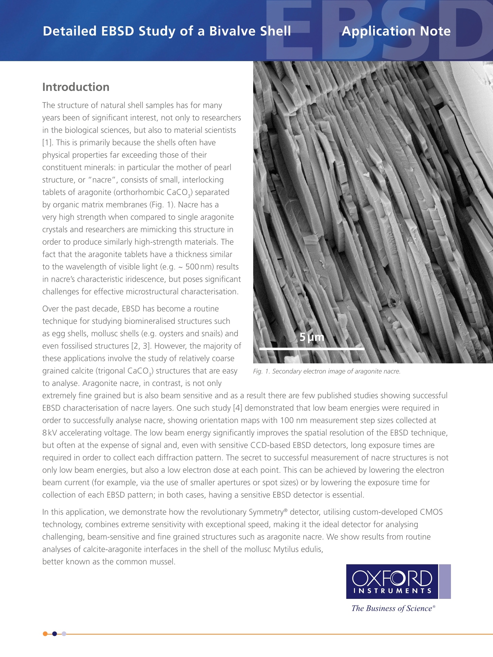
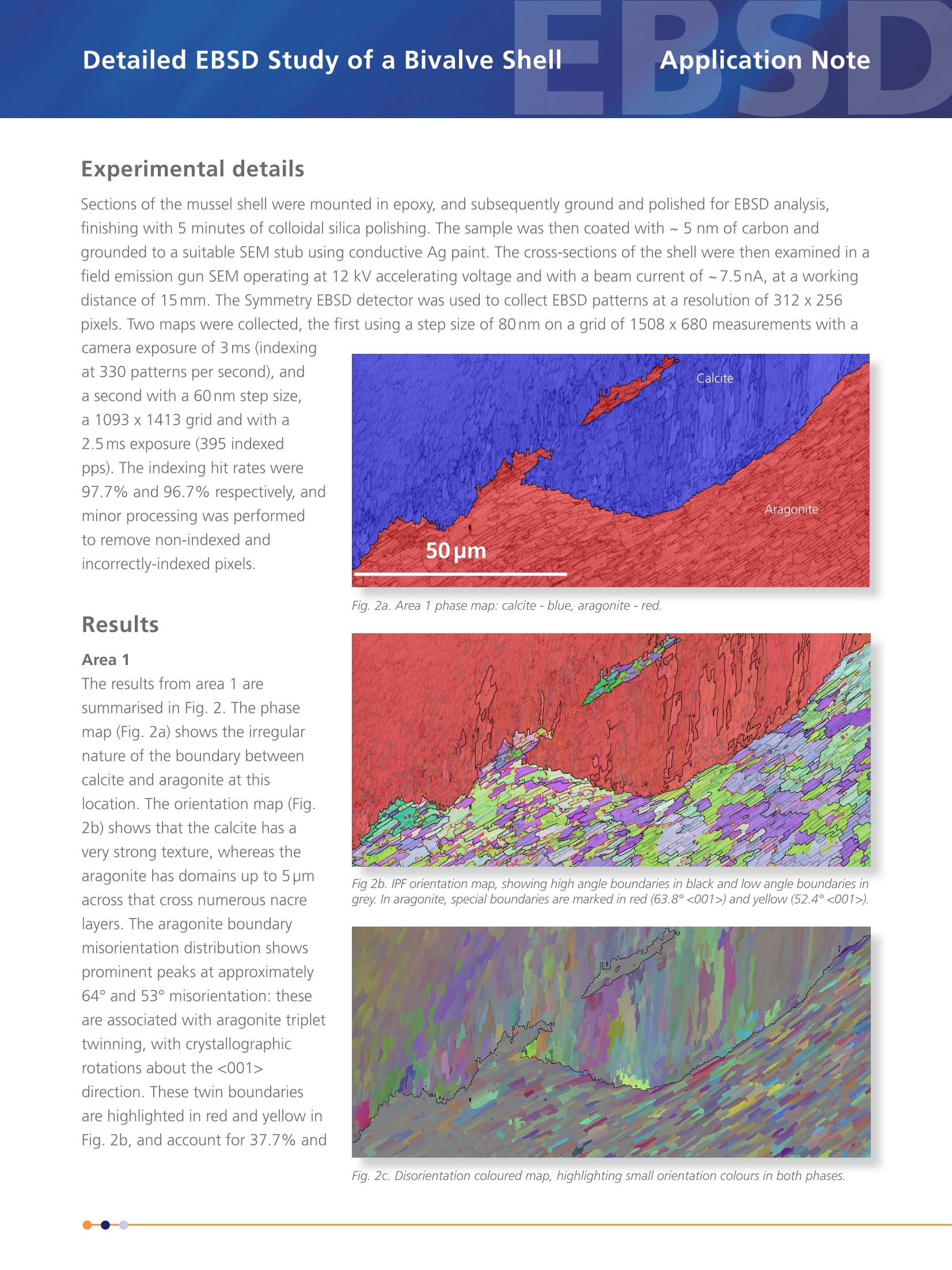
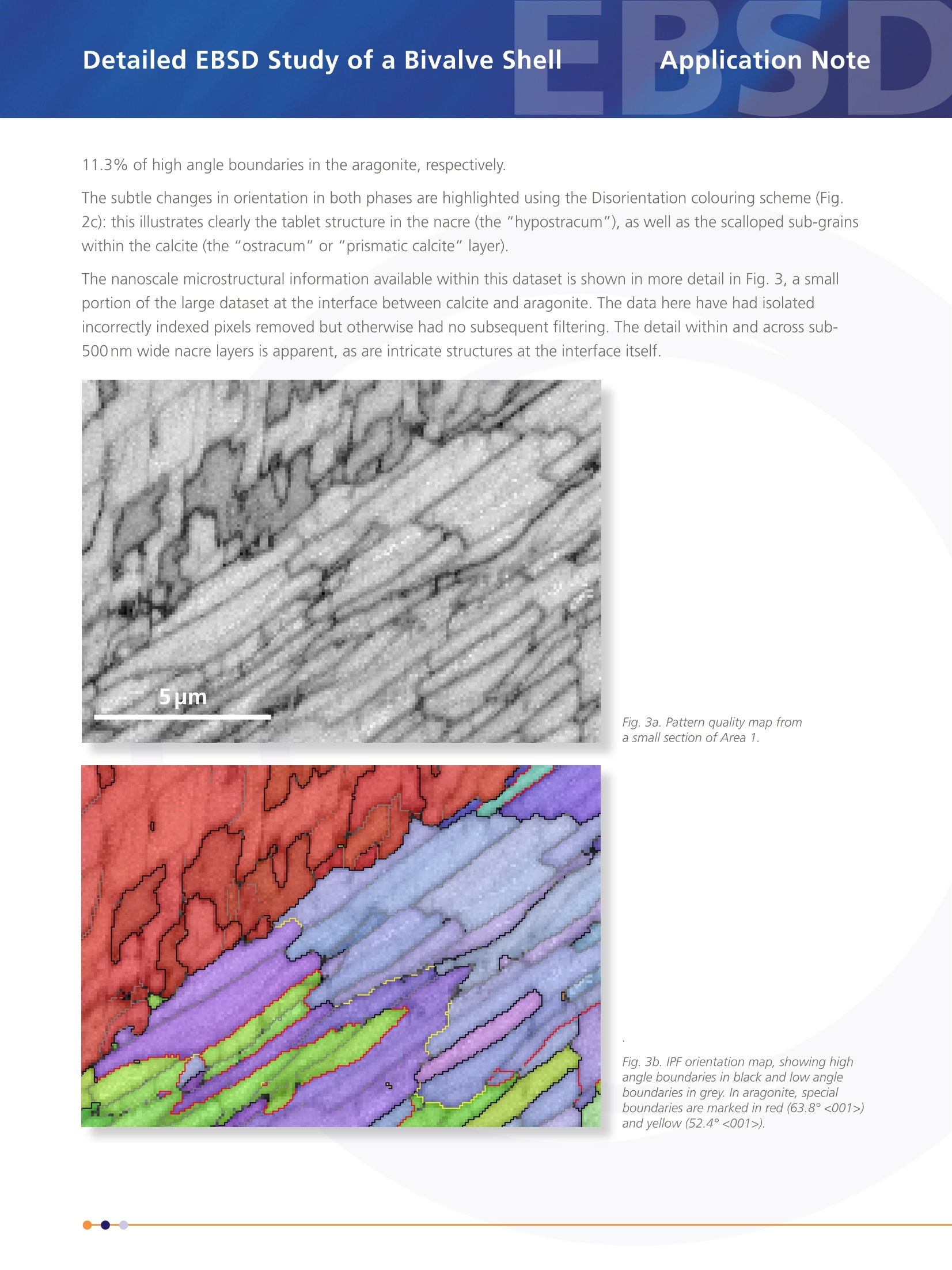
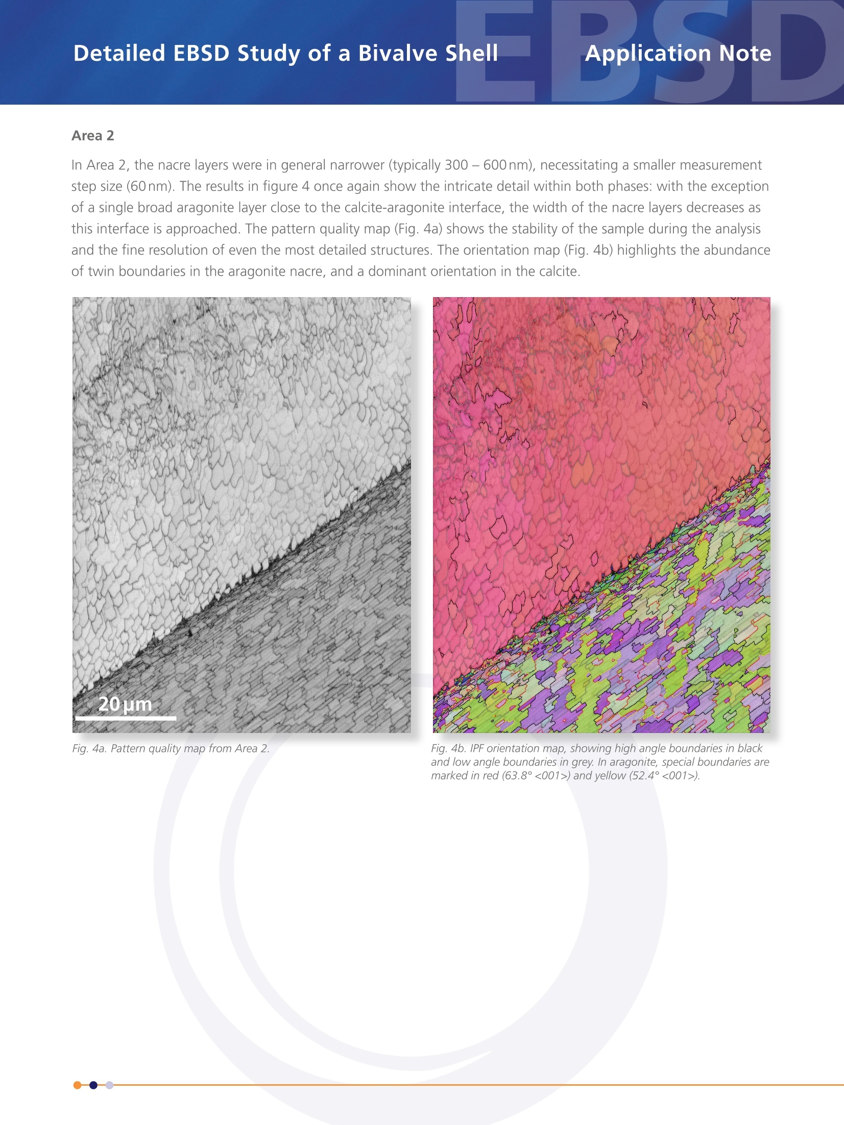
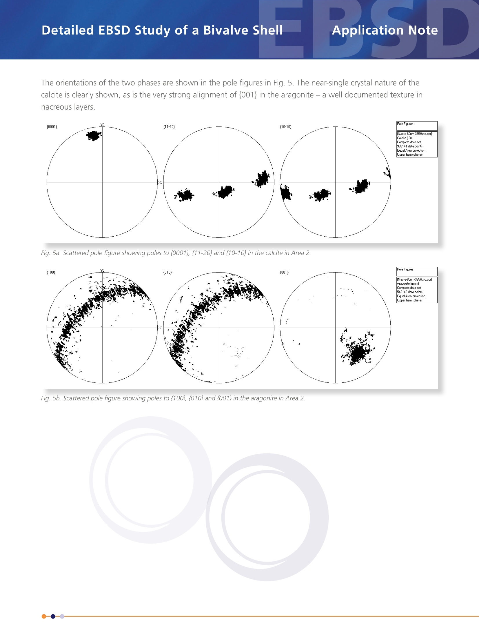
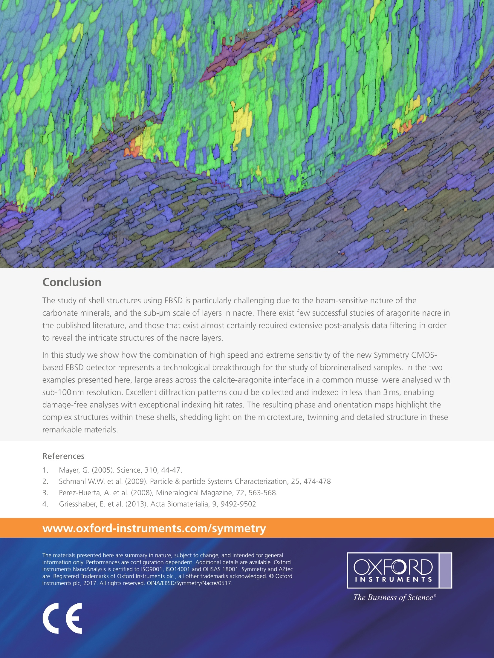
Detailed EBSD Study of a Bivalve Shell Detailed EBSD Study of a Bivalve ShellApplication Note Application Note Fig. 1. Secondary electron image of aragonite nacre. to analyse. Aragonite nacre, in contrast, is not only extremely fine grained but is also beam sensitive and as a result there are few published studies showing successfulEBSD characterisation of nacre layers. One such study [4] demonstrated that low beam energies were required inorder to successfully analyse nacre, showing orientation maps with 100 nm measurement step sizes collected at8kV accelerating voltage. The low beam energy significantly improves the spatial resolution of the EBSD technique,but often at the expense of signal and, even with sensitive CCD-based EBSD detectors, long exposure times arerequired in order to collect each diffraction pattern. The secret to successful measurement of nacre structures is notonly low beam energies, but also a low electron dose at each point. This can be achieved by lowering the electronbeam current (for example, via the use of smaller apertures or spot sizes) or by lowering the exposure time forcollection of each EBSD pattern; in both cases, having a sensitive EBSD detector is essential. In this application, we demonstrate how the revolutionary Symmetry@ detector, utilising custom-developed CMOStechnology, combines extreme sensitivity with exceptional speed, making it the ideal detector for analysingchallenging, beam-sensitive and fine grained structures such as aragonite nacre. We show results from routineanalyses of calcite-aragonite interfaces in the shell of the mollusc Mytilus edulis, better known as the common mussel. at 330 patterns per second), anda second with a 60nm step size,a 1093 x 1413 grid and with a2.5 ms exposure (395 indexedpps). The indexing hit rates were97.7% and 96.7% respectively, andminor processing was performedto remove non-indexed andincorrectly-indexed pixels. Results Area 1The results from area 1 aresummarised in Fig. 2. The phasemap (Fig. 2a) shows the irregularnature of the boundary betweencalcite and aragonite at thislocation. The orientation map (Fig.2b) shows that the calcite has avery strong texture, whereas thearagonite has domains up to 5 pmacross that cross numerous nacrelayers. The aragonite boundarymisorientation distribution showsprominent peaks at approximately64° and 53°misorientation: theseare associated with aragonite triplettwinning, with crystallographicrotations about the <001>direction. These twin boundariesare highlighted in red and yellow inFig.2b, and account for 37.7% and Sections of the mussel shell were mounted in epoxy, and subsequently ground and polished for EBSD analysis,finishing with 5 minutes of colloidal silica polishing. The sample was then coated with ~ 5 nm of carbon andgrounded to a suitable SEM stub using conductive Ag paint. The cross-sections of the shell were then examined in afield emission gun SEM operating at 12 kV accelerating voltage and with a beam current of ~7.5nA, at a workingdistance of 15 mm. The Symmetry EBSD detector was used to collect EBSD patterns at a resolution of 312 x256pixels. Two maps were collected, the first using a step size of 80nm on a grid of 1508 x680 measurements with acamera exposure of 3 ms (indexing Fig. 2a. Area 1 phase map: calcite-blue, aragonite-red. Fig 2b. IPF orientation map, showing high angle boundaries in black and low angle boundaries ingrey. In aragonite, special boundaries are marked in red (63.8°<001>) and yellow (52.4°<001>). 11.3% of high angle boundaries in the aragonite, respectively. The subtle changes in orientation in both phases are highlighted using the Disorientation colouring scheme (Fig.2c): this illustrates clearly the tablet structure in the nacre (the"hypostracum"), as well as the scalloped sub-grainswithin the calcite (the "ostracum" or"prismatic calcite"layer). The nanoscale microstructural information available within this dataset is shown in more detail in Fig. 3, a smallportion of the large dataset at the interface between calcite and aragonite. The data here have had isolatedincorrectly indexed pixels removed but otherwise had no subsequent filtering. The detail within and across sub-500 nm wide nacre layers is apparent, as are intricate structures at the interface itself. Fig. 3a. Pattern quality map froma small section of Area 1. Fig. 3b. IPForientation map, showing highangle boundaries in black and low angleboundaries in grey. In aragonite, specialboundaries are marked in red (63.8°<001>)and yellow (52.4°<001>). Detailed EBSD Study of a Bivalve Shell Area 2 In Area 2, the nacre layers were in general narrower (typically 300-600 nm), necessitating a smaller measurementstep size (60nm). The results in figure 4 once again show the intricate detail within both phases: with the exceptionof a single broad aragonite layer close to the calcite-aragonite interface, the width of the nacre layers decreases asthis interface is approached. The pattern quality map (Fig. 4a) shows the stability of the sample during the analysisand the fine resolution of even the most detailed structures. The orientation map (Fig. 4b) highlights the abundanceof twin boundaries in the aragonite nacre, and a dominant orientation in the calcite. Fig. 4a. Pattern quality map from Area 2. Fig. 4b. IPF orientation map, showing high angle boundaries in blackand low angle boundaries in grey. In aragonite, special boundaries aremarked in red(63.8°<001>) and yellow (52.4°<001>). The orientations of the two phases are shown in the pole figures in Fig. 5. The near-single crystal nature of thecalcite is clearly shown, as is the very strong alignment of{001} in the aragonite- a well documented texture innacreous layers. Fig. 5a. Scattered pole figure showing poles to {0001},{11-20} and {10-10} in the calcite in Area 2. Fig. 5b. Scattered pole figure showing poles to {100}, {010} and {001} in the aragonite in Area 2. Conclusion The study of shell structures using EBSD is particularly challenging due to the beam-sensitive nature of thecarbonate minerals, and the sub-pm scale of layers in nacre. There exist few successful studies of aragonite nacre inthe published literature, and those that exist almost certainly required extensive post-analysis data filtering in orderto reveal the intricate structures of the nacre layers. In this study we show how the combination of high speed and extreme sensitivity of the new Symmetry CMOS-based EBSD detector represents a technological breakthrough for the study of biomineralised samples. In the twoexamples presented here, large areas across the calcite-aragonite interface in a common mussel were analysed withsub-100nm resolution. Excellent diffraction patterns could be collected and indexed in less than 3 ms, enablingdamage-free analyses with exceptional indexing hit rates. The resulting phase and orientation maps highlight thecomplex structures within these shells, shedding light on the microtexture, twinning and detailed structure in theseremarkable materials. References 1. Mayer, G. (2005). Science, 310, 44-47. 2. Schmahl W.W. et al. (2009). Particle & particle Systems Characterization, 25, 474-478 3. Perez-Huerta, A. et al.(2008), Mineralogical Magazine, 72, 563-568. 4. Griesshaber, E. et al. (2013). Acta Biomaterialia, 9, 9492-9502 www.oxford-instruments.com/symmetry The materials presented here are summary in nature, subject to change, and intended for generalinformation only.Performances are configuration dependent.Additional details are available. OxfordInstruments NanoAnalysis is certified to ISO9001, ISO14001 and OHSAS 18001. Symmetry and AZtecare Registered Trademarks of Oxford Instruments plc , all other trademarks acknowledged.@ OxfordInstruments plc, 2017. All rights reserved. OINA/EBSD/Symmetry/Nacre/0517. The Business ofScience@ IntroductionThe structure of natural shell samples has for manyyears been of significant interest, not only to researchersin the biological sciences, but also to material scientists[1]. This is primarily because the shells often havephysical properties far exceeding those of theirconstituent minerals: in particular the mother of pearlstructure, or “nacre”, consists of small, interlockingtablets of aragonite (orthorhombic CaCO3) separatedby organic matrix membranes (Fig. 1).
确定
还剩4页未读,是否继续阅读?

产品配置单

牛津仪器科技(上海)有限公司为您提供《海贝中元素构成检测方案(EBSD系统)》,该方案主要用于其他中元素构成检测,参考标准--,《海贝中元素构成检测方案(EBSD系统)》用到的仪器有EBSD探测器Symmetry S3 牛津仪器
推荐专场


相关方案
更多
该厂商其他方案
更多