


This application note monitors the existence of an intermediate state by probing the refolding of denatured cytochrome c using a JASCO CD spectrometer and the SFS-492 Stopped-Flow system.
方案详情

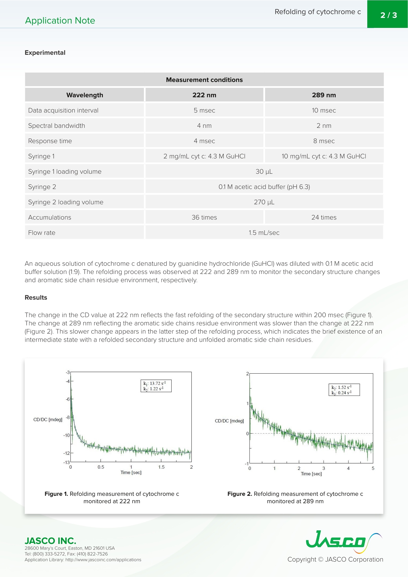
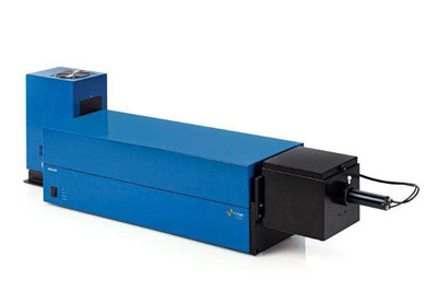
Application NoteCDDSSpectroscopy Refolding of cytochrome c2/3Application Note CD spectra provide information regarding the secondaryand tertiary structure of proteins. While the far-UV regionelucidates structural information of the peptide backbonechain, the near-UV region of the spectrum highlightschanges involving the aromatic amino acid residues.Therefore, coupling a CD spectrometer with a stopped-flowsystem is considered one of the best methods for analysingthe unfolding and refolding of proteins. This system nowprovides not only structural information pertaining to theprotein in question, but also supplies this data on a sub-millisecond time scale. The user can now obtain a moredetailed picture of the time scale for when each proteinunfolds and refolds. This application notes demonstrates the use of the J-1500CD spectrometer and SFS-492 Stopped-Flow system tomonitor the refolding process of cytochrome c. Keywords J-1500, circular dichroism,stopped-flow, SFS-492, proteinfolding, biochemistry, refolding J-1500 CD spectrometer View product information at www.jascoinc.com 28600 Mary’s Court, Easton, MD 21601 USA Experimental Wavelength 222nm 289nm Data acquisition interval 5 msec 10 msec Spectral bandwidth 4nm 2nm Response time 4 msec 8 msec Syringe 1 2 mg/mL cyt c: 4.3 M GuHCI 10 mg/mL cyt c: 4.3 M GuHCI Syringe 1 loading volume 30pL Syringe 2 0.1M acetic acid buffer (pH 6.3) Syringe 2 loading volume 270 uL Accumulations 36 times 24 times Flow rate 1.5 mL/sec An aqueous solution of cytochrome c denatured by guanidine hydrochloride (GuHCI) was diluted with 0.1 M acetic acidbuffer solution (1:9). The refolding process was observed at 222 and 289 nm to monitor the secondary structure changesand aromatic side chain residue environment, respectively. Results The change in the CD value at 222 nm reflects the fast refolding of the secondary structure within 200 msec (Figure 1)The change at 289 nm reflecting the aromatic side chains residue environment was slower than the change at 222 nm(Figure 2). This slower change appears in the latter step of the refolding process, which indicates the brief existence of anintermediate state with a refolded secondary structure and unfolded aromatic side chain residues. Figure 1. Refolding measurement of cytochrome cmonitored at 222 nm Figure 2. Refolding measurement of cytochrome cmonitored at 289 nm 28600 Mary’s Court, Easton, MD 21601 USA Conclusion This application note monitors the existence of an intermediate state by probing the refolding of denatured cytochrome cusing a JASCO CD spectrometer and the SFS-492 Stopped-Flow system. References 1. Elove, G. A., Chaffotte, A. F., Roder, H., and M. E. Goldberg, Biochemistry (1992), 31,6876. 2. Chaffotte, A. F., Guillou,Y., and M. E. Goldberg, Biochemistry (1992), 31,9694. 28600 Mary's Court, Easton, MD 21601 USA JASCO INC.Tel: ( Fax: ( pplication Library: http://www.jascoinc.com/applications This application note monitors the existence of an intermediate state by probing the refolding of denatured cytochrome c using a JASCO CD spectrometer and the SFS-492 Stopped-Flow system.
确定
还剩1页未读,是否继续阅读?

产品配置单

JASCO 公司为您提供《细胞色素C中再折叠检测方案(圆二色光谱仪)》,该方案主要用于其他中再折叠检测,参考标准--,《细胞色素C中再折叠检测方案(圆二色光谱仪)》用到的仪器有JASCO日本分光 圆二色光谱仪/圆二色谱 J-1500
推荐专场
相关方案
更多
该厂商其他方案
更多