


方案详情
文
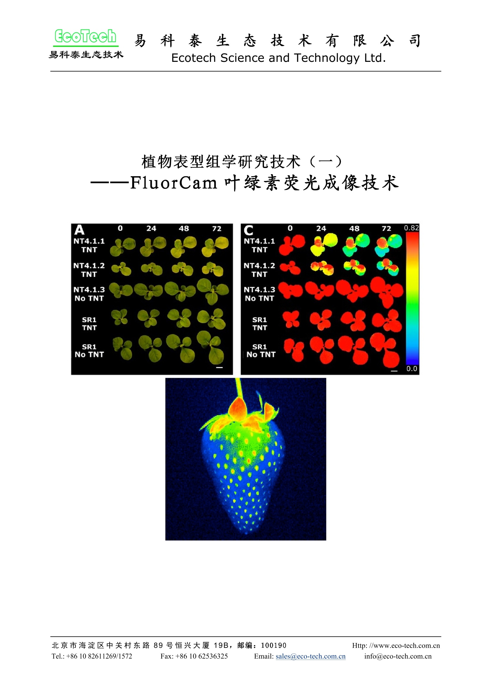
Rousseau等(High throughput quantitative phenotyping of plant resistance using
chlorophyll fluorescence image analysis. Plant Methods, 2013, 9:17),利用 FluorCam开放
式叶绿素荧光成像系统作为高通量表型分析平台,采用图像阈值分割等分析方法,对植物
病原体感染进行了定量分析检测,根据Fv/Fm将感染分为不同阶段/等级,特别是可以将用
其它方法难以分辨出来的感染前期加以分辨,并对5个品种的菜豆对普通细菌性疫病的抗性
进行了定量分析评价。
方案详情

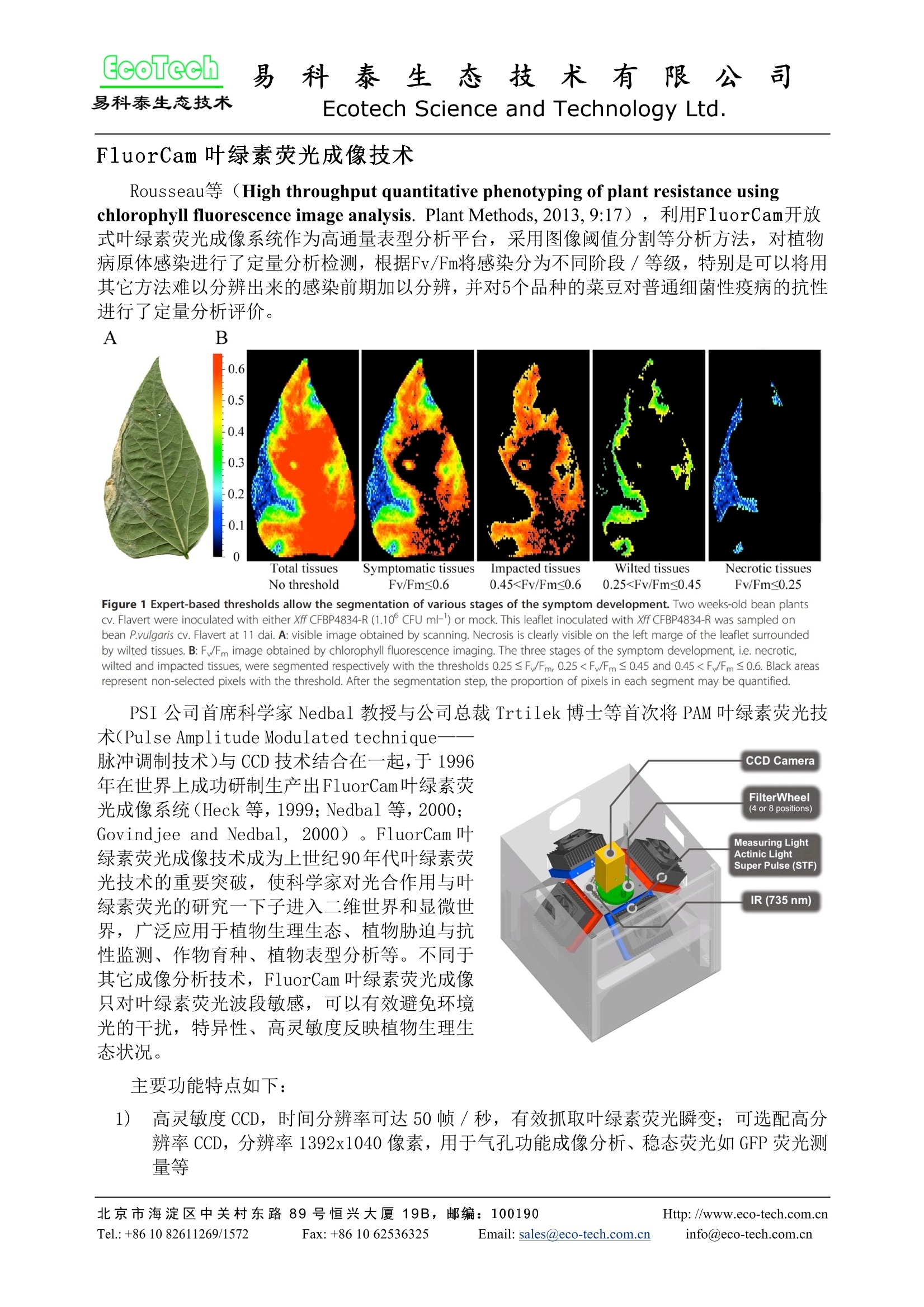
EcoTech易科泰生态技术科泰 生态 技术 有 限 公 司Ecotech Science and Technology Ltd. 易 植物表型组学研究技术(一)——FluorCam叶绿素荧光成像技术 FluorCam 叶绿素荧光成像技术 Rousseau等 (High throughput quantitative phenotyping of plant resistance using chlorophyll fluorescence image analysis. Plant Methods, 2013,9:17),利用FluorCam开放式叶绿素荧光成像系统作为高通量表型分析平台,采用图像阈值分割等分析方法,对植物病原体感染进行了定量分析检测,根据Fv/Fm将感染分为不同阶段/等级,特别是可以将用其它方法难以分辨出来的感染前期加以分辨,并对5个品种的菜豆对普通细菌性疫病的抗性进行了定量分析评价。 A B Figure 1 Expert-based thresholds allow the segmentation of various stages of the symptom development. Two weeks-old bean plantscv. Flavert were inoculated with either Xff CFBP4834-R(1.10° CFU ml-') or mock. This leaflet inoculated with Xff CFBP4834-R was sampled onbean P.vulgaris cv. Flavert at 11 dai. A: visible image obtained by scanning. Necrosis is clearly visible on the left marge of the leaflet surroundedby wilted tissues. B: F/Fm image obtained by chlorophyll fluorescence imaging. The three stages of the symptom development,i.e. necrotic,wilted and impacted tissues, were segmented respectively with the thresholds 0.25 ≤F/Fm, 0.25
确定

还剩6页未读,是否继续阅读?

产品配置单
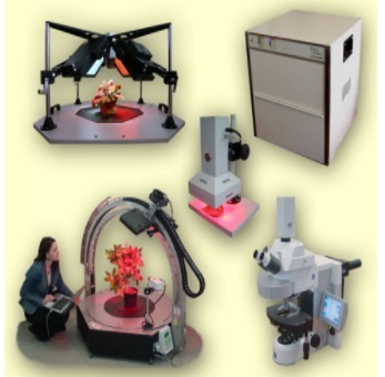
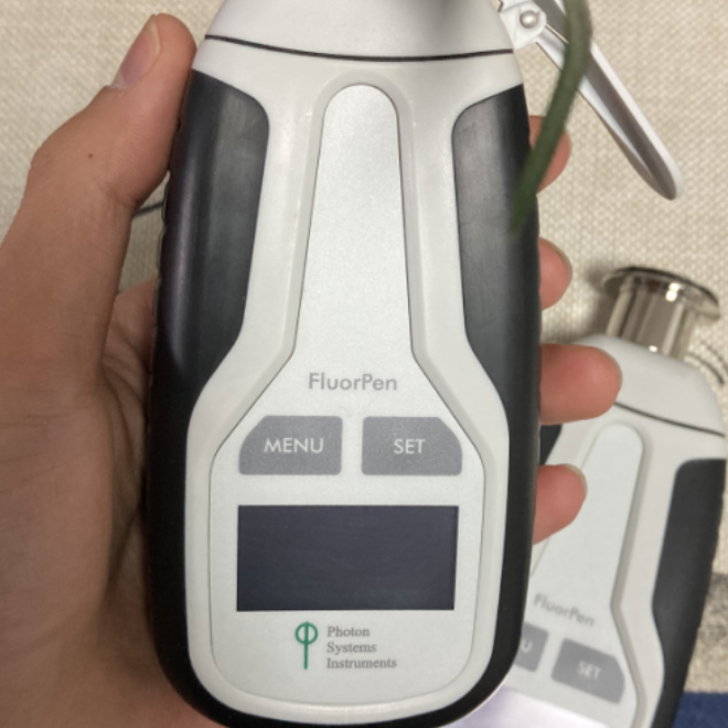
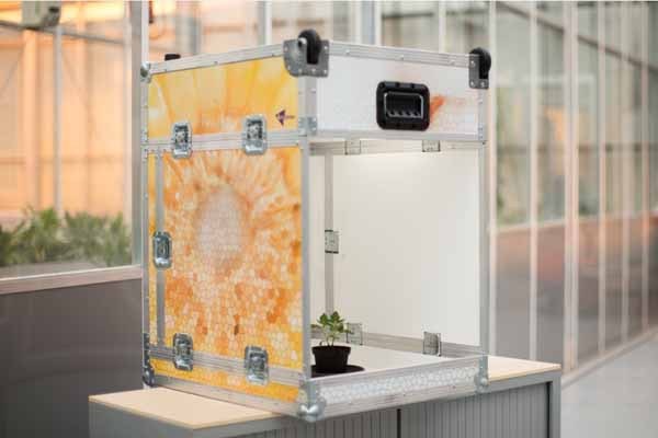
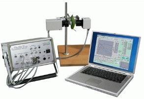
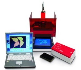
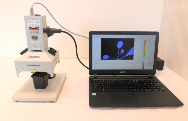
北京易科泰生态技术有限公司为您提供《植物中荧光检测方案(植物荧光成像)》,该方案主要用于豆类中荧光检测,参考标准--,《植物中荧光检测方案(植物荧光成像)》用到的仪器有FluorCam叶绿素荧光技术叶绿素荧光成像系统
推荐专场
相关方案
更多
该厂商其他方案
更多