


方案详情
文
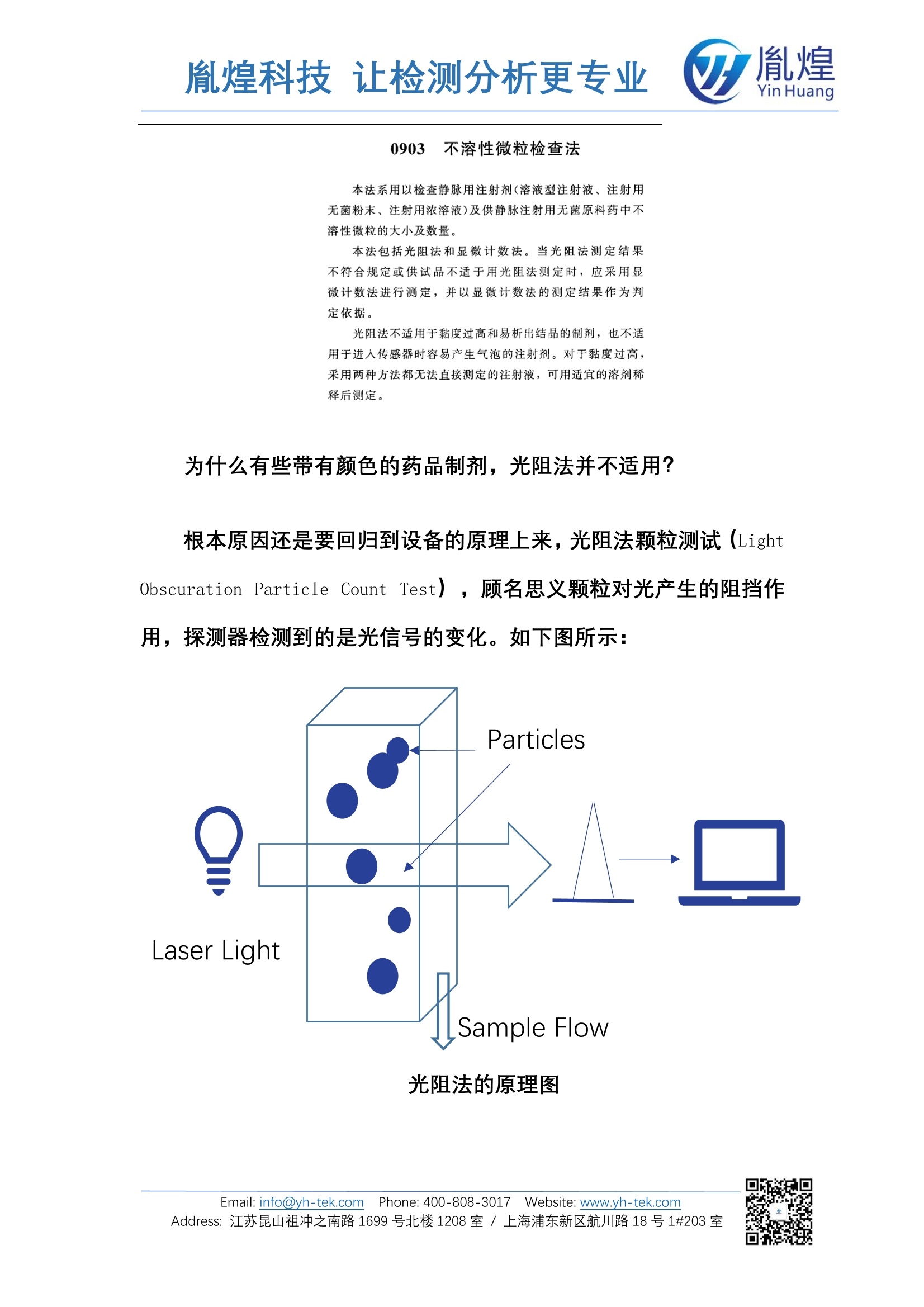
当药品有颜色时,液体介质会对光源有较强的吸收作用,从而降低了接收器接收到的出射光强度,此时获得的光强值变化不准确。
同时,光阻法设备在做标准曲线校准时,常规用的都是聚苯乙烯小球分散在水介质中制备的标准粒子。而水的物理特性参数和带有颜色的药品介质的物理参数有较大差异,因此该校准曲线也并非是有色药品本身的。
从以上两点,均可得出光阻法原理的不溶性微粒仪检测有色样品的准确性需要用显微计数法进行复核验证。且显微计数法得到的结果更准确。因为显微计数法的原理是图像法,有图有真相,眼见为实
方案详情

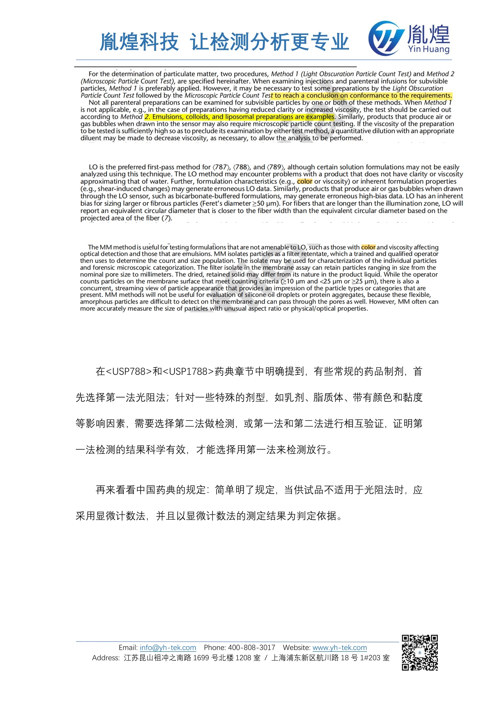
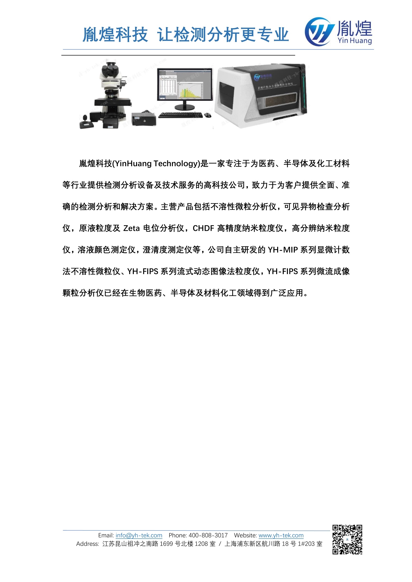
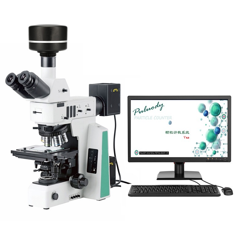
深度解析有色注射液不溶性微粒检测药典规定目前已经在临床上使用的有颜色注射液品种越来越多,如盐酸米托蒽醌注射液,蔗糖铁注射液等。带有颜色样品的实物图针对该类带有颜色的药品制剂,到底该如何检测亚可见微粒及可见异物?先来看看美国药典是如何规定的:和在<USP788>和药典章节中明确提到,有些常规的药品制剂,首先选择第一法光阻法;针对一些特殊的剂型,如乳剂、脂质体、带有颜色和黏度等影响因素,需要选择第二法做检测,或第一法和第二法进行相互验证,证明第一法检测的结果科学有效,才能选择用第一法来检测放行。再来看看中国药典的规定:简单明了规定,当供试品不适用于光阻法时,应采用显微计数法,并且以显微计数法的测定结果为判定依据。为什么有些带有颜色的药品制剂,光阻法并不适用?根本原因还是要回归到设备的原理上来,光阻法颗粒测试(Light Obscuration Particle Count Test),顾名思义颗粒对光产生的阻挡作用,探测器检测到的是光信号的变化。如下图所示:光阻法的原理图当药品有颜色时,液体介质会对光源有较强的吸收作用,从而降低了接收器接收到的出射光强度,此时获得的光强值变化不准确。同时,光阻法设备在做标准曲线校准时,常规用的都是聚苯乙烯小球分散在水介质中制备的标准粒子。而水的物理特性参数和带有颜色的药品介质的物理参数有较大差异,因此该校准曲线也并非是有色药品本身的。从以上两点,均可得出光阻法原理的不溶性微粒仪检测有色样品的准确性需要用显微计数法进行复核验证。且显微计数法得到的结果更准确。因为显微计数法的原理是图像法,有图有真相,眼见为实。检测到的颗粒实物图胤煌科技(YinHuang Technology)是一家专注于为医药、半导体及化工材料等行业提供检测分析设备及技术服务的高科技公司,致力于为客户提供全面、准确的检测分析和解决方案。主营产品包括不溶性微粒分析仪,可见异物检查分析仪,原液粒度及Zeta电位分析仪,CHDF高精度纳米粒度仪,高分辨纳米粒度仪,溶液颜色测定仪,澄清度测定仪等,公司自主研发的YH-MIP系列显微计数法不溶性微粒仪、YH-FIPS系列流式动态图像法粒度仪,YH-FIPS系列微流成像颗粒分析仪已经在生物医药、半导体及材料化工领域得到广泛应用。胤煌Yin Huang胤煌科技支让检测分析更专业 胤煌Yin Huang 深度解析有色注射液不溶性微粒检测药典规定 目前已经在临床上使用的有颜色注射液品种越来越多,如盐酸米托蒽醌注射液,蔗糖铁注射液等。 带有颜色样品的实物图 针对该类带有颜色的药品制剂,到底该如何检测亚可见微粒及可见异物? 先来看看美国药典是如何规定的: 和 胤煌科技支让检测分析更专业 For the determination of particulate matter, two procedures, Method 1 (Light Obscuration Particle Count Test) and Method 2 (Microscopic Particle Count Test), are specified hereinafter. When examining injections and parenteral infusions for subvisible particles, Method 1 is preferably applied. However, it may be necessary to test some preparations by the Light Obscuration Particle Count Test followed by the Microscopic Particle Count Test to reach a conclusion on conformance to the requirements. Not all parenteral preparations can be examined for subvisible particles by one or both of these methods. When Method 1is not applicable, e.g., in the case of preparations having reduced clarity or increased viscosity, the test should be carried outaccording to Method 2. Emulsions, colloids, and liposomal preparations are examples. Similarly, products that produce air orgas bubbles when drawn into the sensor may also require microscopic particle count testing. If the viscosity of the preparationto be tested is sufficiently high so as to preclude its examination by either test method, a quantitative dilution with an appropriatediluent may be made to decrease viscosity, as necessary, to allow the analysis to be performed. LO is the preferred first-pass method for (787),<788), and(789), although certain solution formulations may not be easilyanalyzed using this technique. The LO method may encounter problems with a product that does not have clarity or viscosityapproximating that of water. Further, formulation characteristics (e.g., color or viscosity) or inherent formulation properties(e.g.,shear-induced changes) may generate erroneous LO data. Similarly, products that produce air or gas bubbles when drawnthrough the LO sensor, such as bicarbonate-buffered formulations, may generate erroneous high-bias data. LO has an inherentbias for sizing larger or fibrous particles (Feret’s diameter≥50 pm). For fibers that are longer than the illumination zone, LO willreport an equivalent circular diameter that is closer to the fiber width than the equivalent circular diameter based on theprojected area of the fiber (7). The MM method is useful for testing formulations that are not amenable to LO, such as those with color and viscosity affectingoptical detection and those that are emulsions. MM isolates particles as a filter retentate,which a trained and qualified operatorthen uses to determine the count and size population. The isolate may be used for characterization of the individual particlesand forensic microscopic categorization. The filter isolate in the membrane assay can retain particles ranging in size from thenominal pore size to millimeters. The dried, retained solid may differ from its nature in the product liquid. While the operatorcounts particles on the membrane surface that meet counting criteria (≥10 pm and <25 pm or >25 pm), there is also aconcurrent, streaming view of particle appearance that provides an impression of the particle types or categories that arepresent.MM methods will not be useful for evaluation of silicone oil droplets or protein aggregates, because these flexible,amorphous particles are difficult to detect on the membrane and can pass through the pores as well. However, MM often canmore accurately measure the size of particles with unusual aspect ratio or physical/optical properties. 在和药典章节中明确提到,有些常规的药品制剂,首先选择第一法光阻法;针对一些特殊的剂型,如乳剂、脂质体、带有颜色和黏度等影响因素,需要选择第二法做检测,或第一法和第二法进行相互验证,证明第一法检测的结果科学有效,才能选择用第一法来检测放行。 再来看看中国药典的规定:简单明了规定,当供试品不适用于光阻法时,应采用显微计数法,并且以显微计数法的测定结果为判定依据。 胤煌科技支让检测分析更专业 0903 不溶性微粒检查法 本法系用以检查静脉用注射剂(溶液型注射液、注射用无菌粉末、注射用浓溶液)及供静脉注射用无菌原料药中不溶性微粒的大小及数量。 本法包括光阻法和显微计数法。当光阻法测定结果不符合规定或供试品不适于用光阻法测定时,应采用显微计数法进行测定,并以显微计数法的测定结果作为判定依据。 光阻法不适用于黏度过高和易析出结晶的制剂,也不适用于进人传感器时容易产生气泡的注射剂。对于黏度过高,采用两种方法都无法直接测定的注射液,可用适宜的溶剂稀释后测定。 为什么有些带有颜色的药品制剂,光阻法并不适用? 根本原因还是要回归到设备的原理上来,光阻法颗粒测试(LightObscuration Particle Count Test),顾名思义颗粒对光产生的阻挡作用,探测器检测倒的是光信号的变化。如下图所示: 光阻法的原理图 当药品有颜色时,液体介质会对光源有较强的吸收作用,从而降低了接收器接收到的出射光强度,此时获得的光强值变化不准确。 同时,光阻法设备在做标准曲线校准时,常规用的都是聚苯乙烯小球分散在水介质中制备的标准粒子。而水的物理特性参数和带有颜色的药品介质的物理参数有较大差异,因此该校准曲线也并非是有色药品本身的。 从以上两点,均可得出光阻法原理的不溶性微粒仪检测有色样品的准确性需要用显微计数法进行复核验证。且显微计数法得到的结果更准确。因为显微计数法的原理是图像法,有图有真相,眼见为实。 检测到的颗粒实物图 胤煌科技(YinHuang Technology)是一家专注于为医药、半导体及化工材料等行业提供检测分析设备及技术服务的高科技公司,致力于为客户提供全面、准确的检测分析和解决方案。主营产品包括不溶性微粒分析仪,可见异物检查分析仪,原液粒度及 Zeta 电位分析仪, CHDF 高精度纳米粒度仪,高分辨纳米粒度仪,溶液颜色测定仪,澄清度测定仪等,公司自主研发的 YH-MIP系列显微计数法不溶性微粒仪、YH-FIPS系列流式动态图像法粒度仪, YH-FIPS 系列微流成像颗粒分析仪已经在生物医药、半导体及材料化工领域得到广泛应用。
确定


还剩3页未读,是否继续阅读?

产品配置单

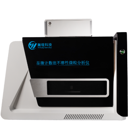
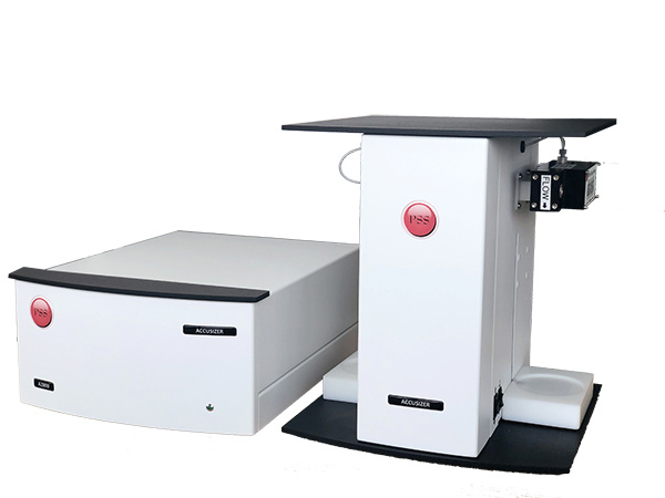
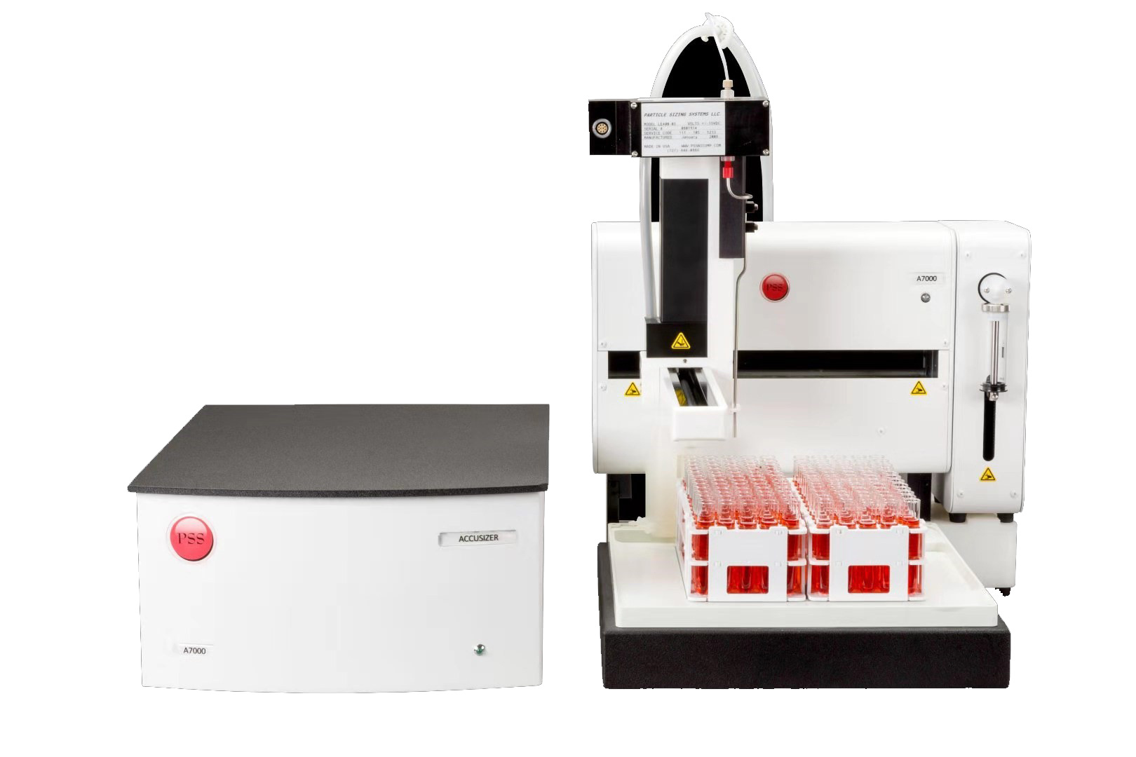
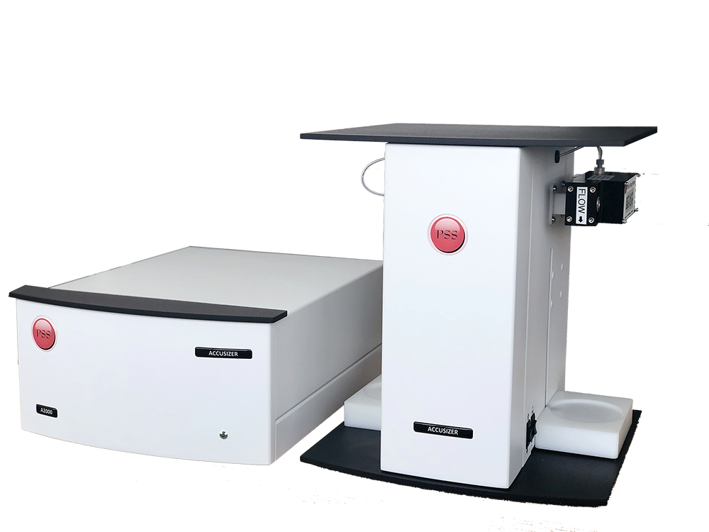
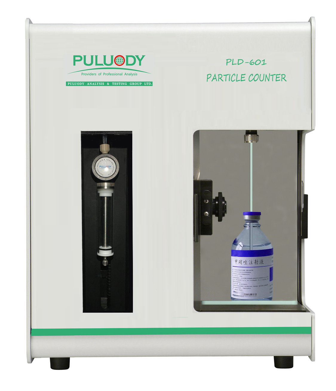
上海胤煌科技有限公司为您提供《深度解析有色注射液不溶性微粒检测药典规定》,该方案主要用于化药制剂中注射剂及特殊剂型相关检测,参考标准--,《深度解析有色注射液不溶性微粒检测药典规定》用到的仪器有全自动显微计数法不溶性微粒分析仪
推荐专场
相关方案
更多
该厂商其他方案
更多