


方案详情
文
本应用文献介绍了半胱氨酸偶联的抗体药物偶联物(ADC)-本妥昔单抗经过疏水作用色谱(HIC)分离后采用基于色谱峰收集的原理对其进行了馏分收集。安捷伦1260生物惰性液相色谱和1260生物惰性馏分收集器不仅能实现馏分收集,同时使生物药物能够在一个完全非金属的流路下完成再分析的工作,且避免HIC高盐体系的腐蚀。安捷伦1260生物惰性馏分收集器做到了高精度的馏分收集,收集的结果通过了相同HIC方法的再分析验证。同时,馏分和ADC的主峰也采用反相色谱进行了再验证。
方案详情

Fraction Analysis of Cysteine-LinkedAntibody-Drug ConjugatesUsing Hydrophobic InteractionChromatography Agilent 1260 Infinity II Bio-Inert System Sonja Schneider Agilent Technologies, Inc Waldbronn, Germany This Application Note describes the peak-based fraction collection of thecysteine-linked antibody-drug conjugate (ADC) brentuximab vedotin after hydro-phobic interaction chromatography (HIC). The combination of the Agilent 1260Infinity II Bio-Inert LC with the Agilent 1260 Infinity II Bio-Inert Fraction Collectorenabled fraction collection as well as re-analysis of biopharmaceuticals in acomplete metal-free flow path while preventing corrosion caused by the highsalt containing buffers used in HIC. The improved features of the 1260 Infinity llBio-Inert Fraction Collector achieved highly precise fraction collection as provedby re-analysis using the same HIC method. The fractionation as well as the iden-tification of the ADC main peaks was confirmed with re-analysis using reversed-phase chromatography. Introduction The availability of possible conjuga-tion sites means that cysteine-linkedantibody-drug conjugates (ADCs) aretypically less heterogenous regarding thedistribution and quantity of conjugatedsmall molecule drugs when compared tolysine-linked ADCs. Different numbers ofdrugs can be conjugated to the mono-l一clonal antibody (mAb) in the conjugationprocess, Therefore, cysteine-linked ADCscontain a mixture of isoforms with 0, 2,4, 6, and 8 drugs attached. In additionto the drug-to-antibody ratio (DAR), theanalysis of the drug distribution is essen-tial to ensure the safety and efficacyof the drug. The reduced complexityenables analysis using high performanceliquid chromatography (HPLC) analyticalmethods with hydrophobic interactionchromatography (HIC), for example, tocharacterize the heterogeneity of theADC. The analysis of ADCs using HICis described in a previous ApplicationNote. The information provided by HICcan be insufficient due to a differentdistribution profile than the expected pro-file with five peaks, representing the DARvalues of0, 2,4,6,and 8. An additionalanalysis might be required. The automation of biomolecule semi-preparative workflows representsmajor challenges in analytical liquidchromatography. Low flow rates andtime-based fraction collection lead toconsiderable limitations in small-scalepreparation. Time-based fractionation isprone to errors due to mistimed fraction-ation points, resulting in potential splitfractions in a single peak. Especially for HIC workflows, where anextremely high amount of salt is used(ammonium sulfate in a concentra-tion of up to 2 M in mobile phase A),the Agilent 1260 Infinity II Bio-Inert LCSystem is an excellent fit to prevent cor-rosion, which is often found in stainlesssteel systems. The 1260 Infinity II Bio-Inert LC in com-bination with the Agilent 1260 Infinity llBio-Inert Fraction Collector allows theuser to perform highly accurate peak-based fraction collection in a com-pletely metal-free flow path. In addition,the 1260 Infinity II Bio-Inert FractionCollector contains improved features toraise peak-based fractionation to a newlevel. This Application Note describes theHIC analysis of brentuximab vedotin,fraction collection of the mainpeaks, and re-analysis using HIC andreversed-phase chromatography. Instrumentation The Agilent 1260 Infinity II Bio-InertLC system consisted of the followingmodules: iAAgilent 1260 Infinity II Bio-InertPump (G5654A) Agilent 1260 Infinity II Bio-InertMultisampler (G5668A) with samplecooler (Option 100) Agilent 1260 Infinity II MulticolumnThermostat (G7116A) with bio-inertheat exchanger (Option 019) Agilent 1260 Infinity II Diode ArrayDetector WR (G7115A) with bio-inertflow cell (Option 028) Agilent 1260 Infinity II Bio-inertFraction Collector (G5664B) Columns Generic HIC column Agilent PLRP-S 300A,2.1×50 mm, 3 um HPLC column(PL1912-1301) Software Agilent OpenLAB CDS ChemStationEdition for LC Systems,version C.01.07 SR3 Sample Brentuximab vedotin (Adcetris) dissolvedin Buffer B at 200 mg/mL Sample PreparationAfter Fractionation The HIC fractions and the pure ADC werereduced with 30 mM tris(2-carboxyethyl)phosphine (TCEP) at 37 C for 1 hourbefore re-analysis by reversed-phasechromatography. Chemicals Trifluoroacetic acid (TFA), sodiumphosphate monobasic and dibasic,ammonium sulfate, and tris(2-carboxy-ethyl)phosphin were purchased fromSigma-Aldrich, St. Louis, Missouri, USA.Fresh ultrapure water was obtainedfrom a Milli-QIntegral system equippedwith an LC-Pak Polisher and a 0.22-ummembrane point-of-use cartridge(Millipak). All solvents used were LCgrade. Acetonitrile and isopropanol werepurchased from Merck, Germany. Results and Discussion Brentuximab vedotin was analyzed usingHIC, revealing five main peaks that corre-spond to the mAb containing 0, 2, 4, 6, or8 MMAE drugs (see Figure 1). The peakswere identified by comparing the HICchromatogram to those for brentuximabvedotin found in literature1.2 Chromatographic conditions Mobile phase A) 2 M ammonium sulfate in 100 mM sodium phosphate pH 7B) 100 mM sodium phosphate pH 7 C) Isopropanol Flow rate 0.4 mL/min Gradient 0minutes-55%A,45%B,0 %C 15 minutes -0 %A,80 %B,20%C20 minutes -0 %A, 80 %B,20%C 21 minutes - 55%A,45%B,0%C Stop time 31 minutes Needle wash mode Standard Wash Injection volume 20 pL Column temperature 30°C Diode array detection 280nm/4nm >0.025 minutes (0.5 seconds response time) (10 Hz) Table 2. Chromatographic conditions for reversed-phase re-analysis of HIC fractions. Chromatographic conditions Mobile phase A) Water+0.1 % TFAB) ACN+ 0.1 % TFA Flow rate 0.3 mL/min Gradient 0 minutes-75%A,25%B1 minutes-60%A,40%B15 minutes -50 %A, 50 %B 16 minutes -20 %A,80 %B Stop time 16 minutes Post time 5 minutes Needle wash mode Standard Wash Injection volume 30 pL Column temperature 90°C Diode array detection 280nm/4nm >0.025 minutes (0.5 seconds response time) (10 Hz) Figure 1. Analysis of brentuximab vedotin on the Agilent 1260 Infinity II Bio-inert LC. D0-D8 refers todifferent DAR species. To enable further analysis, the mainpeaks were collected using the1260 Infinity II Bio-Inert FractionCollector. To find the optimal collectionparameters, the HIC chromatogram ofbrentuximab vedotin was loaded in themethod settings of Agilent OpenLABCDS ChemStation Edition in the FractionPreview option. Figure 2 displays thefraction collection method setup with theloaded HIC chromatogram.Based on thepreviously recorded chromatogram, thefollowing fraction options were chosen: Peak-based fractionation was chosenas the fraction trigger mode. With thismode, the user can estimate and enterbasic parameters for peak thresholdand slope based on experience andthe expected sample concentration. Acombination of threshold and slope waschosen for peak detection,whereas thethresholds (three different thresholds...were chosen), as well as the slopes, weredirectly determined in the previouslyrecorded chromatogram in the fractionpreview. Volume slice recovery fraction mode: Whenever the signal rises above theentered threshold or slope parameters,the fraction mode switches to peak-based, and collects distinct fractionsdepending on the signal. With this option,the compounds of interest are collectedin single fractions, whereas the volumeslice recovery collection prevents anysample loss in case threshold/slopeparameters are improperly set. Fractioncollection starts with volume slice recov-ery at the time specified in the timetable. Figure 2. Fraction collector method settings in Agilent OpenLAB CDS ChemStation Edition, displaying collection timetable, peak trigger modes, and fractionpreview. The latter enables the user to review how changes of threshold,slope, and time settings affect the fraction collection. Figure 3 shows the resulting chromato-gram displaying the collected fractions inthe data analysis. To confirm successful fraction collec-tion, the fractions were re-analyzedusing the same HIC method that wasused for the separation of brentuximabvedotin but disabling fraction collectionin the method setup (Figure 2 shows thisoption). Figure 4 shows the re-analysisusing HIC for brentuximab vedotin aswell as the collected main peaks. There-analysis using HIC demonstratessuccessful fractionation. To confirm the identification foundin literature, additional analysis wasrequired. We used a reversed-phaseanalytical method for additional confir-mation. Reversed-phase HPLC can beapplied to separate and qualify light- andheavy-chain species carrying differentdrug loads. Figure 5 shows a separationof TCEP-reduced brentuximab vedotin,enabling a determination of light chain(LC), light chain plus one small moleculedrug load (LC+1), heavy chain (HC),and heavy chain plus one, two, andthree drug loads (HC+1, HC+2, HC+3)compared to literature3. Figure 3. Collected fraction after HIC with a combination of peak-based fraction detection and volumeslice recover fraction collectionmode. mAU Figure 4. Overlay of the HIC analysis of brentuximab vedotin in blue (lowest chromatogram) as well as thecollected fractions. Figure 6 shows the re-analysis ofthe collected and reduced HIC frac-tions using reversed-phase LC withthe Agilent PLRP-S column, which isspecially suited for very hydrophobicsamples such as ADCs. The re-analysisof the fractions is shown in the smallchromatograms below the HIC frac-tionation. The reversed-phase analysisof the single fractions is in completeaccordance with the HIC separations.The first peak in the HIC chromatogram(DO, fraction C12) contains LC and HCwith no drug load, confirmed by the twomain peaks representing LC and HCin the reversed-phase chromatogram.The re-analysis of the second peak (D2,fraction D3) contains four main peaks,representing LC, LC+1, HC,and HC+1.The re-analysis of the third peak (D4,fraction D7) contains five main peaks,representing LC,LC+1,HC,HC+1, andHC+2. The re-analysis of the fourth peak(D6, fraction D9) contains LC, LC+1,HC+2,HC+3,enabling a total drug loadof six small molecules. In the re-analysis of D8(fraction D11), there are only twomain peaks visible: LC+1 and HC+3,which makes a drug load of eight smallmolecules. In addition to the re-anal-ysis using HIC, the re-analysis using reversed-phase analysis confirms notonly the correct fractionation after theHIC separation of brentuximab vedotin,but also the correct peak labeling in theHIC chromatogram. Figure 5. Separation of Brentuximab vedotin using the Agilent PLRP-Scolumn. Figure 6. Fraction collection of Brentuximab vedotin after HIC and re-analysis using reversed-phase with Agilent PLRP-S column. Conclusion Peak-based fraction collection wasperformed after the HIC separation ofbrentuximab vedotin. The peak-basedfraction trigger mode was enhanced withimproved features for optimal and exactfraction collection. The correct fraction-ation as well as the identification of theADC main peaks was confirmed usingHIC and reversed-phase re-analysis. The Agilent 1260 Infinity II Bio-Inert LCwith the Agilent 1260 Infinity Il Bio-InertFraction Collector was shown to be anoptimal combination for the fractionationand re-analysis of ADCs. 1.Schneider, S. Analysis ofCysteine-linked Antibody DrugConjugates using HydrophobicInteraction ChromatographytyⅡ1on the Agilent 1260 Infinity IlBio-Inert LC, Agilent TechnologiesApplication Note, publication number5991-8493EN,2017. 2. Younes; et al.BrentuximabVedotin (SGN-35) for RelapsedCD30-Positive Lymphomas,N. Engl.J. Med. 2010,363,1812-1821. 3. Wakankar, A.; et al. Analytical meth-ods for physiochemical character-ization of antibody drug conjugates,Landes BioScience, mAbs 2011, 3:2,161-1722. www.agilent.com/chem For Research Use Only. Not for use in diagnostic procedures. 本应用文献介绍了半胱氨酸偶联的抗体药物偶联物(ADC)-本妥昔单抗经过疏水作用色谱(HIC)分离后采用基于色谱峰收集的原理对其进行了馏分收集。Agilent 1260 生物惰性液相色谱和1260生物惰性馏分收集器不仅能实现馏分收集,同时使生物药物能够在一个完全非金属的流路下完成再分析的工作,且避免HIC高盐体系的腐蚀。安捷伦1260生物惰性馏分收集器做到了高精度的馏分收集,收集的结果通过了相同HIC方法的再分析验证。同时,馏分和ADC的主峰也采用反相色谱进行了再验证。
确定


还剩6页未读,是否继续阅读?

产品配置单
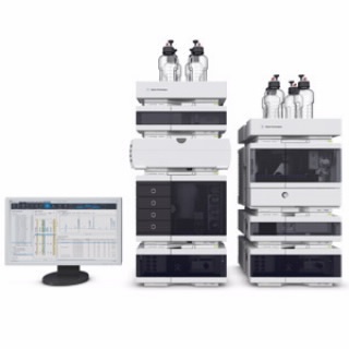
安捷伦科技(中国)有限公司为您提供《半胱氨酸偶联中抗体药物偶联物检测方案(液相色谱仪)》,该方案主要用于其他中含量测定检测,参考标准--,《半胱氨酸偶联中抗体药物偶联物检测方案(液相色谱仪)》用到的仪器有Agilent 1260 Infinity II 液相色谱系统
推荐专场
相关方案
更多
该厂商其他方案
更多