


方案详情
文
分子信标作为一种荧光标记的分子探针,具有极强的特异性和较高的灵敏度,目前已经成为基础医学和生物学的重要研究工具。基于HORIBA Scientific(Jobin Yvon光谱技术)设计生产的荧光光谱仪,讨论了水解ssDNA荧光发射光谱、杂交ssDNA荧光发射光谱以及退火过程中荧光光谱的变化。
方案详情

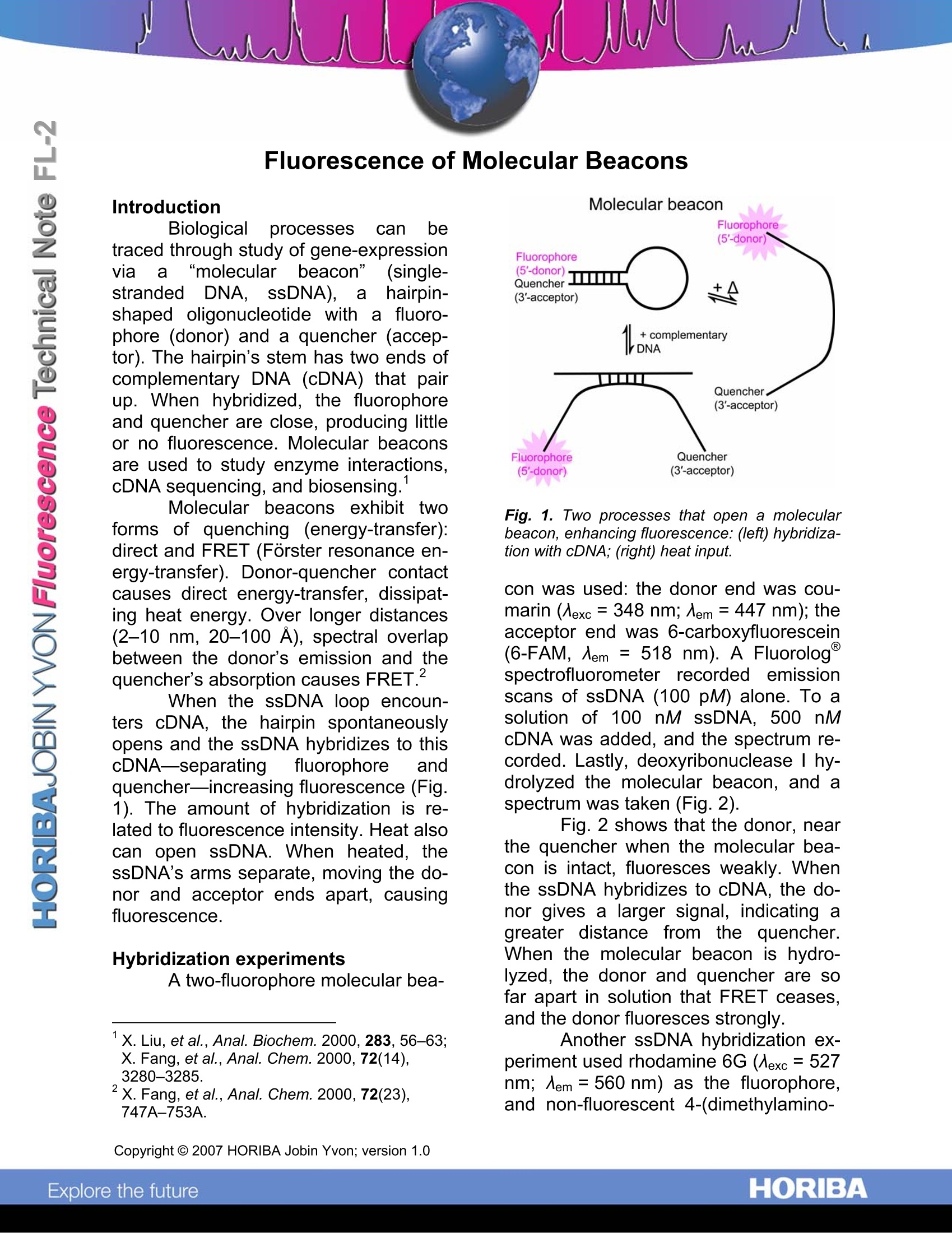
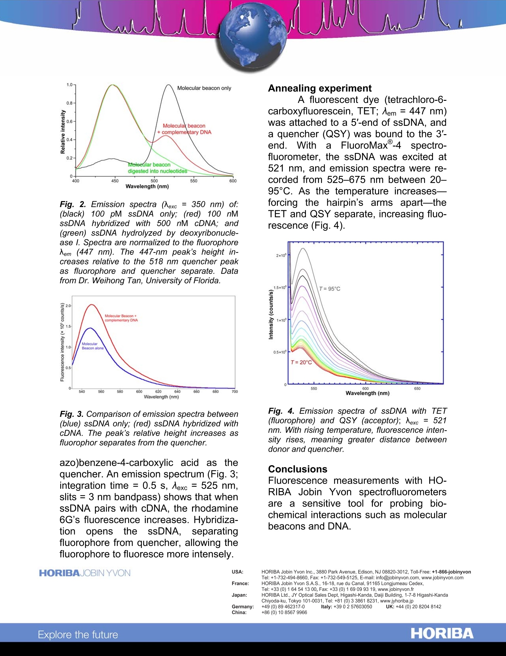
Introduction Biologicalprocesses can betraced through study of gene-expressionvia a “molecularbeacon”(single-strandeDdNA,ssDNA),ahairpin-shaped oligonucleotide with a fluoro-phore (donor) and a quencher (accep-tor). The hairpin’s stem has two ends ofcomplementary DNA (cDNA) that pairup. When hybridized, the fluorophoreand quencher are close, producing littleor no fluorescence. Molecular beaconsare used to study enzyme interactions,cDNA sequencing, and biosensing. Molecular beacons exhibit tytwoforms of quenching (energy-transfer):direct and FRET (Forster resonance en-ergy-transfer). Donor-quencher contactcauses direct energy-transfer, dissipat-ing heat energy. Over longer distances(2-10 nm, 20-100 A), spectral overlapbetween the donor's emission and thequencher’'s absorption causes FRET. When the ssDNA loop encoun-ters cDNA, the hairpin spontaneouslyopens and the ssDNA hybridizes to thiscDNA—separating fluorophore andquencher—increasing fluorescence (Fig.1). The amount of hybridization is re-lated to fluorescence intensity. Heat alsocan open ssDNA. When heated, thessDNA’s arms separate, moving the do-nor and acceptor ends apart, causingtluorescence. Hybridization experimentsAtwo-fluorophore molecular bea- X. Liu, et al.,Anal. Biochem. 2000, 283, 56-63;X. Fang, et al., Anal. Chem.2000,72(14),3280-3285. X. Fang, et al., Anal. Chem. 2000,72(23),747A-753A. Molecular beacon Fig. 1. Two processes that open a molecularbeacon, enhancing fluorescence: (left) hybridiza-tion with cDNA; (right) heat input. con was used: the donor end was cou-marin (Aexc=348 nm; Aem =447 nm); theacceptor end was 6-carboxyfluorescein(6-FAM, Aem = 518 nm). A Fluorologspectrofluorometer recorded emissionscans of ssDNA (100 pM) alone. To asolution of 100 nM ssDNA, 500 nMcDNA was added, and the spectrum re-corded. Lastly, deoxyribonuclease I hy-drolyzed the molecular beacon, and aspectrum was taken (Fig. 2). Fig. 2 shows that the donor, nearthe quencher when the molecular bea-con is intact, fluoresces weakly. Whenthe ssDNA hybridizes to cDNA, the do-nor gives a larger signal, indicating agreater distance from the quencher.When the molecular beacon is hydro-lyzed, the donor and quencher are sofar apart in solution that FRET ceases,and the donor fluoresces strongly. Another ssDNA hybridization ex-periment used rhodamine 6G (Aexc = 527nm; Aem=560 nm) as the fluorophore,and non-fluorescent 4-(dimethylamino- Fig. 2. Emission spectra (Nexc = 350 nm)of:(black)01100 pM ssDNA only; (red)100 nMsSDNAhybridized with 500 nM cDNA;and(green) ssDNA hydrolyzed by deoxyribonucle-ase l. Spectra are normalized to the fluorophoreNem (447 nm). The 447-nm peak’s height in-creases relative to the 518 nm quencher peakas fluorophore and quencher separate. Datafrom Dr. Weihong Tan, University of Florida. Fig. 3. Comparison of emission spectra between(blue) ssDNA only; (red) ssDNA hybridized withcDNA. The peak’s relative height increases asfluorophor separates from the quencher. azo)benzene-4-carboxylic acid asS tthequencher. An emission spectrum (Fig. 3;integration time = 0.5 s, Aexc = 525 nm,slits = 3 nm bandpass) shows that whenssDNA pairs with cDNA, the rhodamine6G's fluorescence increases. Hybridiza-tioni opens1thessDNA, separatingfluorophore from quencher, allowing thefluorophore to fluoresce more intensely. Annealing experiment A fluorescent dye (tetrachloro-6-carboxyfluorescein, TET; 入em= 447 nm)was attached to a 5'-end of ssDNA, anda quencher (QSY) was bound to the 3'-end.WWitha FluoroMax@-4 spectro-fluorometer, the ssDNA was excited at521 nm, and emission spectra were re-corded from 525-675 nm between 20-95C. As the temperature increases-forcing the hairpin’s arms apart-theTET and QSY separate, increasing fluo-rescence (Fig.4). Fig. 4. Emission spectra of ssDNA with TET(fluorophore) and QSY (acceptor); Nexc = 521nm. With rising temperature, fluorescence inten-sity rises, meaning greater distance betweendonor and quencher. Conclusions Fluorescence measurements with HO-RIBA Jobin Yvon spectrofluorometersare a sensitive tool for probing bio-chemical interactions such as molecularbeacons and DNA. Copyright C HORIBA Jobin Yvon; version .HORIBAExplore the future HORIBAJOBIN YVONChina: + ORIBAExplore the future
确定
还剩1页未读,是否继续阅读?

产品配置单

天津东方科捷科技有限公司为您提供《基于Fluoromax-4系统对分子荧光信标的研究》,该方案主要用于其他中--检测,参考标准--,《基于Fluoromax-4系统对分子荧光信标的研究》用到的仪器有HORIBA 科研级荧光光谱仪 FluoroMax+
推荐专场
相关方案
更多