


果皮和果叶中酵母对选定的合成纺织品染料的脱色Decolorization of Selected Synthetic Textile Dyes by Yeasts from Leaves and Fruit Peels
方案详情

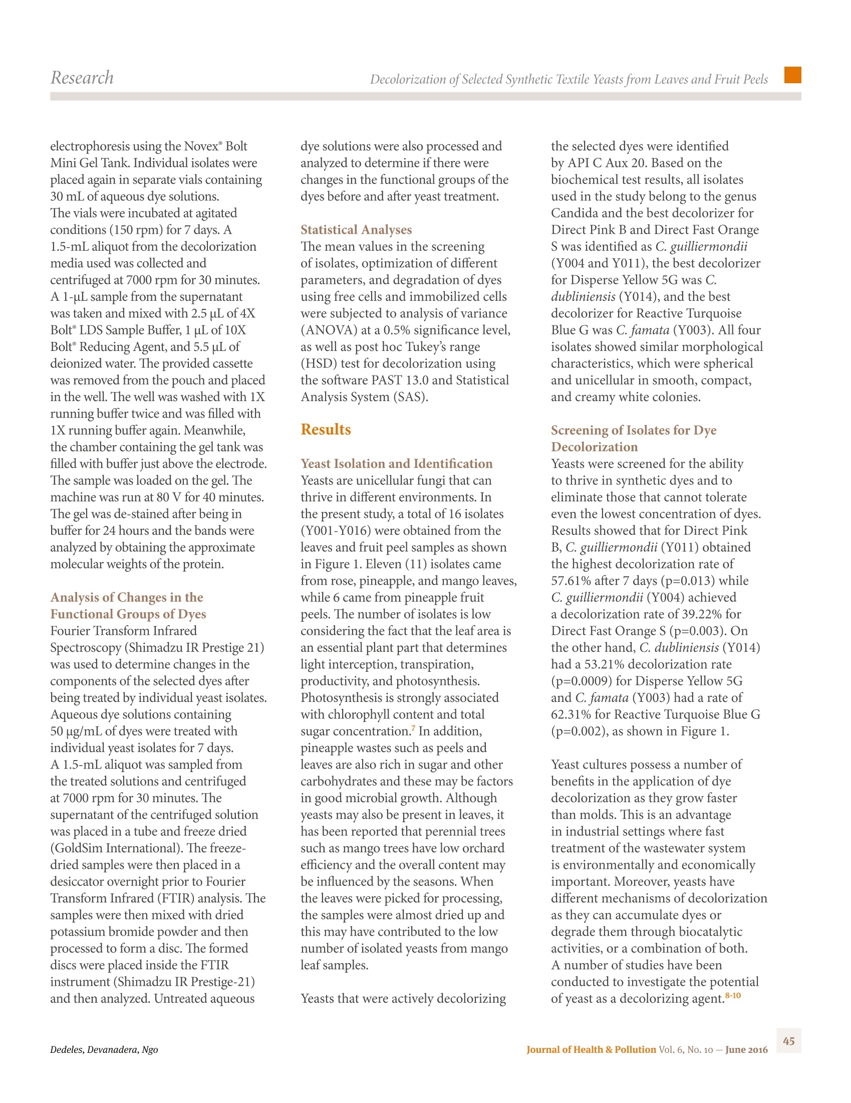
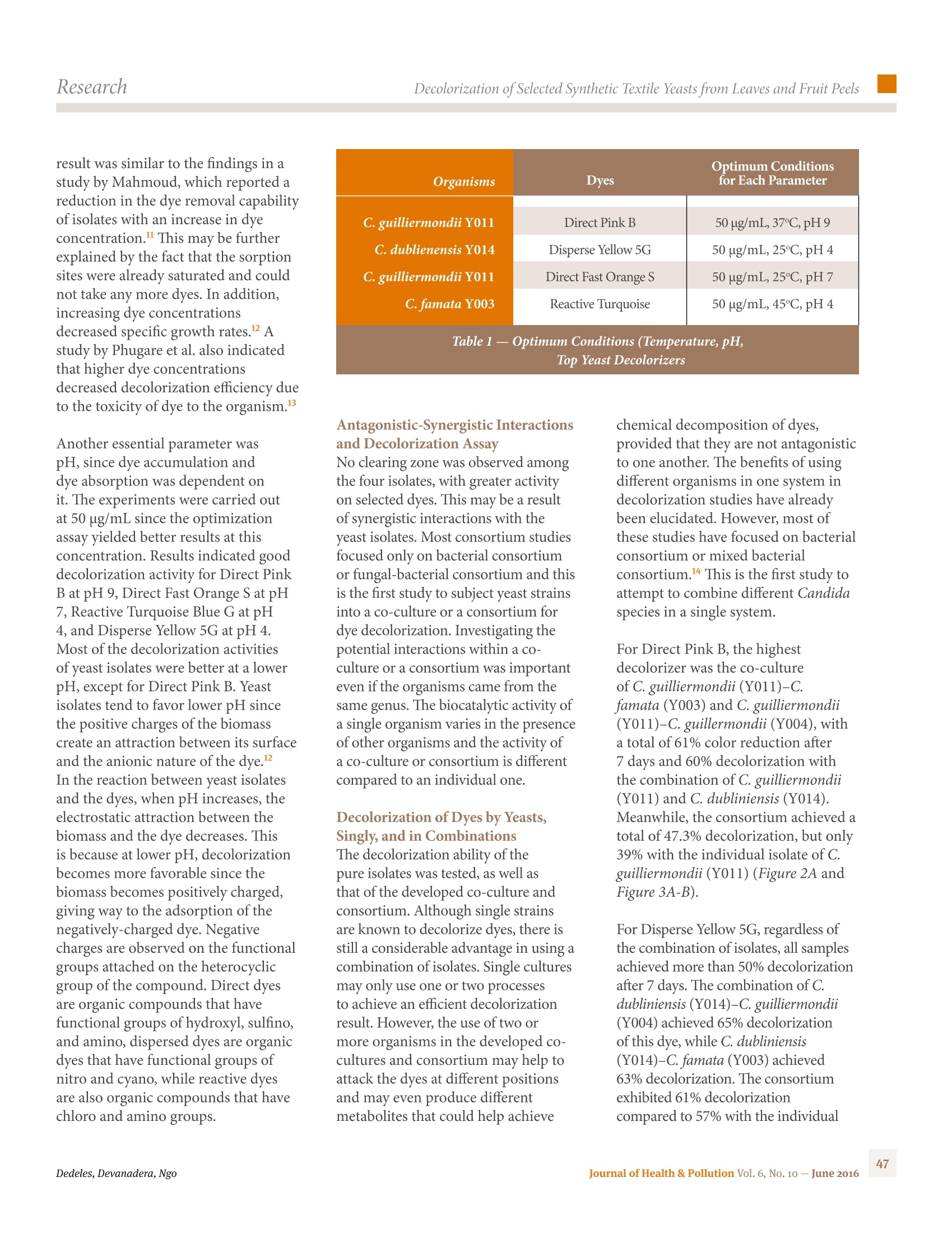
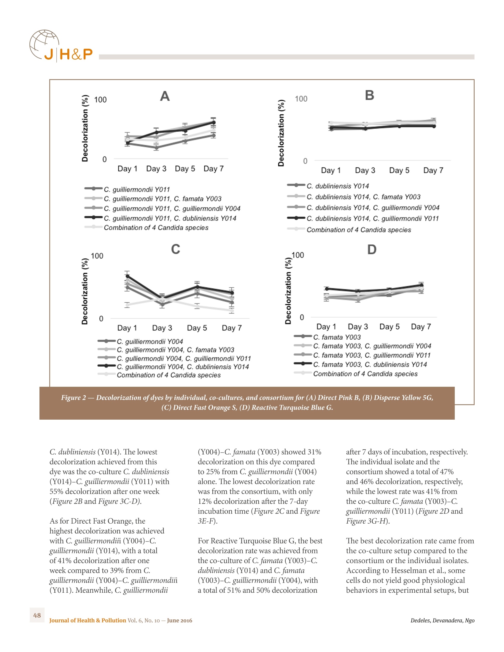
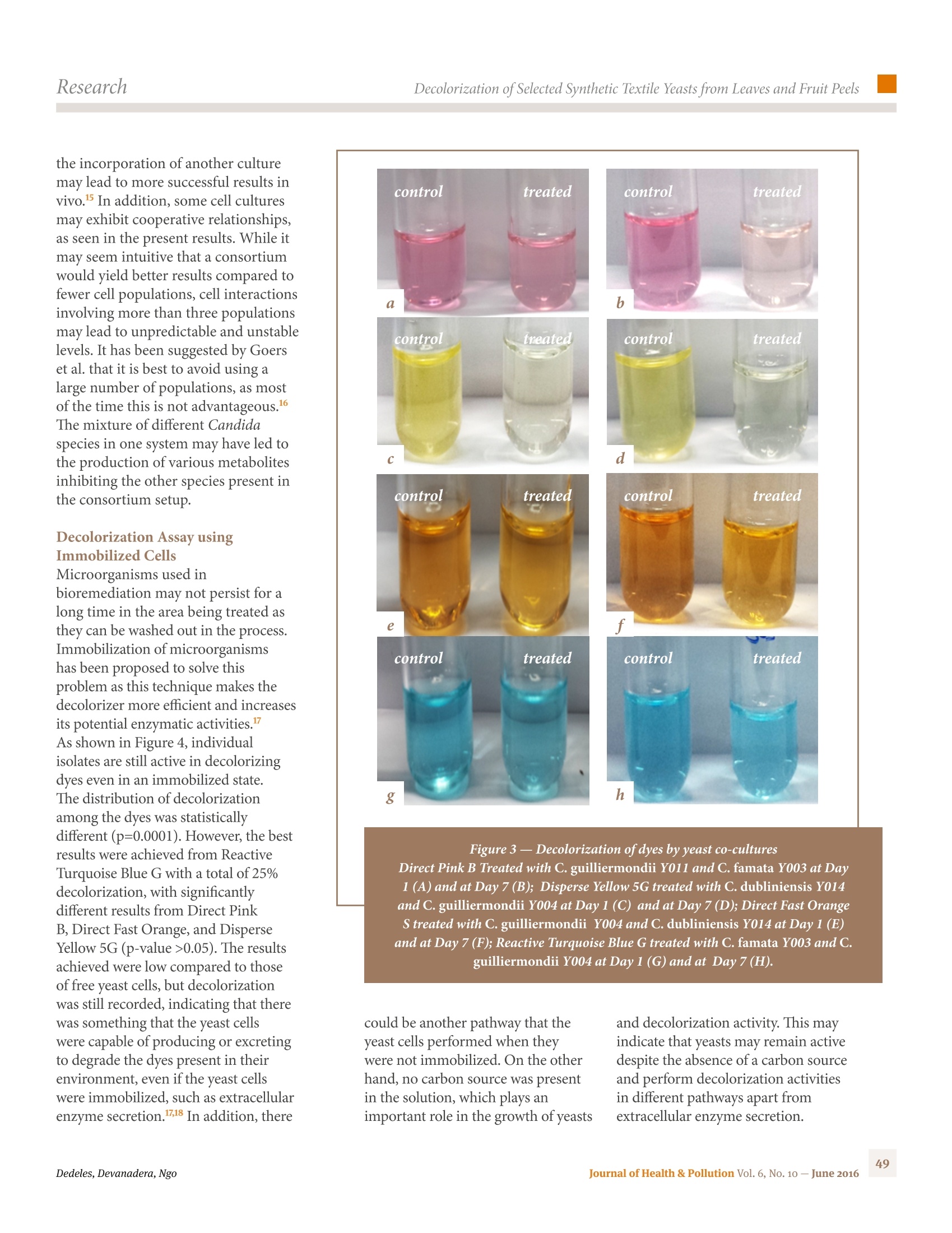
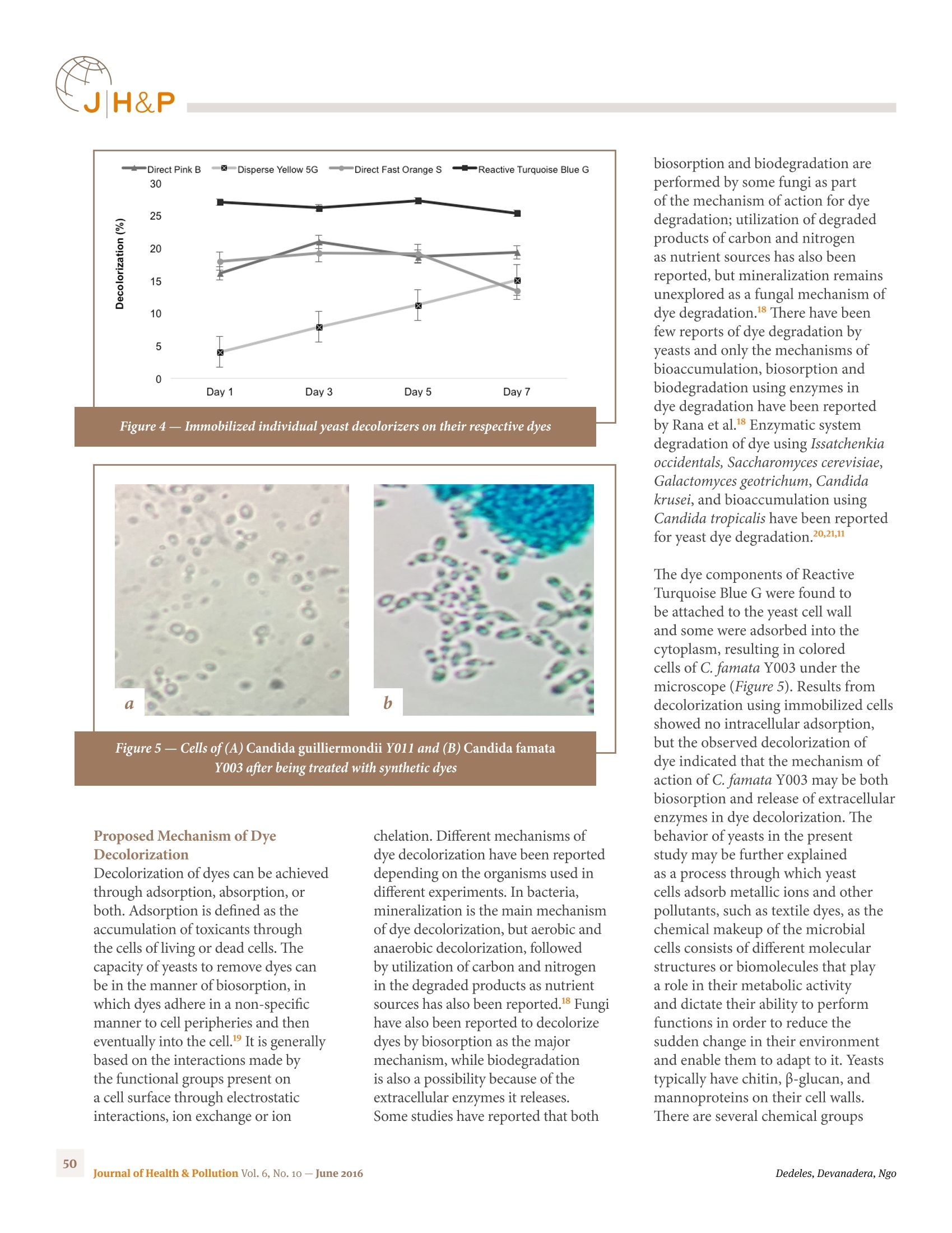
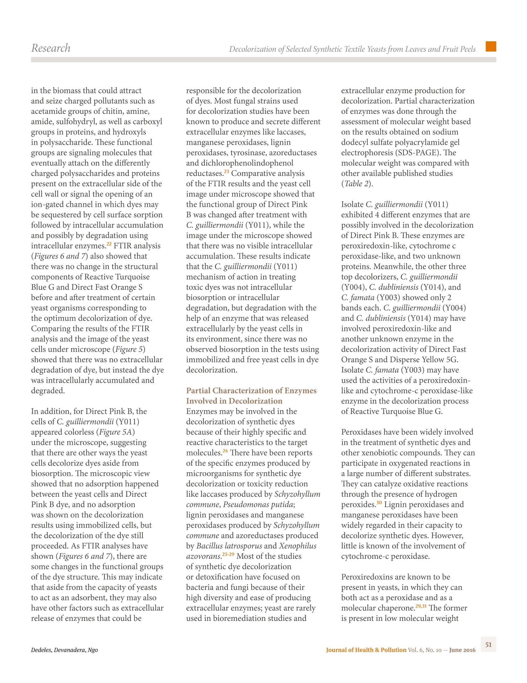
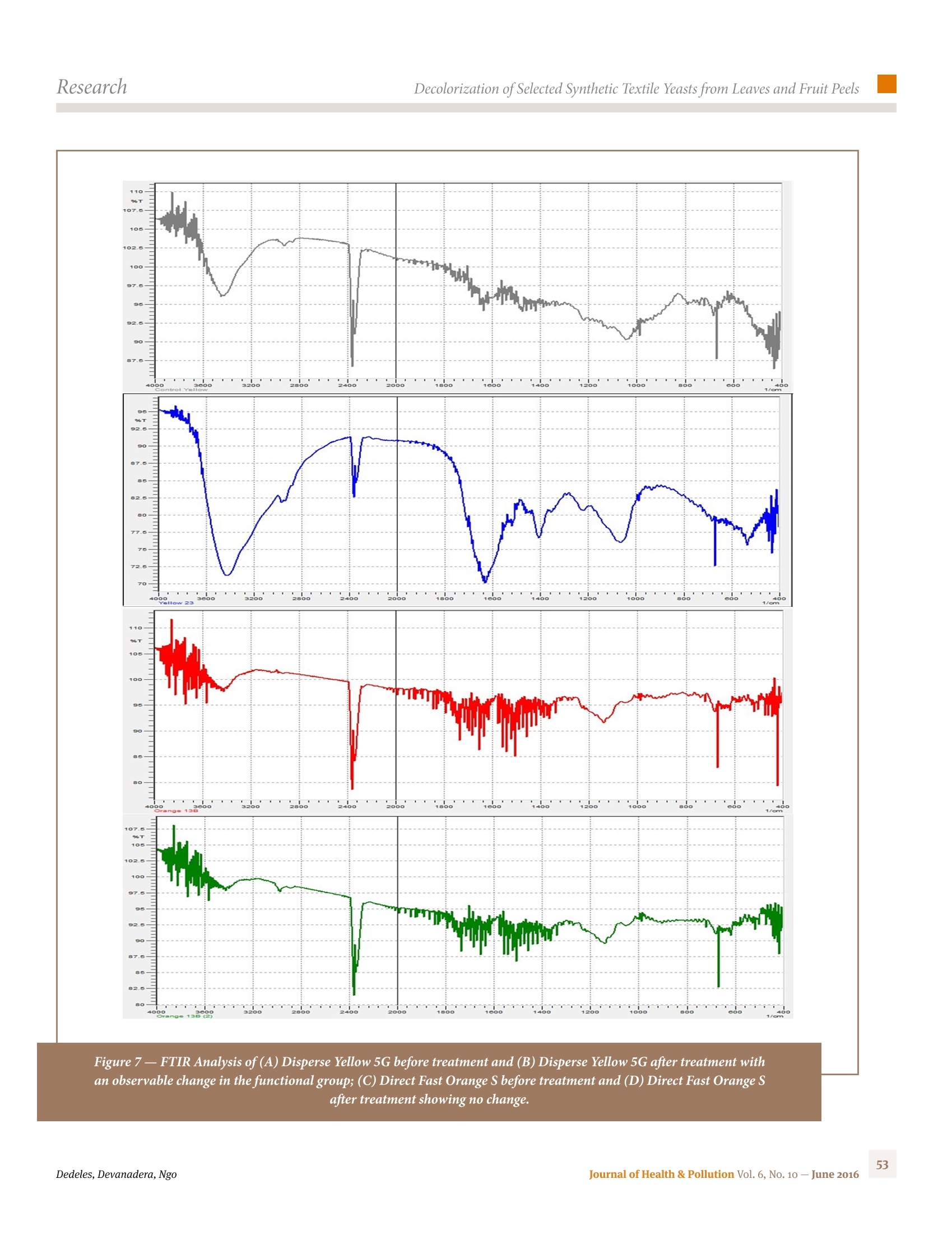
果皮和果叶中酵母对选定的合成纺织品染料的脱色Decolorization of Selected Synthetic Textile Dyes by Yeasts from Leaves and Fruit Peels果皮和果叶中酵母对选定的合成纺织品染料的脱色Research ResearchDecolorization of Selected Synthetic Textile Yeasts from Leaves and Fruit Peels Decolorization of Selected Synthetic Textile Dyes by Yeasts from Leaves and Fruit Peels Anna Christina R. Ngo,1,2 MarkKevin P. Devanadera,1,2 Gina R.Dedeles1,2,3 1 Te hGraduate School, University of Santo Tomas 2 Research Center for the Natural and Applied Sciences, , University of Santo Tomas 3 Department of Biological Sciences,College of Science, University of Santo Tomas, España, Manila,1015 Philippines 圣托马斯大学理学院生物科学系 Corresponding Author:Gina Rio Dedeles Laboratory of Pure and Applied Microbiology, Research Center for the Natural and Applied Sciences, Tohmas Aquinas Research Complex University of Santo Tomas, Manila, 1008 Philippines Tel. +63-(2)-4061611 loc. 8297grdedeles@yahoo.com Introduction Teh textile industry has flourishedalong with the rapid increase in modernization and urbanization ofindustries in terms of yearly production.Azo and anthraquinone dyes are majororganic (synthetic) textile dyes that are commonly used and ofetn preferredin the textile industry because they are more stable, easier to produce, andhave a wider variety of colors than natural dyes. However, discharge orwaste disposal of these dyes into the environment poses a signifciant threatdue to their poor degradability. Tehse dyes are also reported to be toxic and Background. Discharge of textile dyes into the environment poses a signifciant threat.Tehy are poorly biodegradable and toxic due to their complex composition and aromatic structures. In the search for alternatives to physical and chemical treatments, biodegradation of synthetic dyes by various microbes is emerging as an efefctive and promising approach.Objectives. Te hdecolorization of synthetic dyes by yeast co-cultures and consortia from leaves and fruit peels was assessed at a 50 µg/mL dye concentration. Methods. Yeasts isolates from leaves and fruit peels were screened for potential decolorization of synthetic dyes at 25–50 µg/mL. Decolorization parameters were optimized for synergistic properties and development of yeast co-cultures and consortium. Possible decolorization reactions were initially assessed by cell immobilization, sodium dodecyl sulfate polyacrylamide gel electrophoresis (SDS-PAGE), and Fourier transform infrared spectroscopy (FTIR) analysis. Results. A total of 16 organisms were isolated from rose, mango, and pineapple leaves and pineapple fruit peels. Only 4 organisms showed high decolorization of four synthetic dyes: Direct Pink B, Disperse Yellow 5G, Direct Fast Orange S, and Reactive Turquoise Blue G. Te hoptimum condition for best decolorizers of selected dyes at 50 µg/mL were Candida guilliermondii (Y011)for Direct Pink B at pH 9, 37oC; C. dubliniensis (Y014) for Disperse Yellow 5G at pH 4, 25oC; C.guilliermondii (Y004) for Direct Fast Orange S at pH 7, 25oC, and C. famata (Y003) for Reactive Turquoise Blue G at pH 4, 35oC. None of the 4 yeast isolates showed any antagonistic activity when subjected to the lawn-spotting method for the formation of co-cultures and consortium.Te hbest co-cultures obtained 61% decolorization of Direct Pink B, 65% decolorization of Disperse Yellow 5G, 41% decolorization of Direct Fast Orange S, and 50-51% decolorization of Reactive Turquoise Blue G. Immobilized yeast cells were active in decolorizing the dyes and SDS-PAGE analysis confrimed the presence of an extracellular protein. Te hresults of FTIR also showed changes in the functional group of Direct Pink B, but minimal changes in the functional groups of Reactive Turquoise Blue G, indicating a difefrent decolorization pathway.Conclusions. Yeasts in co-cultures and consortia can decolorize toxic synthetic dyes through difefrent decolorization pathways such as enzyme degradation and bioaccumulation. Tish technique may have a use in the treatment of wastewater systems. Competing Interests. Te hauthors declare no competing financial interests. Keywords. dyes, biodegradation, decolorization, microbial consortia J Health Pollution 10: 42–55 (2016) carcinogenic due to their complexchemical composition and aromatic structures.. When these colored efufenltsenter the human system, they may inhibit biological activity and can evencause illnesses such as nausea, skin ulceration, and dermatitis.. Tehrefore,treatments for dye-containing efufenlts are needed. Tehre are several chemicaland physical methods that are used for the removal of dyes in waste watersystems such as adsorption, oxidation,and electrochemical methods. However,these processes lead to high-energy costs, sludge production, and formationof toxic by-products.. Microorganisms are now being tappedas potential agents for the decolorization and degradation of dyes because of their environmentally friendlyprocess and cost-efefctive treatment capability. Studies have reportedthe capability of microorganisms to decolorize a wide range of dyes. In thecourse of the biological treatment of industrial efufenlts, a diverse microbialcommunity is also ofetn noted. Difefrent microorganisms living in the sameenvironment may have variations in their physiological attributes, whereinthey may either compliment or inhibit one another. Microbial strains thatare complimentary can lead to a more efcfieint biodegradation due totheir synergistic interactions.I In the Philippines and elsewhere, however,few studies have been conducted to date to elaborate on the efefctiveness ofmicrobial consortia to decolorize and detoxify hazardous synthetic dyes. Tuhs, this study provides new insights onpollution management in textile plants.It focuses on the use of solitary andcombinations of yeast isolates from leaves and fruit peels as potential decolorizers ofthe synthetic dyes Direct Pink B, Direct Fast Orange S, Disperse Yellow 5G, andReactive Turquoise Blue G. Methods Culture Media and Dyes Sabouraud dextrose agar (SDA) andSaboraud dextrose broth (SDB) used for the isolation and maintenance ofyeasts was purchased from Belman Laboratories (Philippines). Textile dyes Direct Pink B, DirectFast Orange S, Disperse Yellow 5G,and Reactive Turquoise Blue G wereobtained from L.G. Atkimson Import-Export, Inc., (Quezon City, Philippines).Unfortunately, no information was available on either the exact compositionor purity of these specifci dyes. Dye Solution inDecolorization Studies Teh decolorization medium was plain Abbreviations ANOVA Analysis of variance SDA Sabouraud dextrose agar API Analytical proflie index SDB Sabouraud dextrose broth FTIR Fourier transform infrared SDS-PAGE Sodium dodecyl sulfate polyacrylamide gel electrophoresis OD Optical density distilled water, to which the dyes wereadded to arrive at aqueous dye solutions.Tish solution and the media for yeastswere autoclaved at 1210C, 15 pound-force per square inch for 20 minutes. Isolation and Identifciation ofTop Decolorizer Yeasts Source substrata of yeasts such asfresh leaves of rose, pineapple and mango, and pineapple fruit peelswere collected from market waste.Tehse substrates, even in the formof wastes, may be a good source of microorganisms. Tehse sampleswere cut into appropriate sizes,washed three times in sterile distilledwater, and then imprinted (upper surface of leaf pieces) onto platedSDA for 1 minute. Isolation plates were incubated for 24-48 hoursat room temperature. Resulting yeast colonies were confrimed bymicroscopic examination in wet-mount preparations. Confrimed yeastcells were re-streaked onto new SDA for purifciation and eventual isolationinto pure axenic culture in SDA slants maintained in a refrigerated condition. Only the four yeast isolates (onefor each of the four dyes) with the highest decolorization percentageswere identifeid using the analytical proflie index (API) system 20 C Aux(BioMérieux, Marcy l'Etoile, France).Each isolate was grown by streaking onSDA for 24 hours and one colony was inoculated in normal saline solution. Cell suspension (1 mL) from thenormal saline solution was transferred to API C medium and 200 µL fromthe latter was then seeded to each cupule in the identifciation kit. Allidentifciation kits were read afetr 48and 72 hours of incubation at roomtemperature. Binomial identifciations of chosen isolates were provided inthe API database. In addition, the general colonial characteristics andcell morphology of these isolates were studied based on growth in SDA plates. Screening of Yeasts for Growth Tolerance to andDecolorization of Dye Growth tolerance. All yeast isolates(16) were initially screened for growth tolerance by streaking in distilledwater agar (1.5% agar) with 25 µg/mL of the selected dyes Direct PinkB, Direct Fast Orange S, Dispersed Yellow 5G, and Reactive TurquoiseBlue G. Presence or absence of growth was recorded afetr 24-hour incubationof culture plates at room temperature.Isolates with positive growth intriplicate plates for each of the four dyes under study were screened below fordecolorization and used in subsequent experiments; those with no growth wereexcluded. Decolorization Included isolates were screened in 50µg/mL aqueous solutions of the dyes.Cell suspensions enriched for 24 hoursin SDB under agitation condition were centrifuged at 7000 rpm. Pellets ofbiomass were washed two times with distilled water and 0.1 g of each peryeast isolate was added in test vials with 30 mL aqueous dye solution. Controlvials with dye solutions were not seeded.All vials were incubated with agitation(150 rpm, Gerhadt Laboshake) at room temperature and 1.5 mL aliquotsamples were taken afetr 7 days. Cell biomass was removed from all samplesby centrifugation at 7000 rpm for 30minutes and the supernate in test vialsand dye solution in control vials were pipetted to corresponding wells in a96-well microtiter plate for ultraviolet-visible spectrophotometry at difefrentwavelengths of 343, 376, 399, and 400nm, respectively, for Disperse Yellow5G, Direct Pink B, Reactive Turquoise Blue G, and Direct Fast Orange S.Decolorization percentages afetr 7 days were computed as follows: Percent (%) decolorization = Initial optical density (OD)(0h)– Final OD(t)x 100 Initial OD(0h) where, initial OD(0h)and Final OD(t)are absorbance readings at 0 and 48hours of incubation, respectively. Optimization of Assays forDecolorization by the Best Decolorizers Only the top four decolorizer yeastisolates in the assay above were re-assayed for optimum decolorization,each tried with only the specifci dye it decolorized most in the decolorizationassay described above. Triplicate sets of test and control vials were preparedfor each yeast. First-set vials were for optimization of pH (4, 7, and 9)at room temperature, the 2nd set for optimization of temperature (25,37, and 450C), and the 3rd set for optimization of dye concentration(50, 100, and 150 µg/mL), likewise in aqueous solution. Aliquot samples fromtest and control vials were processed as for the initial decolorization assay.Ultraviolet-visible spectrophotometry for optical density readings of processedsamples in 96-well microtiter plates was performed on days 1, 3, 5, and7. Decolorization percentages were calculated as described above. Assay for Synergistic and AntagonisticInteractions Tish assay was done prior to testsfor decolorization and degradation of the dyes by the yeasts, singly or incombinations (dual and in consortia of all four top isolates). Te h“lawn-spotting”assay by Long and Azam was adapted with modifciation because not all theyeasts could be homogenized completely in cell suspension in normal salinesolution. SDA plates were used for all possible dual combinations (“antagonist”and “antagonized”, and roles in reverse).Teh “antagonized” sample (0.1 g biomassin 10 mL normal saline solution) was swabbed on the agar medium, and the“antagonist” sample (1.0 g in 10 mL normal saline solution), spotted onthe lawn of the “antagonized” sample.Presence or absence of zone of inhibitionwas noted afetr 24 hours incubation of assay plates at room temperature.Absence of zone of inhibition indicated possible synergistic interaction, and itspresence, antagonism. Decolorization of Dyes by Yeasts,Singly, and in Combinations Isolates which did not exhibit anyantagonistic interactions to one another in dual combinations weresubjected again to decolorization assay.Teh top four individual isolates, co-cultures, and consortium were used.Teh co-cultures were composed oftwo decolorizers for each dye, while the yeast consortia contained all thefour isolates together in one medium.Teh isolates were pre-enrichedindividually in falsks containing SDB.Teh falsks were placed in a rotaryshaker (150 rpm) for 24 hours at room temperature. Afetr incubation, the cells were harvested at 7000 rpm for30 minutes and washed twice with distilled water. For single isolates, theorganism was standardized to 0.1 g biomass and then inoculated in thedistilled water containing 50 µg/mL of the selected dye. For co-culturesand consortia, the isolates were also standardized to 0.1 g biomass and theninoculated as well in dye solution. Teh vials containing the dye solutions wereplaced again in a rotary shaker (150rpm) at room temperature for oneweek. Control vials with dye solutions were not seeded. Decolorizations weremonitored on days 1, 3, 5, and 7. Decolorization withImmobilized Yeasts Teh method used in the present studywas adapted from a study by Tan et al. Calcium alginate was used for theentrapment of yeast cells. Individual isolates were pre-enriched in separatefalsks containing SDB under agitation condition (150 rpm) for 24 hours. Cellsuspensions were centrifuged at 7000rpm for 30 minutes. Pellets of biomasswere washed twice with distilled water and 0.1 g of each per yeast isolate wasused. Teh cell suspension was mixed with an equal volume of calciumalginate solution. Teh mixture was dropped by means of a syringe intoa calcium chloride solution to form the beads. Teh beads were washedwith sterile normal saline solution and then placed in vials containing30 mL of aqueous dye solutions. All vials were incubated with agitation(150 rpm, Gerhadt Laboshake) at room temperature and 1.5-mL aliquotsamples were monitored likewise with readings on the 1st, 3rd, 5th, and 7thday of incubation. For control tests,another batch of beads was prepared,but without the cell suspension. Partial Characterization ofExtracellular Enzymes Teh extracellular enzymes of each topdecolorizer were assessed through electrophoresis using the Novex® BoltMini Gel Tank. Individual isolates were placed again in separate vials containing30 mL of aqueous dye solutions.Teh vials were incubated at agitatedconditions (150 rpm) for 7 days. A 1.5-mL aliquot from the decolorizationmedia used was collected and centrifuged at 7000 rpm for 30 minutes.A 1-µL sample from the supernatant was taken and mixed with 2.5 µL of 4XBolt® LDS Sample Bufefr, 1 µL of 10X Bolt® Reducing Agent, and 5.5 µL ofdeionized water. Te hprovided cassette was removed from the pouch and placedin the well. Te hwell was washed with 1X running bufefr twice and was fliled with1X running bufefr again. Meanwhile,the chamber containing the gel tank wasfliled with bufefr just above the electrode.Teh sample was loaded on the gel. Tehmachine was run at 80 V for 40 minutes.Teh gel was de-stained afetr being inbufefr for 24 hours and the bands were analyzed by obtaining the approximatemolecular weights of the protein. Analysis of Changes in theFunctional Groups of Dyes Fourier Transform InfraredSpectroscopy (Shimadzu IR Prestige 21)was used to determine changes in thecomponents of the selected dyes afetr being treated by individual yeast isolates.Aqueous dye solutions containing 50 µg/mL of dyes were treated withindividual yeast isolates for 7 days.A 1.5-mL aliquot was sampled fromthe treated solutions and centrifuged at 7000 rpm for 30 minutes. Tehsupernatant of the centrifuged solution was placed in a tube and freeze dried(GoldSim International). Te hfreeze-dried samples were then placed in adesiccator overnight prior to Fourier Transform Infrared (FTIR) analysis. Tehsamples were then mixed with dried potassium bromide powder and thenprocessed to form a disc. Teh formed discs were placed inside the FTIRinstrument (Shimadzu IR Prestige-21)and then analyzed. Untreated aqueous dye solutions were also processed andanalyzed to determine if there were changes in the functional groups of thedyes before and afetr yeast treatment. Statistical Analyses Teh mean values in the screeningof isolates, optimization of difefrent parameters, and degradation of dyesusing free cells and immobilized cells were subjected to analysis of variance(ANOVA) at a 0.5% signifciance level,as well as post hoc Tukey’s range(HSD) test for decolorization using the software PAST 13.0 and StatisticalAnalysis System (SAS). Results Yeast Isolation and Identifciation Yeasts are unicellular fungi that can thrive in difefrent environments. Inthe present study, a total of 16 isolates (Y001-Y016) were obtained from theleaves and fruit peel samples as shown in Figure 1. Eleven (11) isolates camefrom rose, pineapple, and mango leaves,while 6 came from pineapple fruitpeels. Teh number of isolates is low considering the fact that the leaf area isan essential plant part that determines light interception, transpiration,productivity, and photosynthesis.Photosynthesis is strongly associatedwith chlorophyll content and total sugar concentration. In addition,pineapple wastes such as peels and leaves are also rich in sugar and othercarbohydrates and these may be factors in good microbial growth. Althoughyeasts may also be present in leaves, it has been reported that perennial treessuch as mango trees have low orchard efcfieincy and the overall content maybe influenced by the seasons. When the leaves were picked for processing,the samples were almost dried up and this may have contributed to the lownumber of isolated yeasts from mango leaf samples. Yeasts that were actively decolorizing the selected dyes were identifeidby API C Aux 20. Based on the biochemical test results, all isolatesused in the study belong to the genus Candida and the best decolorizer forDirect Pink B and Direct Fast Orange S was identifeid as C. guilliermondii(Y004 and Y011), the best decolorizer for Disperse Yellow 5G was C.dubliniensis (Y014), and the best decolorizer for Reactive TurquoiseBlue G was C. famata (Y003). All four isolates showed similar morphologicalcharacteristics, which were spherical and unicellular in smooth, compact,and creamy white colonies. Screening of Isolates for DyeDecolorization Yeasts were screened for the abilityto thrive in synthetic dyes and to eliminate those that cannot tolerateeven the lowest concentration of dyes.Results showed that for Direct PinkB, C. guilliermondii (Y011) obtained the highest decolorization rate of57.61% afetr 7 days (p=0.013) while C. guilliermondii (Y004) achieveda decolorization rate of 39.22% for Direct Fast Orange S (p=0.003). Onthe other hand, C. dubliniensis (Y014)had a 53.21% decolorization rate(p=0.0009) for Disperse Yellow 5G and C. famata (Y003) had a rate of62.31% for Reactive Turquoise Blue G (p=0.002), as shown in Figure 1. Yeast cultures possess a number ofbeneftis in the application of dye decolorization as they grow fasterthan molds. Tish is an advantage in industrial settings where fasttreatment of the wastewater system is environmentally and economicallyimportant. Moreover, yeasts have difefrent mechanisms of decolorizationas they can accumulate dyes or degrade them through biocatalyticactivities, or a combination of both.A number of studies have beenconducted to investigate the potential of yeast as a decolorizing agent.8-10 Figure 1 — Screening of yeast isolates to difefrent dyes. (A) Direct Pink B, (B) Disperse Yellow 5G, (C) Direct Fast Orange S,(D) Reactive Turquoise Blue G. Blank spaces indicate that the corresponding isolate did not grow on agar with 25 µg/mL of dye and was not included on the seconding screening at 50 µg/mL of dye. (Note: * signifeis that data was statistically signifciant) Some studies have examined whethermicroorganisms can use dye as their sole carbon source. It has also beenreported that yeast cannot grow without the presence of a carbon orenergy source. However, the results obtained from the screening assays inthe present study proved that yeasts can thrive in the presence of dyeseven without the addition of glucose or another easily metabolized carbonsource since their metabolic activities may change and adapt to anothersource of carbon in the screening medium. Optimization of DecolorizationActivity Teh culture conditions wereoptimized to determine the best parameters for the decolorizationof dyes or degradation condition of the isolates, as summarized in Table1. First, incubation temperature was an important parameter asit contributes to the growth of microorganisms, enzyme production,and rate of dye decolorization by the microorganisms. Direct Pink B dyewith C. guilliermondii (Y011) had high decolorization at 370C, Disperse Yellow 5G with C. dubliniensisshowed the highest decolorization at 250C, Direct Fast Orange S showedhighest decolorization at 250C and for Reactive Turquoise Blue G, the highestdecolorization of dye was obtained at 450C. Second, varying dye concentrationshave an efefct on the activity of a microorganism to decolorizeand degrade them. All isolates in the present study yielded betterdecolorization at 50 µg/mL compared to higher dye concentrations. Tish result was similar to the findings in astudy by Mahmoud, which reported a reduction in the dye removal capabilityof isolates with an increase in dye concentration."" Tish may be furtherexplained by the fact that the sorption sites were already saturated and couldnot take any more dyes. In addition,increasing dye concentrationsdecreased specifci growth rates."" A study by Phugare et al. also indicatedthat higher dye concentrations decreased decolorization efcfieincy dueto the toxicity of dye to the organism.33 Another essential parameter waspH, since dye accumulation and dye absorption was dependent onit. Teh experiments were carried out at 50 µg/mL since the optimizationassay yielded better results at this concentration. Results indicated gooddecolorization activity for Direct Pink B at pH 9, Direct Fast Orange S at pH7, Reactive Turquoise Blue G at pH 4, and Disperse Yellow 5G at pH 4.Most of the decolorization activities of yeast isolates were better at a lowerpH, except for Direct Pink B. Yeast isolates tend to favor lower pH sincethe positive charges of the biomass create an attraction between its surfaceand the anionic nature of the dye.22In the reaction between yeast isolatesand the dyes, when pH increases, the electrostatic attraction between thebiomass and the dye decreases. Tish is because at lower pH, decolorizationbecomes more favorable since the biomass becomes positively charged,giving way to the adsorption of the negatively-charged dye. Negativecharges are observed on the functional groups attached on the heterocyclicgroup of the compound. Direct dyes are organic compounds that havefunctional groups of hydroxyl, sulfino,and amino, dispersed dyes are organicdyes that have functional groups of nitro and cyano, while reactive dyesare also organic compounds that have chloro and amino groups. and Decolorization Assay No clearing zone was observed among the four isolates, with greater activityon selected dyes. Tish may be a result of synergistic interactions with theyeast isolates. Most consortium studies focused only on bacterial consortiumor fungal-bacterial consortium and this is the frist study to subject yeast strainsinto a co-culture or a consortium for dye decolorization. Investigating thepotential interactions within a co-culture or a consortium was importanteven if the organisms came from the same genus. Teh biocatalytic activity ofa single organism varies in the presence of other organisms and the activity ofa co-culture or consortium is difefrent compared to an individual one. Decolorization of Dyes by Yeasts,Singly, and in Combinations Teh decolorization ability of thepure isolates was tested, as well as that of the developed co-culture andconsortium. Although single strains are known to decolorize dyes, there isstill a considerable advantage in using a combination of isolates. Single culturesmay only use one or two processes to achieve an efcfieint decolorizationresult. However, the use of two or more organisms in the developed co-cultures and consortium may help to attack the dyes at difefrent positionsand may even produce difefrent metabolites that could help achieve chemical decomposition of dyes,provided that they are not antagonistic to one another. Teh beneftis of usingdifefrent organisms in one system in decolorization studies have alreadybeen elucidated. However, most of these studies have focused on bacterialconsortium or mixed bacterial consortium.or Tish is the frist study toattempt to combine difefrent Candida species in a single system. For Direct Pink B, the highest decolorizer was the co-cultureof C. guilliermondii (Y011)–C.famata (Y003) and C. guilliermondii(Y011)–C. guillermondii (Y004), with a total of 61% color reduction afetr7 days and 60% decolorization with the combination of C. guilliermondii(Y011) and C. dubliniensis (Y014).Meanwhile, the consortium achieved atotal of 47.3% decolorization, but only 39% with the individual isolate of C.guilliermondii (Y011) (Figure 2A and Figure 3A-B). For Disperse Yellow 5G, regardless ofthe combination of isolates, all samples achieved more than 50% decolorizationafetr 7 days. Te hcombination of C.dubliniensis (Y014)–C. guilliermondii(Y004) achieved 65% decolorization of this dye, while C. dubliniensis(Y014)–C. famata (Y003) achieved 63% decolorization. Te hconsortiumexhibited 61% decolorization compared to 57% with the individual C. dubliniensis (Y014). Te hlowestdecolorization achieved from this dye was the co-culture C. dubliniensis(Y014)–C. guilliermondii (Y011) with 55% decolorization afetr one week(Figure 2B and Figure 3C-D). As for Direct Fast Orange, thehighest decolorization was achieved with C. guilliermondiii (Y004)–C.guilliermondii (Y014), with a total of 41% decolorization afetr oneweek compared to 39% from C.guilliermondii (Y004)–C. guilliermondiii(Y011). Meanwhile, C. guilliermondii (Y004)–C. famata (Y003) showed 31%decolorization on this dye compared to 25% from C. guilliermondii (Y004)alone. Te hlowest decolorization rate was from the consortium, with only12% decolorization afetr the 7-day incubation time (Figure 2C and Figure3E-F). For Reactive Turquoise Blue G, the bestdecolorization rate was achieved from the co-culture of C. famata (Y003)–C.dubliniensis (Y014) and C. famata (Y003)–C. guilliermondii (Y004), witha total of 51% and 50% decolorization afetr 7 days of incubation, respectively.Teh individual isolate and the consortium showed a total of 47%and 46% decolorization, respectively,while the lowest rate was 41% fromthe co-culture C. famata (Y003)–C.guilliermondii (Y011) (Figure 2D andFigure 3G-H). Teh best decolorization rate came fromthe co-culture setup compared to the consortium or the individual isolates.According to Hesselman et al., some cells do not yield good physiologicalbehaviors in experimental setups, but the incorporation of another culturemay lead to more successful results in vivo.. In addition, some cell culturesmay exhibit cooperative relationships,as seen in the present results. While itmay seem intuitive that a consortium would yield better results compared tofewer cell populations, cell interactions involving more than three populationsmay lead to unpredictable and unstable levels. It has been suggested by Goerset al. that it is best to avoid using a large number of populations, as mostof the time this is not advantageous.16Teh mixture of difefrent Candidaspecies in one system may have led to the production of various metabolitesinhibiting the other species present in the consortium setup. Decolorization Assay usingImmobilized Cells Microorganisms used inbioremediation may not persist for a long time in the area being treated asthey can be washed out in the process.Immobilization of microorganismshas been proposed to solve this problem as this technique makes thedecolorizer more efcfieint and increases its potential enzymatic activities.""As shown in Figure 4, individual isolates are still active in decolorizingdyes even in an immobilized state.Teh distribution of decolorizationamong the dyes was statistically difefrent (p=0.0001). However, the bestresults were achieved from Reactive Turquoise Blue G with a total of 25%decolorization, with signifciantly difefrent results from Direct PinkB, Direct Fast Orange, and Disperse Yellow 5G (p-value >0.05). Teh resultsachieved were low compared to those of free yeast cells, but decolorizationwas still recorded, indicating that there was something that the yeast cellswere capable of producing or excreting to degrade the dyes present in theirenvironment, even if the yeast cells were immobilized, such as extracellularenzyme secretion."",18 In addition, there could be another pathway that theyeast cells performed when they were not immobilized. On the otherhand, no carbon source was present in the solution, which plays animportant role in the growth of yeasts and decolorization activity. Tish mayindicate that yeasts may remain active despite the absence of a carbon sourceand perform decolorization activities in difefrent pathways apart fromextracellular enzyme secretion. Figure 4 - Im m obilized individual yeast decolorizers on their res p ective dyes Fi g ure 5 - Cells of (A) Candida guil li e r m o nd i i Y011 and (B) Cand i da famata Y003 af t er being treated with synthetic dyes Pr op o s e d M e c h a n i s m o f D y e De c olori z a t ion Decolori za tion of dyes c an be a chi ev ed thr o ugh a ds o rption, a bsorp t i o n , or both. Adsorption is d ef i n ed a s t h e acc u mulation o f toxic a nt s through t h e ce l ls of l iving or de a d c ells . The capacity of yeast s to remove d y e s c a n w h ich dye s a dh e re in a n on-spe ci f i c manner to cell peripheries and then manner to cell per i pheries a nd then event u ally i nto t he c e l l .1 I t is gener al l y ba s e d on t h e inte r ac t ions m ad e b y the f unc t ional groups pre s ent on a c e l l surface th rough elec t ros ta tic interac ti o ns, i on exchange or ion chel a tion . Dif f ere n t mec h a n isms of dye decolor i z a tion h a ve been r e ported depe n ding on the org a nisms u sed in dif f ere n t e x periments . In b ac teri a ,miner a lizati o n i s th e main mechanism of d y e d ecolor i z at ion, but a erobic a nd anaerobic de c o lor i z a t i on, foll o wed Dy u t ili z ation o f ca r b o n and ni tr ogen in the degraded pr o ducts as nutrient s ources h a s als o been rep o rted.18 Fungi ha v e a lso been repor t e d t o dec o lorize d y es b y bios o rption a s the major me ch a n ism, w h ile b io degrad at ion is a lso a possibilit y b e cause o f th e e x tr acel lul a r enz y mes it re lea s es .Some s t u dies ha v e r eported that both biosorption a nd bi o degr a d a tion are pe rf ormed by some fungi as par t of the mecha n ism of a c t i o n f o r d y e degrad a tion; utili z at i on o f degraded produc ts of c a rbon a nd nit r o g en a s nutrient sources ha s al s o b een reporte d , but mineralizat i on remains unexpl or ed a s a fungal mec h anism o f dye degr a da t ion.18 T h er e have b een few rep o r t s of dy e de g r a d a tion b y y e a s t s a nd only t h e mechani s m s of bi o accumul a tion, biosorption a nd bi o degr a dation u s ing enzyme s in dy e degrad at ion hav e been re p orted by R ana et a l. 8 E n zymat i c s y s tem degrad a tion of d y e using Issatche n k i a oc cid ent a ls, Sac c h aromyce s cerevisiae ,G a lact o myc e s ge o t ri chum , Candida krus e i , and b i oaccumul a t i on u sing C a ndida tropi ca lis have b een rep o rted for yeast dye degr a dation.20,21,11 T he d y e comp o nent s of Re a c t ive T urq u oi s e Blue G were f ou nd to be a t tac hed t o the yeast ce l l wa l l an d some were adsorbed i n to the c y topl a sm, res u lti n g i n c o lored cells of C . famat a Y003 u n der the microscope (F i gure 5). Re s ult s fr o m dec o loriz a tion using immobili z ed c e ll s s howed no i nt r a c e l lul a r a dsorption,but t he obser v ed d ecoloriz a tion of dy e indic a ted t h a t the mecha n ism of a c tion of C. f am a t a Y003 may be both bi o sorp t i o n a nd release of extr a ce l l ul a r enzymes i n dy e decoloriz a tion. The be h avi o r of y e a sts in t h e present s tudy m ay be f urt h er expl a i n ed as a process through w hich yeast cells ads o rb metallic i o ns a n d othe r pollut a n t s , such a s tex ti l e d y e s , a s the chemic a l make u p of the microb i al cells c o n s i sts o f dif f eren t molecular s tr u cture s or b i omolecules t h at p l ay a r ole i n their me t aboli c acti vity a n d dict a te t h e i r a bi li ty to per f or m f unct i ons in ord e r t o redu c e the s udden change i n t heir env i ronment and enable them to ad a p t t o it. Yeasts ty p ic a lly have chitin, B-gl u c a n, a nd mann o proteins on their ce l l w a ll s T here a re sever a l chemi c al gr o ups in the biomass that could attractand seize charged pollutants such as acetamide groups of chitin, amine,amide, sulfohydryl, as well as carboxyl groups in proteins, and hydroxylsin polysaccharide. Tehse functional groups are signaling molecules thateventually attach on the difefrently charged polysaccharides and proteinspresent on the extracellular side of the cell wall or signal the opening of anion-gated channel in which dyes may be sequestered by cell surface sorptionfollowed by intracellular accumulation and possibly by degradation usingintracellular enzymes.eg FTIR analysis (Figures 6 and 7) also showed thatthere was no change in the structural components of Reactive TurquoiseBlue G and Direct Fast Orange S before and afetr treatment of certainyeast organisms corresponding to the optimum decolorization of dye.Comparing the results of the FTIR analysis and the image of the yeastcells under microscope (Figure 5)showed that there was no extracellulardegradation of dye, but instead the dye was intracellularly accumulated anddegraded. In addition, for Direct Pink B, thecells of C. guilliermondii (Y011)appeared colorless (Figure 5A)under the microscope, suggesting that there are other ways the yeastcells decolorize dyes aside from biosorption. Teh microscopic viewshowed that no adsorption happened between the yeast cells and DirectPink B dye, and no adsorption was shown on the decolorizationresults using immobilized cells, but the decolorization of the dye stillproceeded. As FTIR analyses have shown (Figures 6 and 7), there aresome changes in the functional groups of the dye structure. Tish may indicatethat aside from the capacity of yeasts to act as an adsorbent, they may alsohave other factors such as extracellular release of enzymes that could be responsible for the decolorizationof dyes. Most fungal strains used for decolorization studies have beenknown to produce and secrete difefrent extracellular enzymes like laccases,manganese peroxidases, lignin peroxidases, tyrosinase, azoreductasesand dichlorophenolindophenol reductases..3 Comparative analysisof the FTIR results and the yeast cell image under microscope showed thatthe functional group of Direct Pink B was changed afetr treatment withC. guilliermondii (Y011), while the image under the microscope showedthat there was no visible intracellular accumulation. Tehse results indicatethat the C. guilliermondii (Y011)mechanism of action in treatingtoxic dyes was not intracellular biosorption or intracellulardegradation, but degradation with the help of an enzyme that was releasedextracellularly by the yeast cells in its environment, since there was noobserved biosorption in the tests using immobilized and free yeast cells in dyedecolorization. Partial Characterization of Enzymes Involved in Decolorization Enzymes may be involved in the decolorization of synthetic dyesbecause of their highly specifci and reactive characteristics to the targetmolecules.24 Tehre have been reports of the specifci enzymes produced bymicroorganisms for synthetic dye decolorization or toxicity reductionlike laccases produced by Schyzohyllum commune, Pseudomonas putida;lignin peroxidases and manganese peroxidases produced by Schyzohyllumcommune and azoreductases produced by Bacillus latrosporus and Xenophilusazovorans.25-29 Most of the studies of synthetic dye decolorizationor detoxifciation have focused on bacteria and fungi because of theirhigh diversity and ease of producing extracellular enzymes; yeast are rarelyused in bioremediation studies and extracellular enzyme production fordecolorization. Partial characterization of enzymes was done through theassessment of molecular weight based on the results obtained on sodiumdodecyl sulfate polyacrylamide gel electrophoresis (SDS-PAGE). Tehmolecular weight was compared with other available published studies(Table 2). Isolate C. guilliermondii (Y011)exhibited 4 difefrent enzymes that are possibly involved in the decolorizationof Direct Pink B. Tehse enzymes are peroxiredoxin-like, cytochrome cperoxidase-like, and two unknown proteins. Meanwhile, the other threetop decolorizers, C. guilliermondii (Y004), C. dubliniensis (Y014), andC. famata (Y003) showed only 2bands each. C. guilliermondii (Y004)and C. dubliniensis (Y014) may have involved peroxiredoxin-like andanother unknown enzyme in the decolorization activity of Direct FastOrange S and Disperse Yellow 5G.Isolate C. famata (Y003) may haveused the activities of a peroxiredoxin-like and cytochrome-c peroxidase-likeenzyme in the decolorization process of Reactive Turquoise Blue G. Peroxidases have been widely involvedin the treatment of synthetic dyes and other xenobiotic compounds. Tehy canparticipate in oxygenated reactions in a large number of difefrent substrates.Tehy can catalyze oxidative reactions through the presence of hydrogenperoxides. Lignin peroxidases and manganese peroxidases have beenwidely regarded in their capacity to decolorize synthetic dyes. However,little is known of the involvement of cytochrome-c peroxidase. Peroxiredoxins are known to bepresent in yeasts, in which they can both act as a peroxidase and as amolecular chaperone.29,31 Teh former is present in low molecular weight Figure 6 — FTIR Analysis of (A) Direct Pink B before treatment and (B) Direct Pink B afetr treatment with an observable change in the functional groups; (C) Reactive Turquoise Blue G before treatment and (D) Reactive Turquoise Blue G afetr treatment showing no change in the functional group. Fi gure 7 - FTIR Analysis o f (A) Dis p e r se Yellow 5G befor e treatme n t and (B) D is pe rse Yellow 5G after treatment with an obse r vable c h a ng e in th e f unctional group; (C) Dir e ct Fast Oran ge S before treatmen t and (D) Direct Fas t Orange S af t er treatm e nt showing no change. complexes, while the latter forms inhigh molecular weight complexes.As reactive oxygen species can beformed through aerobic and stressful conditions, these proteins may help toprotect cells from oxidative stress and other damage. With all yeast isolatesbeing exposed to aerobic conditions and producing a 126.45 kDa complex,it could be concluded that these enzymes help yeast cells survive whensubjected to synthetic dyes. However,there have been no further studiesof the capacity of peroxiredoxins to decolorize dyes. Meanwhile, two unknown possibleenzymes have been produced by C.guilliermondii (Y011), C. guilliermondii(Y004), and C. dubliniensis (Y014).It is difcfulit to identify the possibleenzyme involved since there are no published studies of these two proteinsand no study focusing on the possible enzyme decolorization of dyes usingyeasts, especially in the genus Candida. Synthetic organic dyes such as azo andanthraquinone dyes have been known to be generally toxic, carcinogenic,and mutagenic. Teh results of the present study showed that dyes weretransformed to a lesser toxic form,except for Disperse Yellow 5G, afetrbeing treated aerobically by difefrent yeast co-cultures and consortium. Conclusions Teh top four yeast isolates obtainedfrom rose, mango and pineapple leaves and pineapple fruit peels havethe ability to decolorize synthetic textile dyes such as Direct PinkB, Direct Fast Orange S, Disperse Yellow 5G, and Reactive TurquoiseBlue G. Optimized conditions of dye decolorization were assessed ata 50 µg/mL dye concentration at pH 4, 7, and 9, with varying incubationtemperatures of 25 to 45oC. Teh four individual isolates did not exhibit any antagonistic interactions and thebest decolorization using co-cultures and consortium were determined tobe 41 to 65% decolorization. FTIR results also revealed that there werechanges in the functional groups for Direct Pink B and minimal changesfor Reactive Turquoise Blue G, which might have been decolorized throughextracellular activities. Immobilization of yeast cells confrimed thatdecolorization of Direct Pink B was due to the help of extracellularenzymes and accompanied by the result of SDS-PAGE analysis, whichshowed the possible molecular weight of the secreted enzyme. Immobilizedcells that were not able to decolorize the dyes were assumed to degradethe dyes through biosorption and internal degradation, which was alsosupported by the microscopic analysis of the yeast cells. Tish suggests thatyeasts can be used as potential agents of bioremediation, especially in thetreatment of efufelnts with synthetic dyes. Due to the threat of physical andchemical treatments to human and animal health, the use of a biologicalapproach is a promising safer alternative. Acknowledgments We thank the Research Center forthe Natural and Applied Sciences,University of Santo Tomas forproviding the equipment and facilities for this research, the Departmentof Science and Technology-Science Education Institute and theAccelerated Science and Technology Human Resource DevelopmentProgram (ASTHRDP) for the funds needed for this research. References 1. Banat IM, Nigam P, Singh D, Marchant R . Microbial decolorization of textile-dyecontaining efufenlts: a review. Bioresour Technol [Internet]. 1996 Dec [cited 2014 Oct 27];58(3):217-27. Available from: http://www.sciencedirect.com/science/article/pii/S0960852496001137Subscription required to view. 2. Shah V, Garg N, Madamwar D . Exopolysaccharide production by marine cyanobacterium cyanothece sp.:application in dye removal by its gelation phenomenon.Appl Biochem Biotechnol [Internet]. 1999 [cited 2015 Jan 4];82(2): 81-90. Available from: http://eurekamag.com/research/013/122/013122952.php Subscription required to view. 3. Wang H, Su JQ, Zheng XW, Tian Y, Xiong XJ,Zheng TL . Bacterial decolorization and degradation of the reactive dye Reactive Red 180 by Citrobacter sp.CK3. Int Biodeterior Biodegrad [Internet]. 2009 Jun [cited 2014 Oct 27];63(4):395-99. Available from: http://www.sciencedirect.com/science/article/pii/S0964830508001996Subscription required to view. 4. Alkhatib MF, Alam MZ, Muyibi SA, Husain IA . An isolated bacterial consortium for crude oil biodegradation. Afr J Biotechnol [Internet]. 2011 Dec 16[cited 2014 Oct 27];10(81):18763-7. Available from: http://www.academicjournals.org/article/article1380891575_Alkhatib%20et%20al.pdf 5. Long RA, Azam F . Antagonistic interactions among marine pelagic bacteria. Appl Environ Microbiol [Internet].2001 Nov [cited 2015 Jan 4];67(11):4975-83. Available from: http://aem.asm.org/content/67/11/4975.full.pdf+html 6. Tan L, Li H, Ning S, Xu B . Aerobic decolorization and degradation of azo dyes by suspended growing cells and immobilized cells of a newly isolated yeast Magnusiomyces ingens LH-F1. Bioresour Technol [Internet]. 2014 Apr [cited 2014 Oct 27];158:321-8.Available from: http://www.sciencedirect.com/science/article/pii/S096085241400234X Subscription required to view. 7. Rymbai H, Laxman RH, Dinesh MR, Sunoj VS,Ravishankar KV, Jha AK . Diversity in leaf morphology and physiological characteristics among mango (Mangifera indica) cultivars popular in difefrent agro-climatic regions of India. Sci Hortic [Internet]. 2014 Sep 11[cited 2015 Jan 4];176:189-93. Available from: http://www.sciencedirect.com/science/article/pii/S0304423814003550Subscription required to view. 8. Vitor V, Corso CR . Decolorization of textile dye by Candida albicans isolated from industrial efufenlts. J Ind Microbiol Biotechnol [Internet]. 2008 Nov [cited 2014 Oct 27];35(11):1353–7. Available from: http://link.springer.com/article/10.1007%2Fs10295-008-0435-5 Subscription required to view. 9. Gonen F, Aksu Z . Predictive expressions of growth and Remazol Turquoise Blue-G reactive dye bioaccumulation properties of Candida utilis. Enzym Microb Technol [Internet]. 2009 Jul 8 [cited 2015 Jan 4];45(1):15–21. Available from: http://www.sciencedirect.com/science/article/pii/S0141022909000672 Subscription required to view. 10. Charumathi D, Das N . Removal of synthetic dye basic Violet 3 by immobilised Candida tropicalis grown on sugarcane bagasse extract medium. Int J Eng Sci Technol [Internet]. 2010 Jan [cited 2014 Nov 11];2(9):4325-35. Available from: http://www.researchgate.net/publication/50346357_removal_of_synthetic_dye_basic_violet_3_by_immobilised_Candida_tropicalis_grown_on_sugarcane_bagasse_extract_medium 11. Mahmoud MS . Decolorization of certain reactive dye from aqueous solution using Baker’s Yeast (Saccharomyces cerevisiae) strain. HBRC J [Internet]. Forthcoming 2014 [cited 2014 Oct 27]. Available from: http://www.sciencedirect.com/science/article/pii/S168740481400064912. Zulfkiar MA, Setiyanto H . Adsorption of Congo Red from aqueous solution using powdered eggshell.Int J Chem Tech Res [Internet]. 2013 Apr-Jun [cited 2014 Nov 11];5(4):1532-40. Available from: http://www.researchgate.net/proflie/Henry_Setiyanto/publication/236263530_Study_of_the_adsorption_kinetics_and_thermodynamic_for_the_removal_of_Congo_red_from_aqueous_solution_using_powdered_eggshell/links/0deec526875c001962000000.pdf 13. Phugare S, Patil P, Govindwar S, Jadhav J .Exploitation of yeast biomass generated as a waste product of distillery industry for remediation of textile industry efufenlt. Int Biodeterior Biodegrad [Internet].2010 Dec [cited 2014 Oct 28];64(8):716-26. Available from: http://www.sciencedirect.com/science/article/pii/S0964830510001472 Subscription required to view.14. Tony BD, Goyal D, Khanna S . Decolorization of textile azo dyes by aerobic bacterial consortium. Int Biodeterior Biodegrad [Internet]. 2009 Feb [cited 2014 Nov 11];63(4):462-9. Available from: http://www.sciencedirect.com/science/article/pii/S096483050900016X Subscription required to view.15. Hesselman MC, Odoni DI, Ryback BM, Groot S, Heck RG, Keijsers J, Kolkman P, Nieuwenhuijse D, Nuland YM, Sebus E, Spee R, Vries H, Wapenaar MT, Ingham CJ, Schroen K, Santos VA, Spaans SK,Hugenholtz F, Passel MW . A multi-platform folw device for microbial (Co-) cultivation and microscopic analysis.PLOS ONE [Internet]. 2012 May 14 [cited 2014 Nov 11];7(5):e36982. Available from: http://journals.plos.org/plosone/article?id=10.1371/journal.pone.0036982 16. Goers L, Freemont P, Polizzi KM . Co-culture systems and technologies: taking synthetic biology to the next level. J Royal Soc Interface [Internet]. 2014 Jul [cited 2014Nov 11];11(96):20140065. Available from: http://rsif. royalsocietypublishing.org/content/11/96/2014006517. Milovanovic A, Bozic N, Vujcic Z . Cell wall invertase immobilization within calcium alginate beads. Food Chem [Internet]. 2007 [cited 2014 Nov 11]; 104(1):81-6. Available from: http://www.sciencedirect.com/science/article/pii/S0308814606008636 Subscription required to view. 18. Rana S, Sharma R, Chandra S. Microbial degradation of synthetic textile dyes: a cost-efefctive and eco-friendly approach. Afr J Microbiol Res [Internet].2013 Jun 11 [cited 2014 Oct 27];7(24):2983-9. Available from: http://www.academicjournals.org/article/ article1380291977_Rana%20et%20al.pdf 19. Donmez G . Bioaccumulation of the reactive textile dyes by Candida tropicalis growing in molasses medium.Enzym Microb Technol [Internet]. 2002 Mar 13 [cited 2014 Oct 27];30(3):363-6. Available from: http://www.sciencedirect.com/science/article/pii/S0141022901005117Subscription required to view. 20. Ramalho PA, Cardoso MH, Cavaco-Paulo A,Ramalho MT . Characterization of azo reduction activity in a novel ascomycete yeast strain. Appl Environ Microbiol [Internet]. 2004 Apr [cited 2014 Nov 11];70(4):2279-88. Available from: http://aem.asm.org/content/70/4/2279.full.pdf+html 21. Das D, Charumathi D, Das N . Combined efefcts of sugarcane bagasse extract and synthetic dyes on the growth and bioaccumulation properties of Pichia fermentans MTCC 189. J Hazard Mater [Internet]. 2010Nov 15 [cited 2014 Nov 11];183(1-3):497–505. Available from: http://www.sciencedirect.com/science/article/pii/S0304389410009362 Subscription required to view. 22. Dhankhar R, Hooda A . Fungal biosorption – an alternative to meet the challenges of heavy metal pollution in aqueous solutions. Environ Technol [Internet]. 2011 Apr [cited 2014 Oct 27];32(5):467-91. Available from: http://www.tandfonline.com/doi/pdf/10.1080/09593330.2011.572922 23. Kariminiaae-Hamedaani H, Sakurai A, Sakakibara M . Decolorization of synthetic dyes by a new manganese peroxidase-producing white rot fungus. Dyes Pigments [Internet]. 2007 [cited 2014 Dec 18];72(2):157-62. Available from: http://www.sciencedirect.com/science/article/pii/S0143720805002676 Subscription required to view. 24. Raghukumar C, Chandramohan D, Michel FC,Redd CA . Degradation of lignin and decolorization of paper mill bleach plant efufenlt (BPE) by marine fungi.Biotechnol Lett [Internet]. 1996 Jan [cited 2014 Dec 18];18(1):105-6. Available from: http://link.springer.com/article/10.1007%2FBF00137820 Subscription required to view. 25. Asgher M, Yasmeen Q, Iqbal HM . Enhanced decolorization of solar brilliant red 80 textile dye by an indigenous white rot fungus Schizophyllum communa IBL-06. Saudi J Biol Sci [Internet]. 2013 Oct [cited 2014Dec 18];20(4):347–52. Available from: http://www.sciencedirect.com/science/article/pii/S1319562X13000375 26. Wang Z, Xue M, Huang K, Liu Z . Textile dyeing wastewater treatment. In: Hauser PJ, editor. Advances in treating textile efufenlt [Internet]. Rijeka, Croatia: InTech;2011 Oct 26 [cited 2015 Jan 3]. Chapter 5. Available from:http://www.intechopen.com/books/advances-in-treating-textile-efufenlt/textile-dyeing-wastewater-treatment 27. Sandhya S, Sarayu K, Uma B, Swaminathan K .Decolorizing kinetics of a recombinant Escherichia coli SS125 strain harboring azoreductase gene from Bacillus latrosporus RRK1. Bioresour Technol [Internet]. 2008 May [cited 2015 Jan 3];99(7):2187-91. Available from: http://www.sciencedirect.com/science/article/pii/S0960852407004488 Subscription required to view.28. Blumel S, Knackmuss HJ, Stolz A . Molecular cloning and characterization of the gene coding for the aerobic azoreductase from Xenophilus azovorans KF46FF. Appl Environ Microbiol [Internet]. 2002 Aug [cited 2015 Jan 3];68(8):3948-55. Available from: http://aem.asm.org/content/68/8/3948.full.pdf+html 29. Jang HH, Lee KO, Chi YH, Jung BG, Park SK, Park JH, Lee JR, Lee SS, Moon JC, Yun JW, Choi YO, Kim WY,Kang JS, Cheong GW, Yun DJ, Rhee SG, Cho MJ, Lee SY .Two enzymes in one; two yeast peroxiredoxins display oxidative stress-dependent switching from a peroxidase to a molecular chaperone function. Cell [Internet].2004 May 28 [cited 2015 Jan 3];117(5):625-35. Available from: http://www.sciencedirect.com/science/article/pii/ S0092867404004878 30. Conesa A, Punt PJ, Hondel CA . Fungal peroxidases:molecular aspects and applications. J Biotechnol [Internet].2002 Feb 14 [cited 2015 Jan 4];93(2),143-58. Available from: http://www.sciencedirect.com/science/article/pii/S0168165601003947 Subscription required to view.31. Munhoz DC, Netto LE . Cytosolic thioredoxin peroxidase I and II are important defenses of yeast against organic hydroperoxide insult: catalases and peroxiredoxins cooperate in the decomposition of H2O2by yeast. J Biol Chem [Internet]. 2004 Aug 20 [cited 2015Jan 4];279(34):35219-27. Available from: http://www.jbc.org/content/279/34/35219.full.pdf+html
确定

还剩12页未读,是否继续阅读?

产品配置单
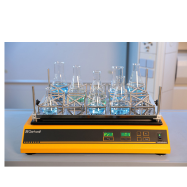
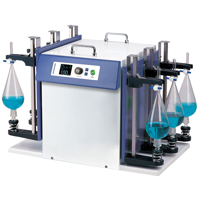
中国格哈特为您提供《果皮和果叶中酵母对选定的合成纺织品染料的脱色振荡》,该方案主要用于废水中染料检测,参考标准--,《果皮和果叶中酵母对选定的合成纺织品染料的脱色振荡》用到的仪器有格哈特强力高重现振荡器LS500/RO500、格哈特快速干燥仪STL56、德国移液器MM
推荐专场
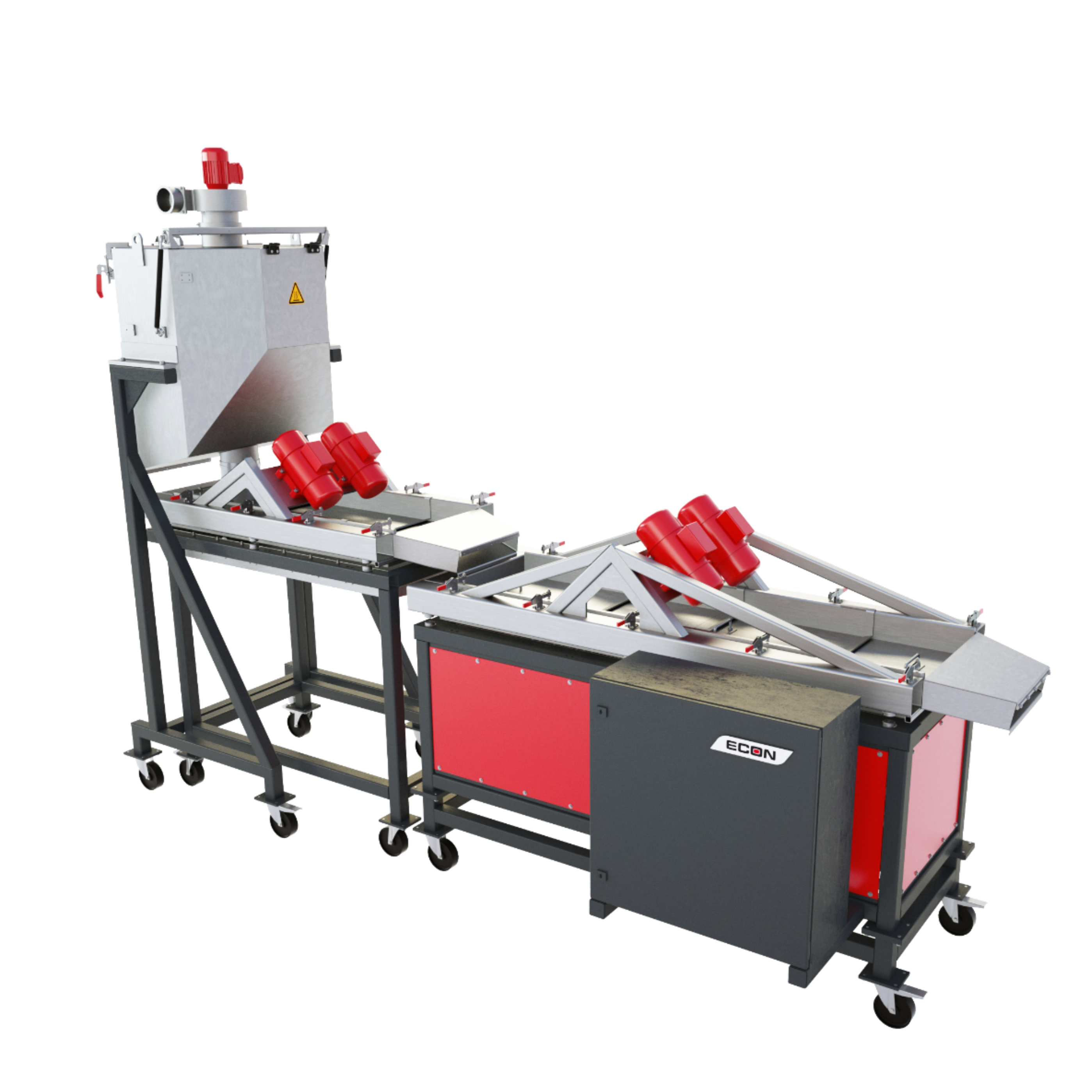
快速干燥仪
更多
相关方案
更多
该厂商其他方案
更多