


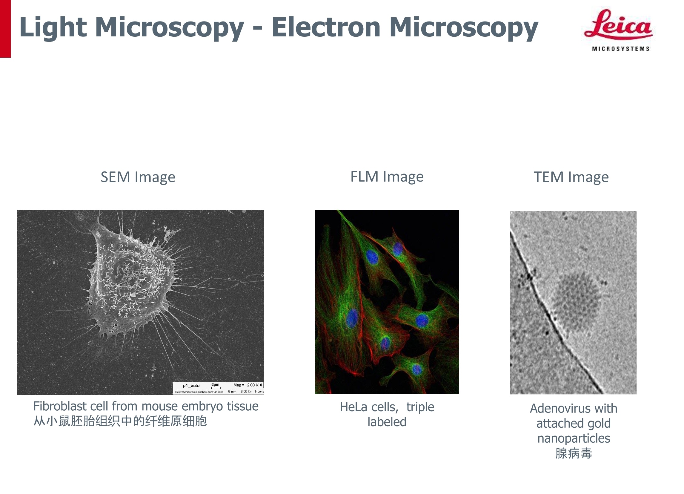
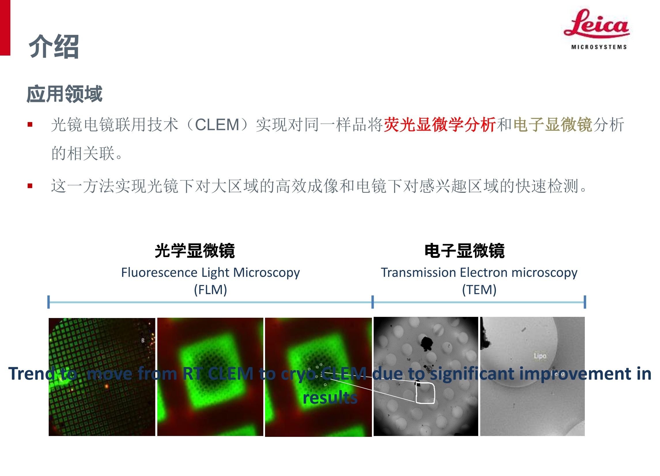
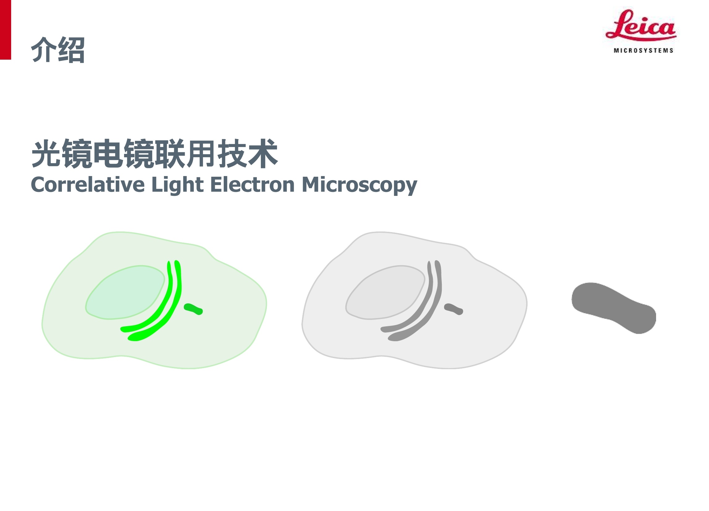
光镜电镜联用技术(CLEM)实现对同一样品将 荧光显微学分析和 电子显微镜分析
的相关联。这一方法实现光镜下对大区域的高效成像和电镜下对感兴趣区域的快速检测。
方案详情

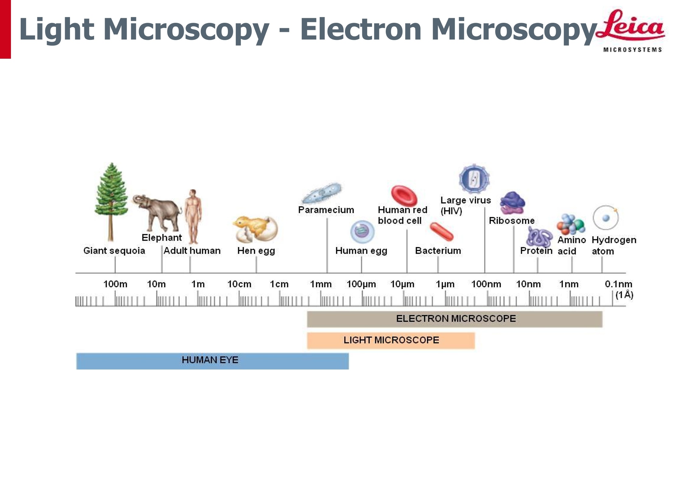
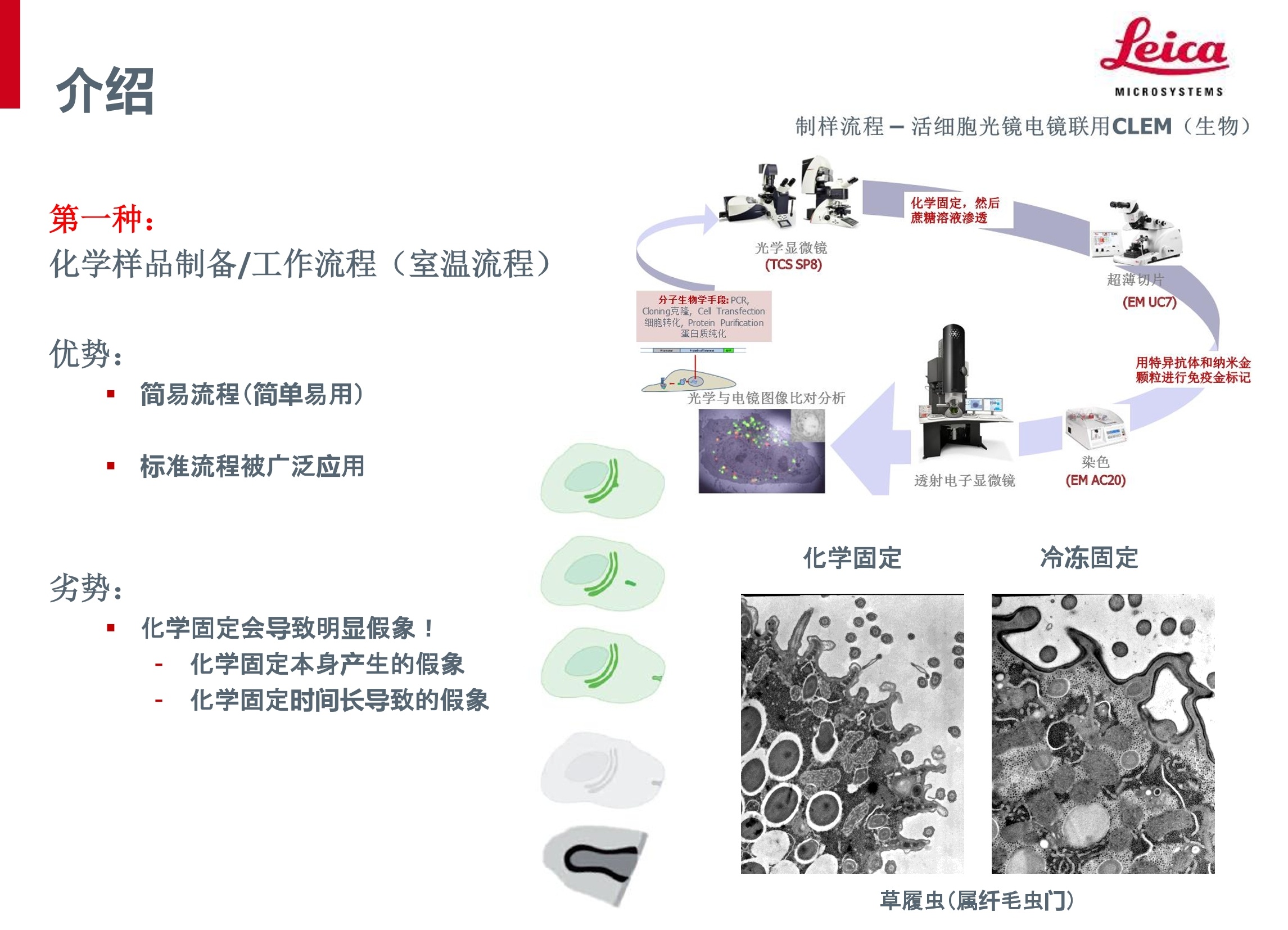
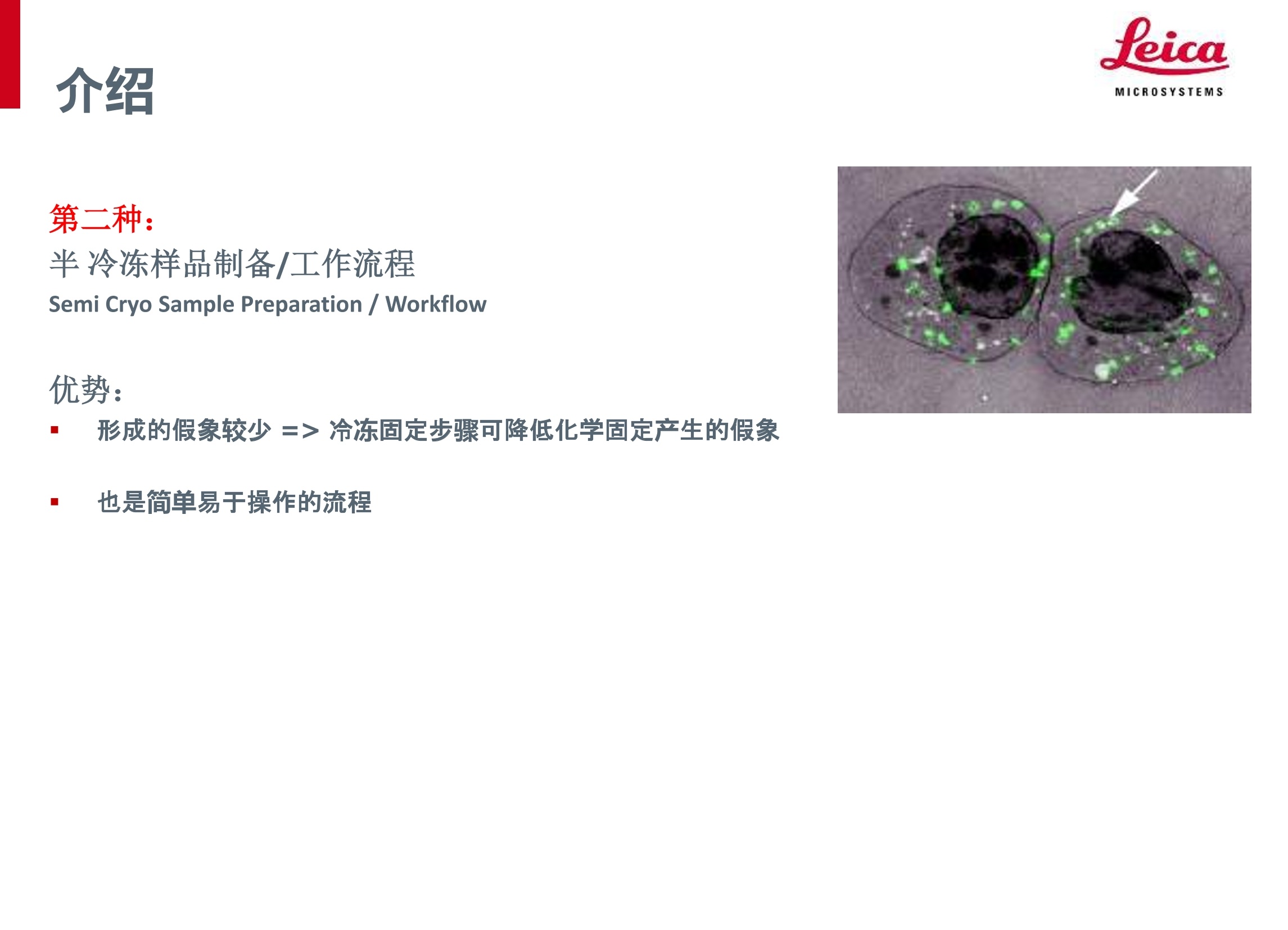
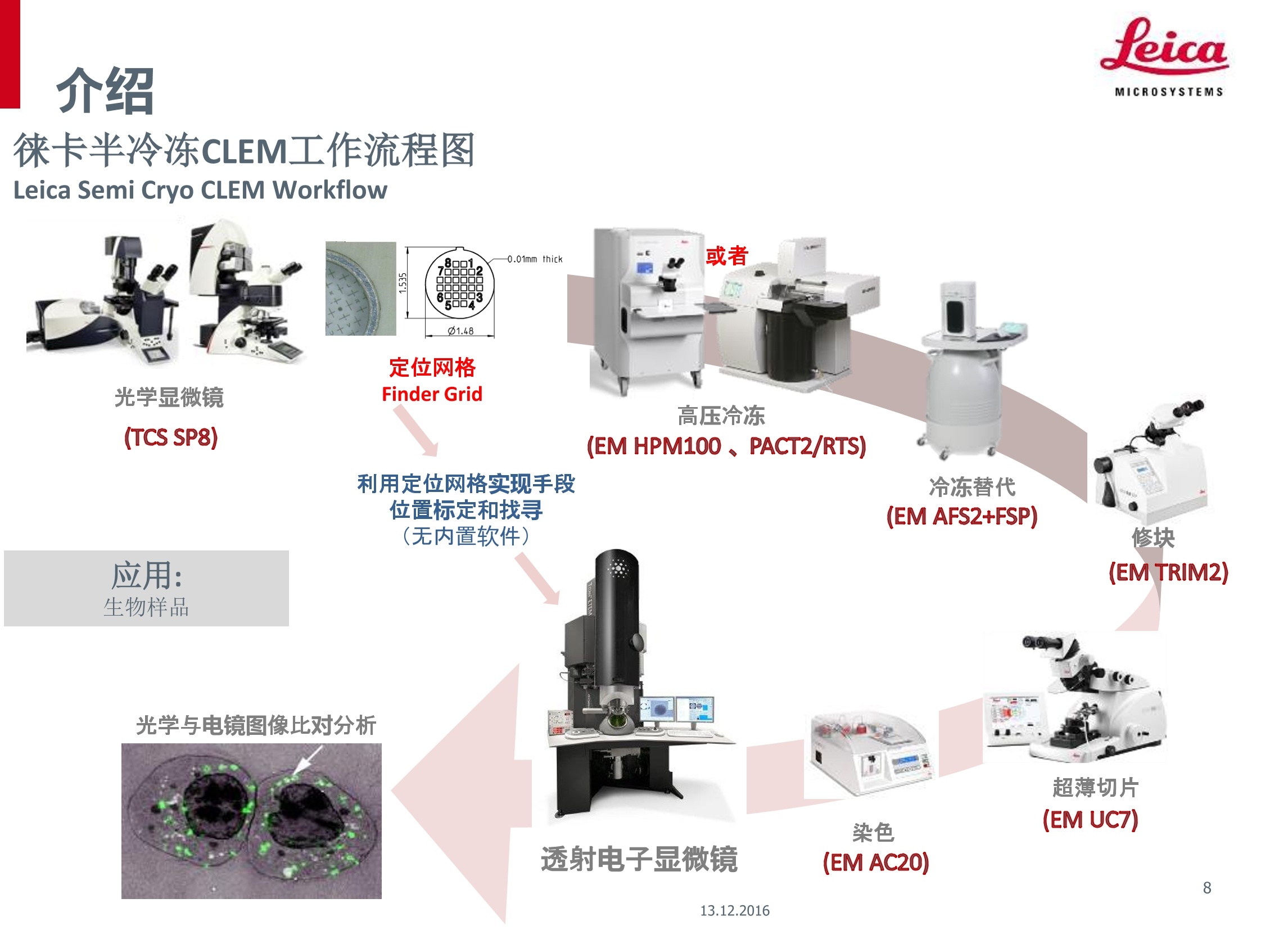
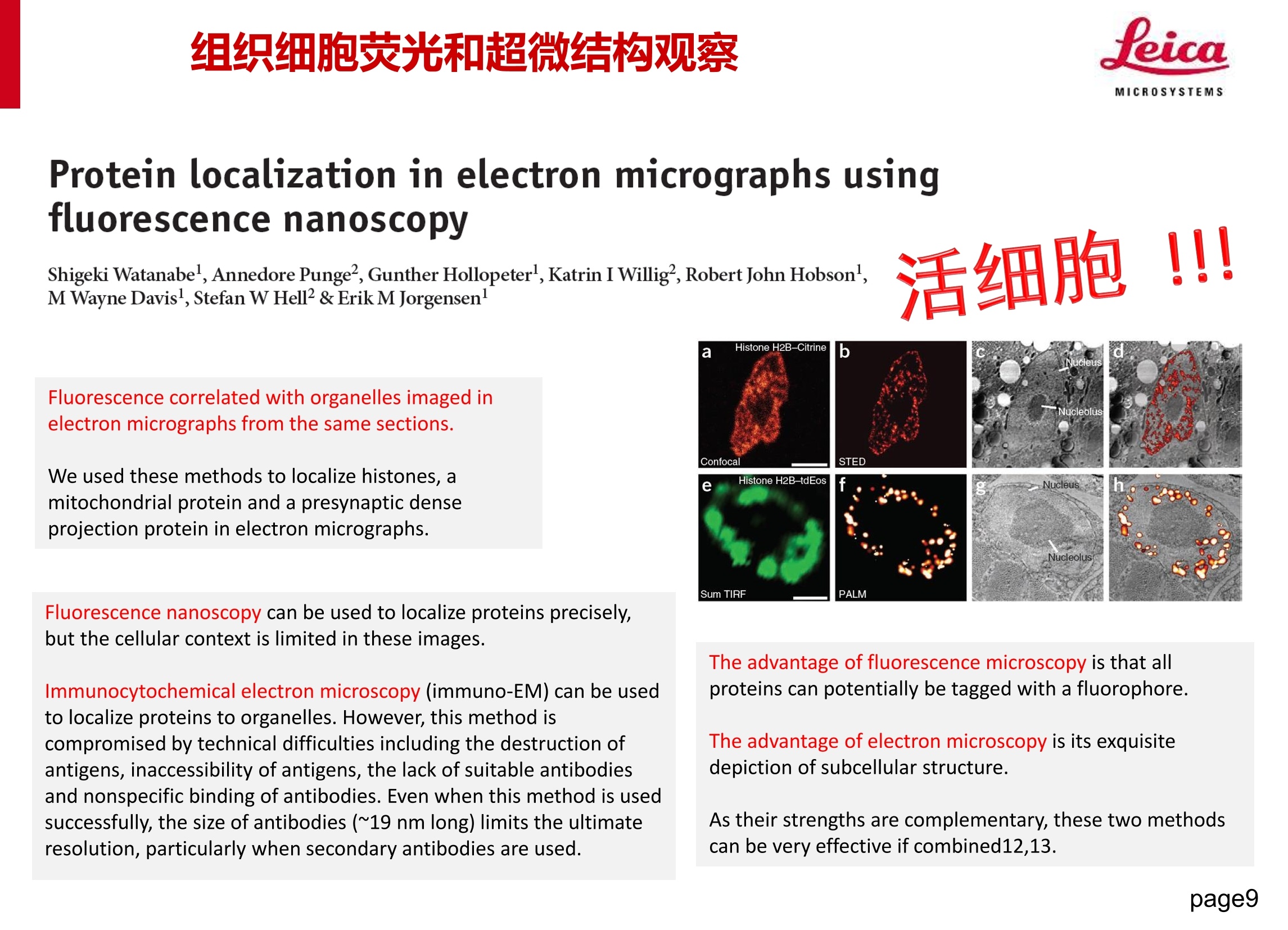
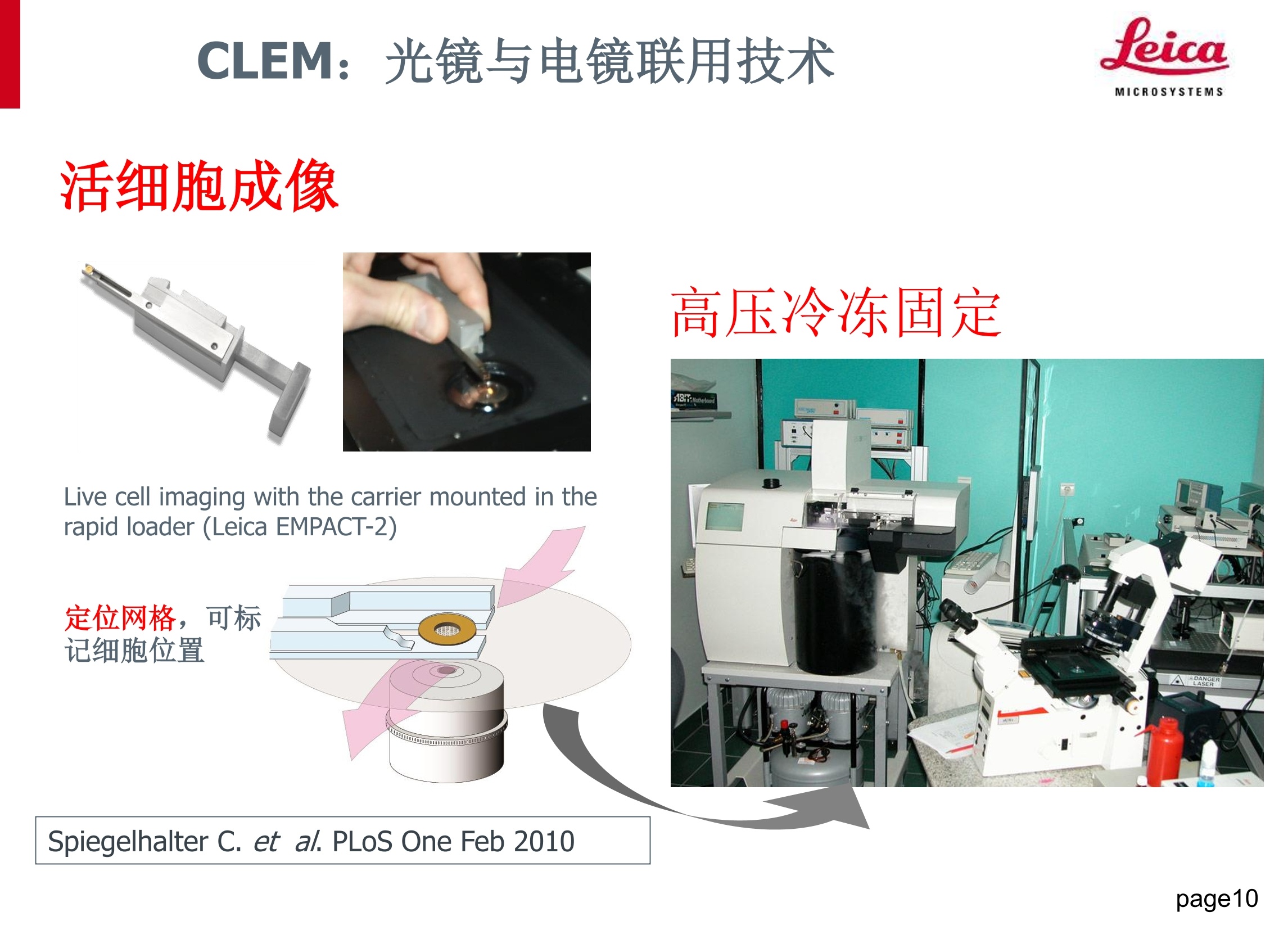
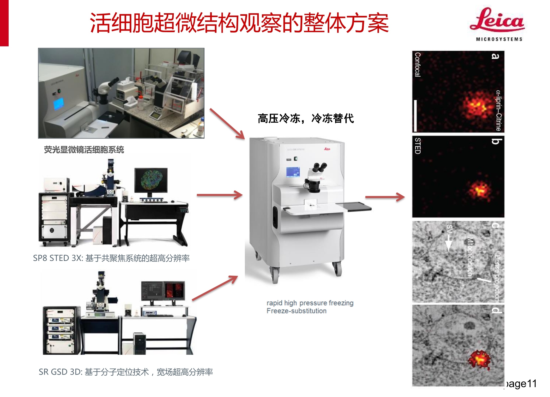
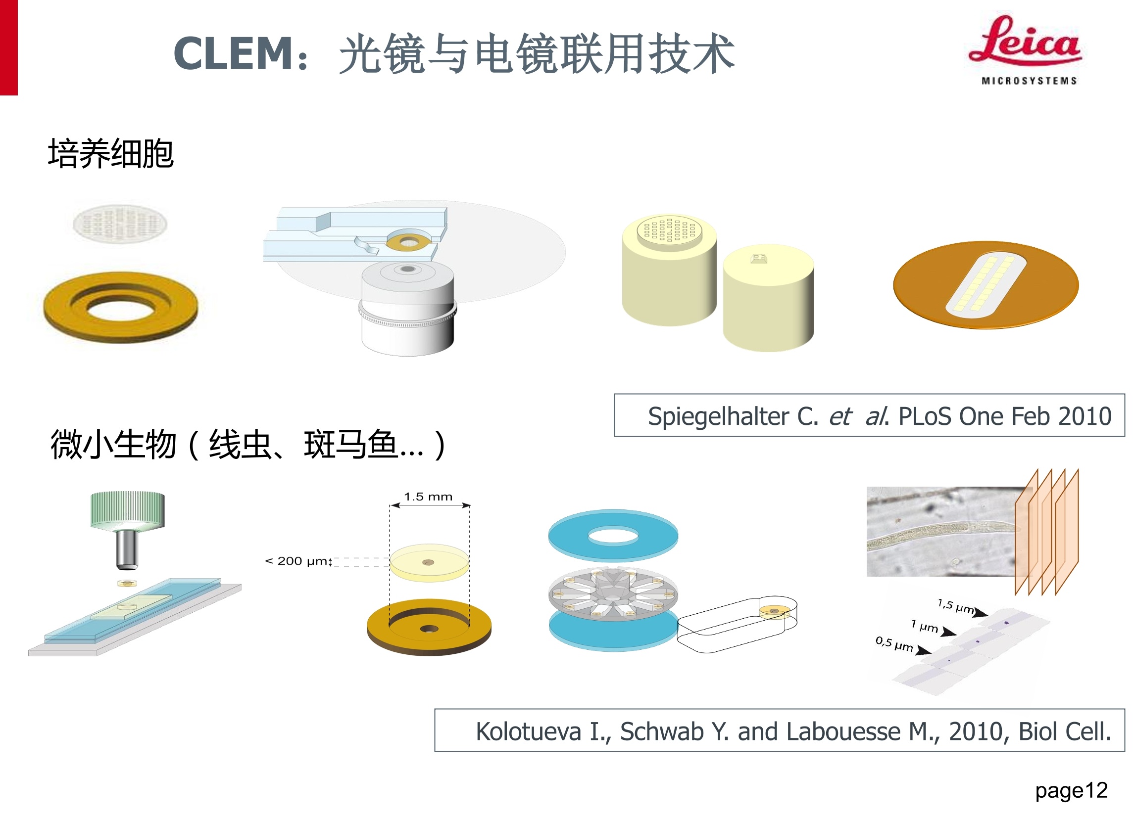
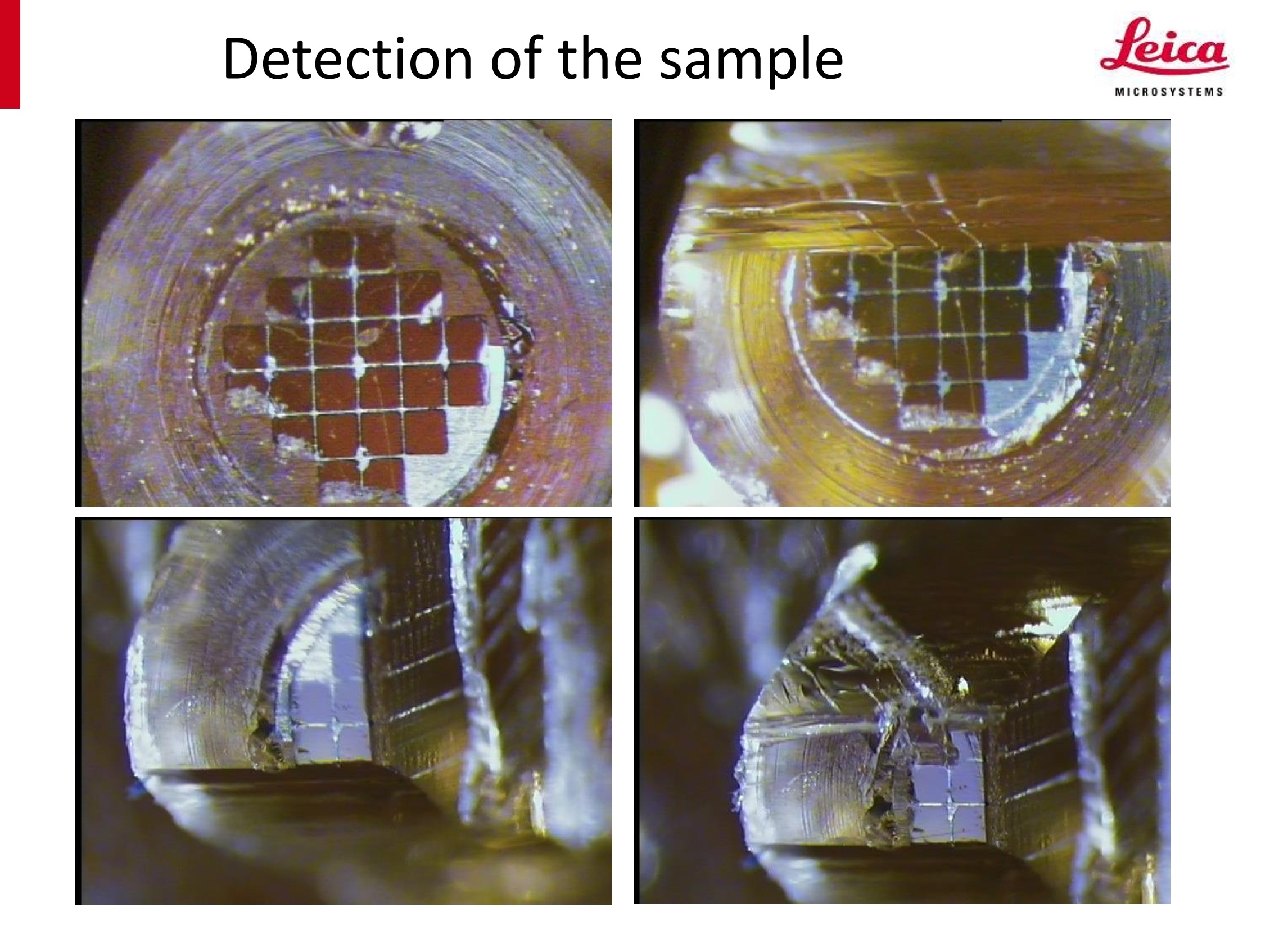
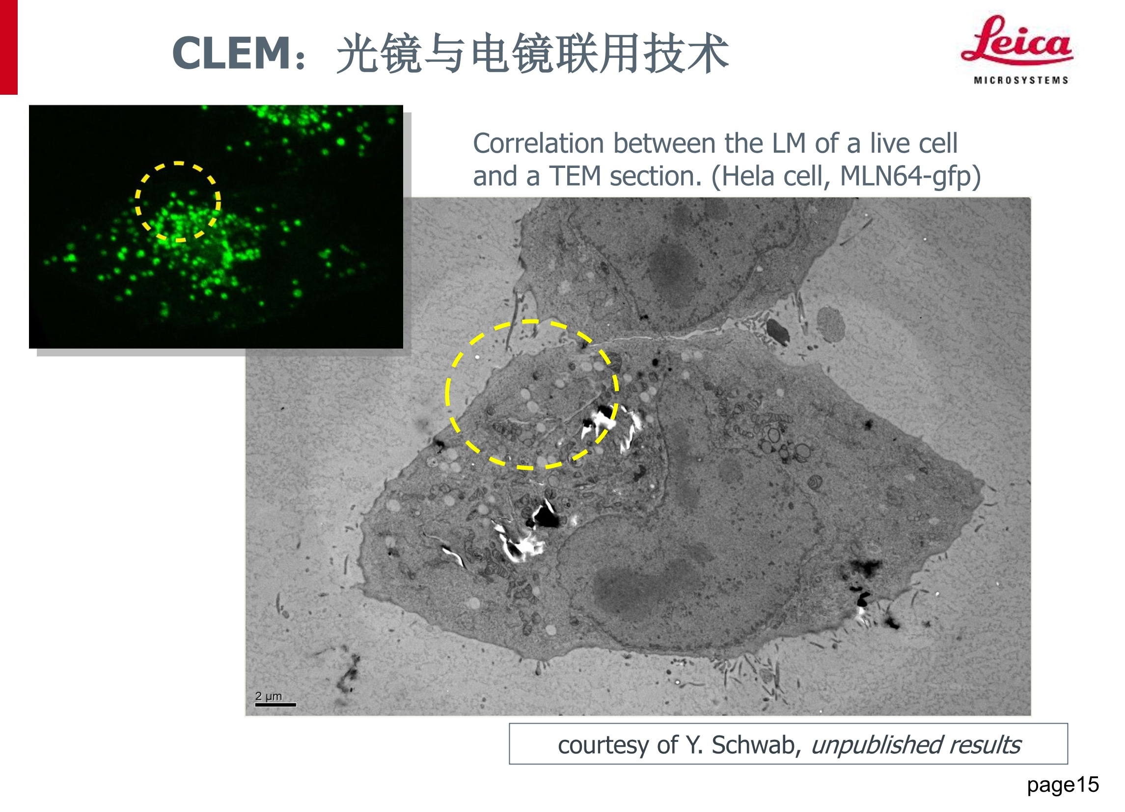
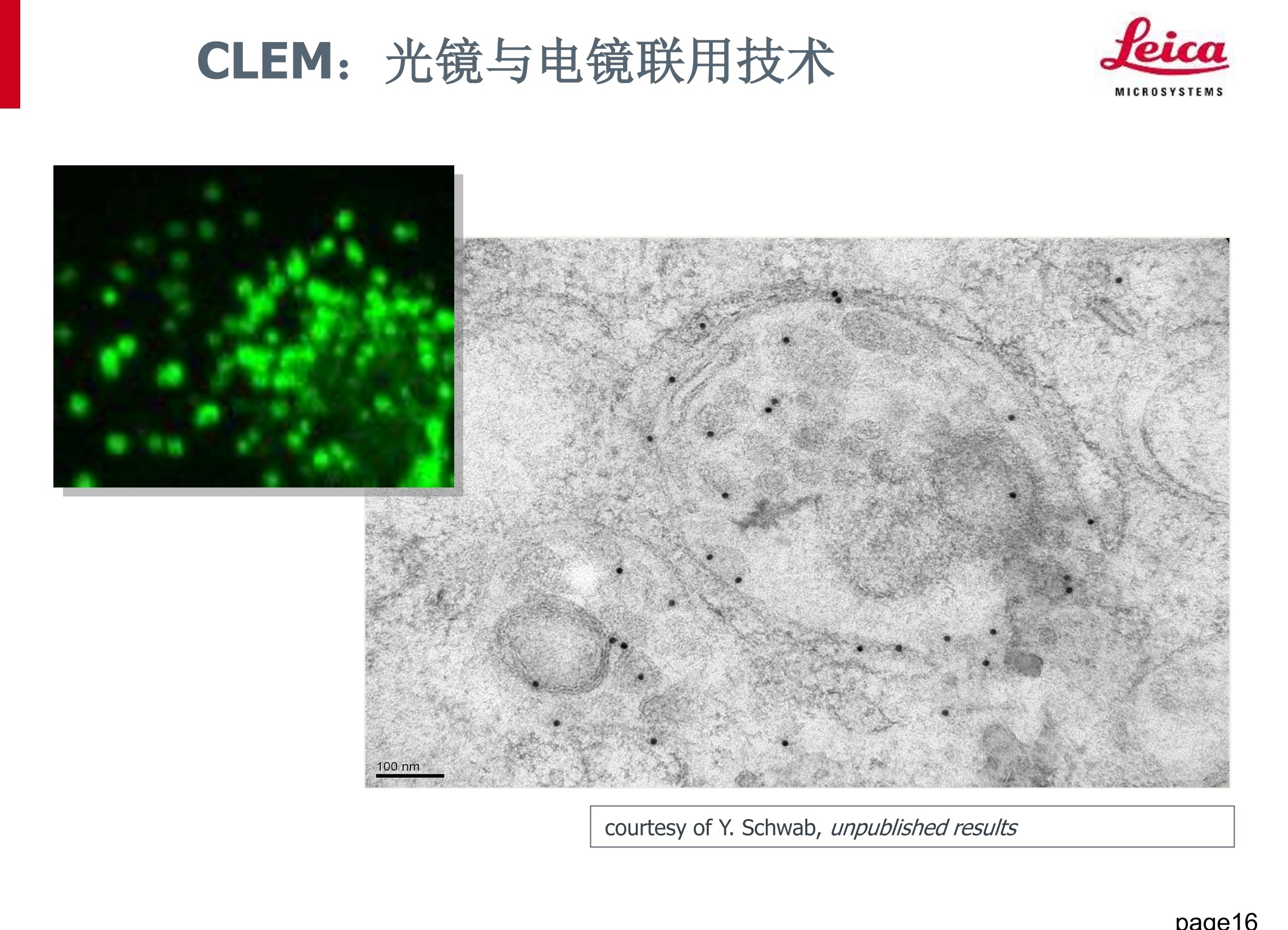
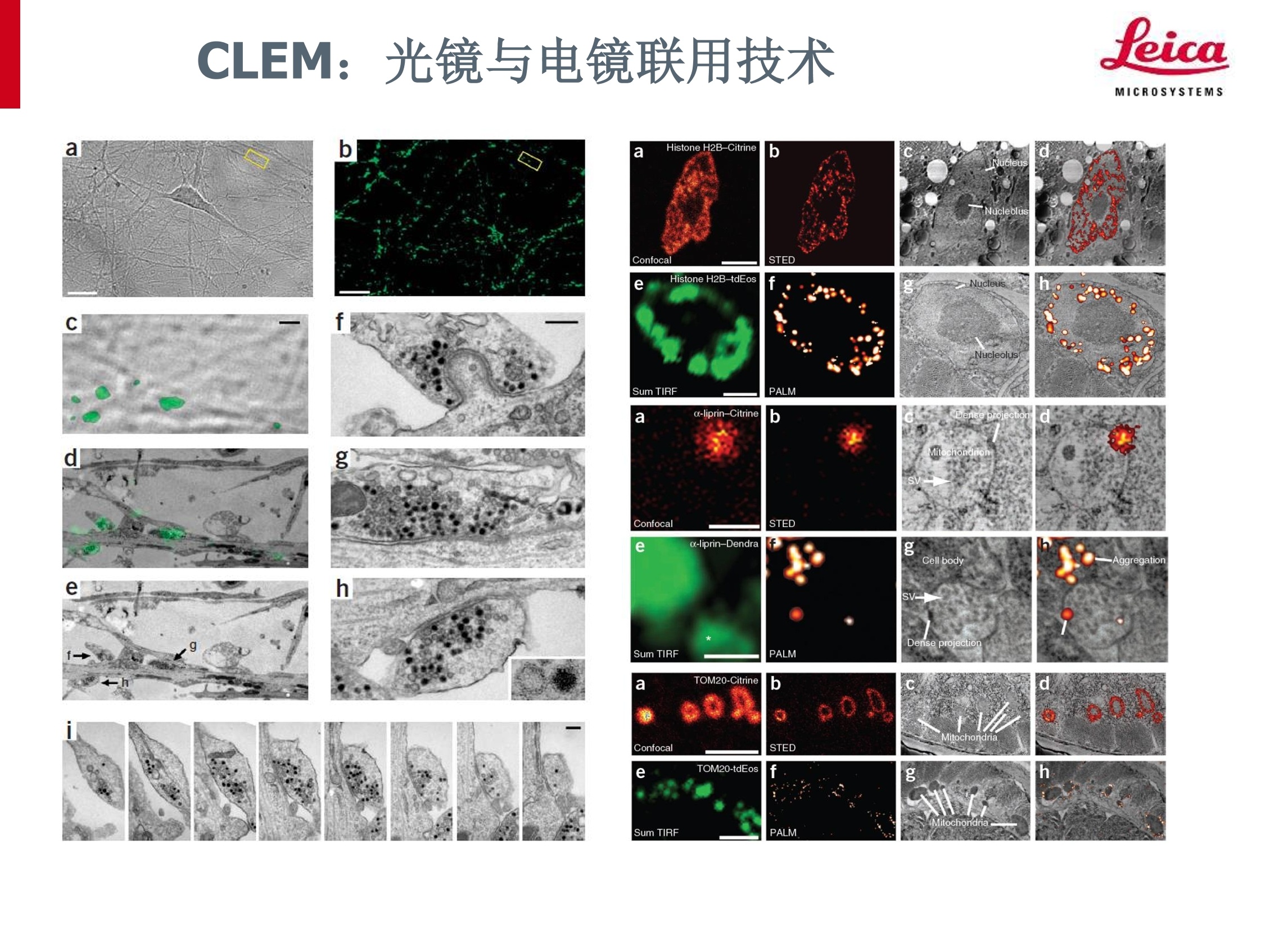
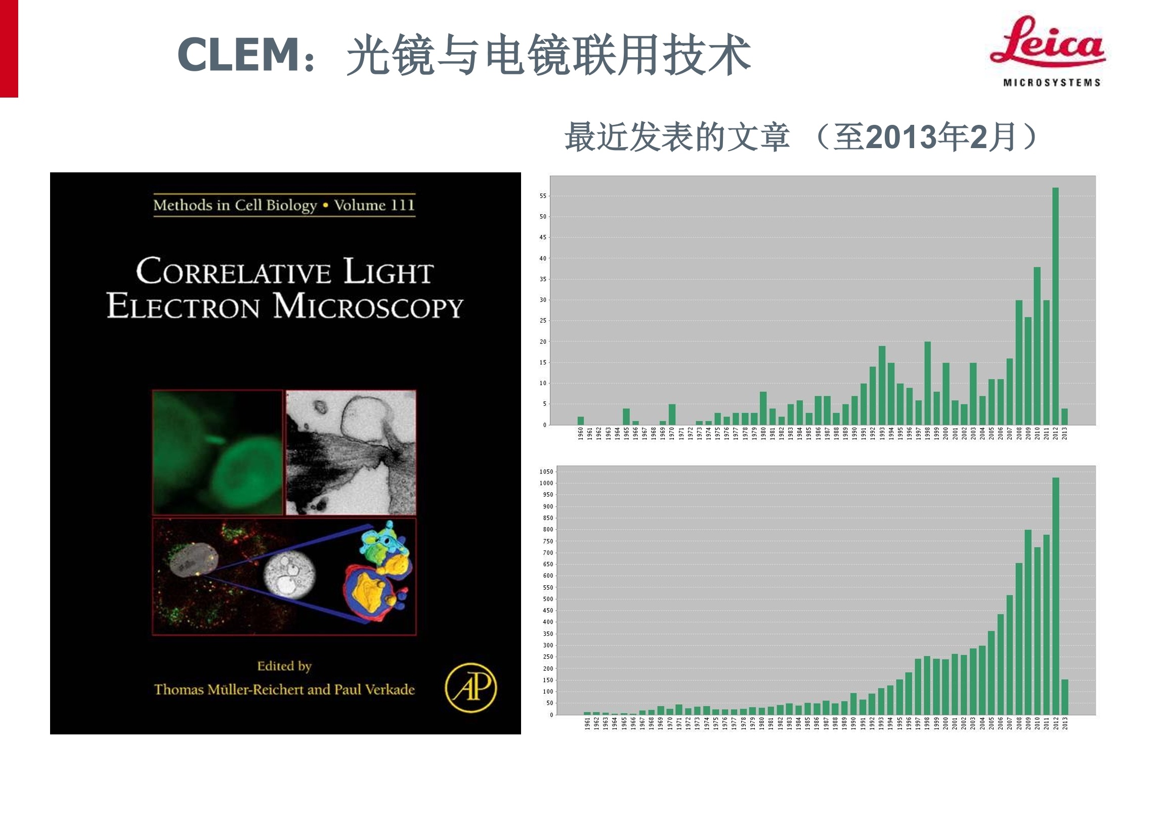
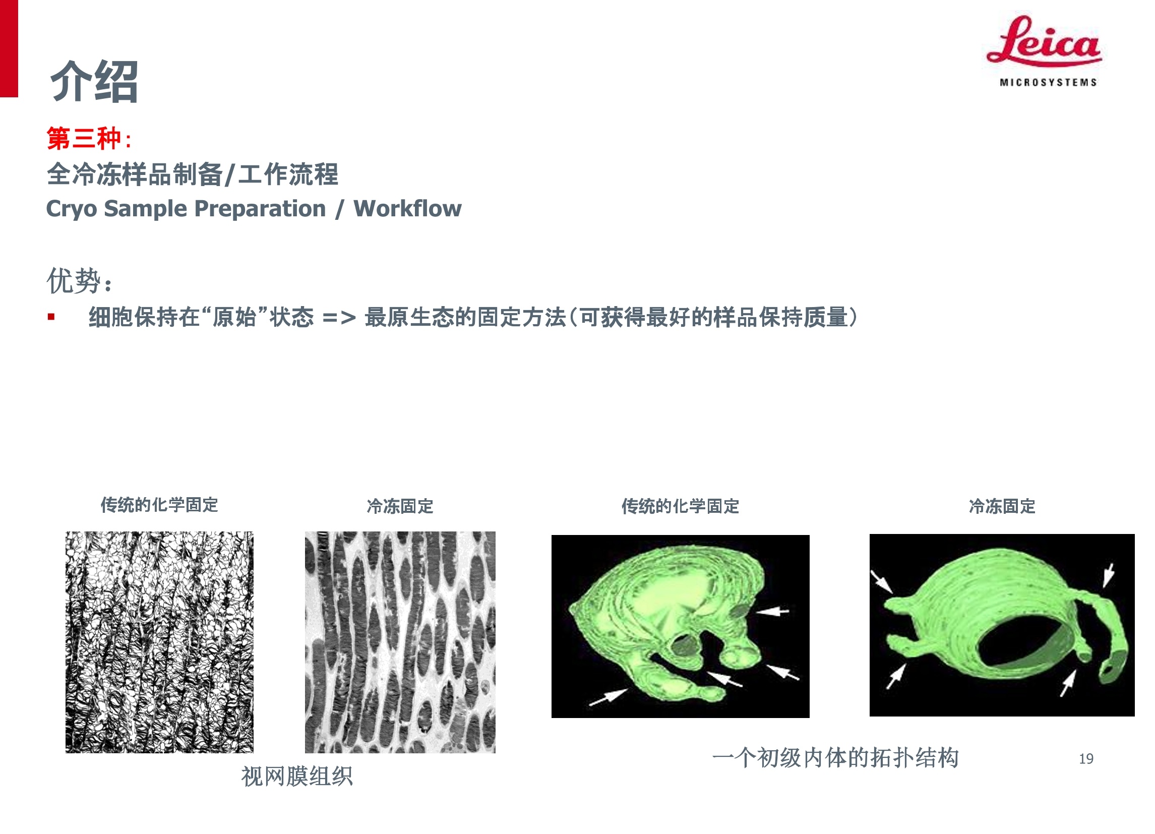
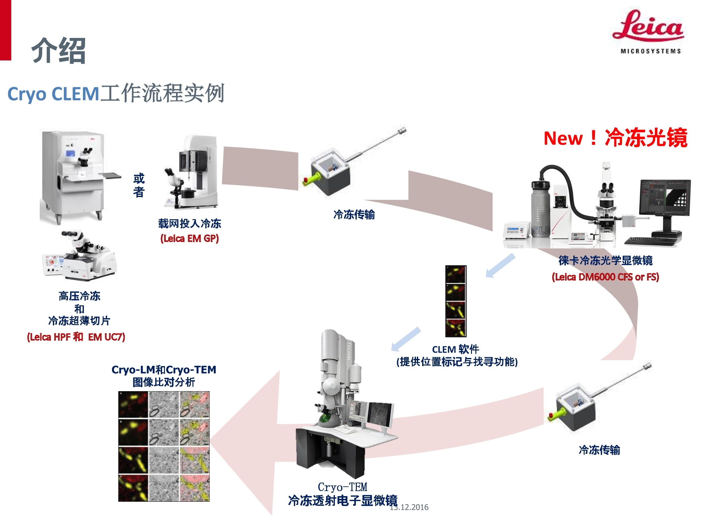
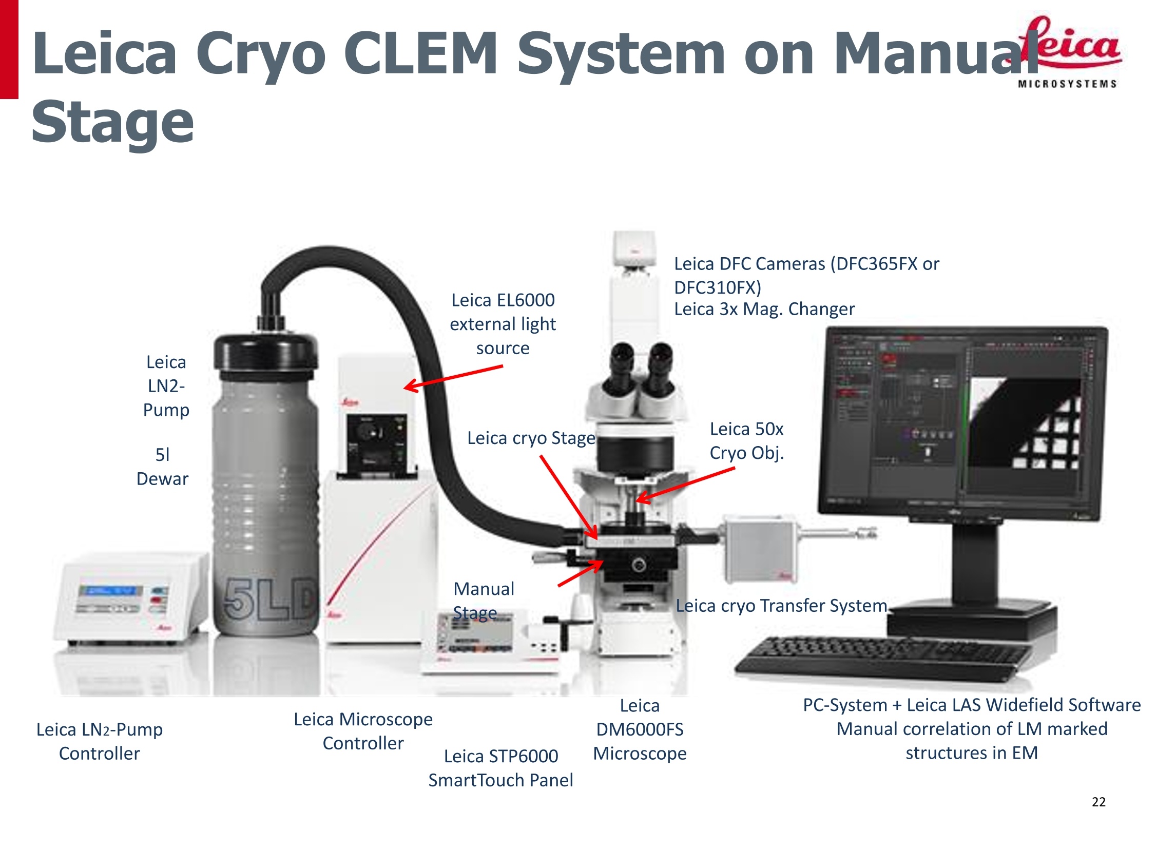
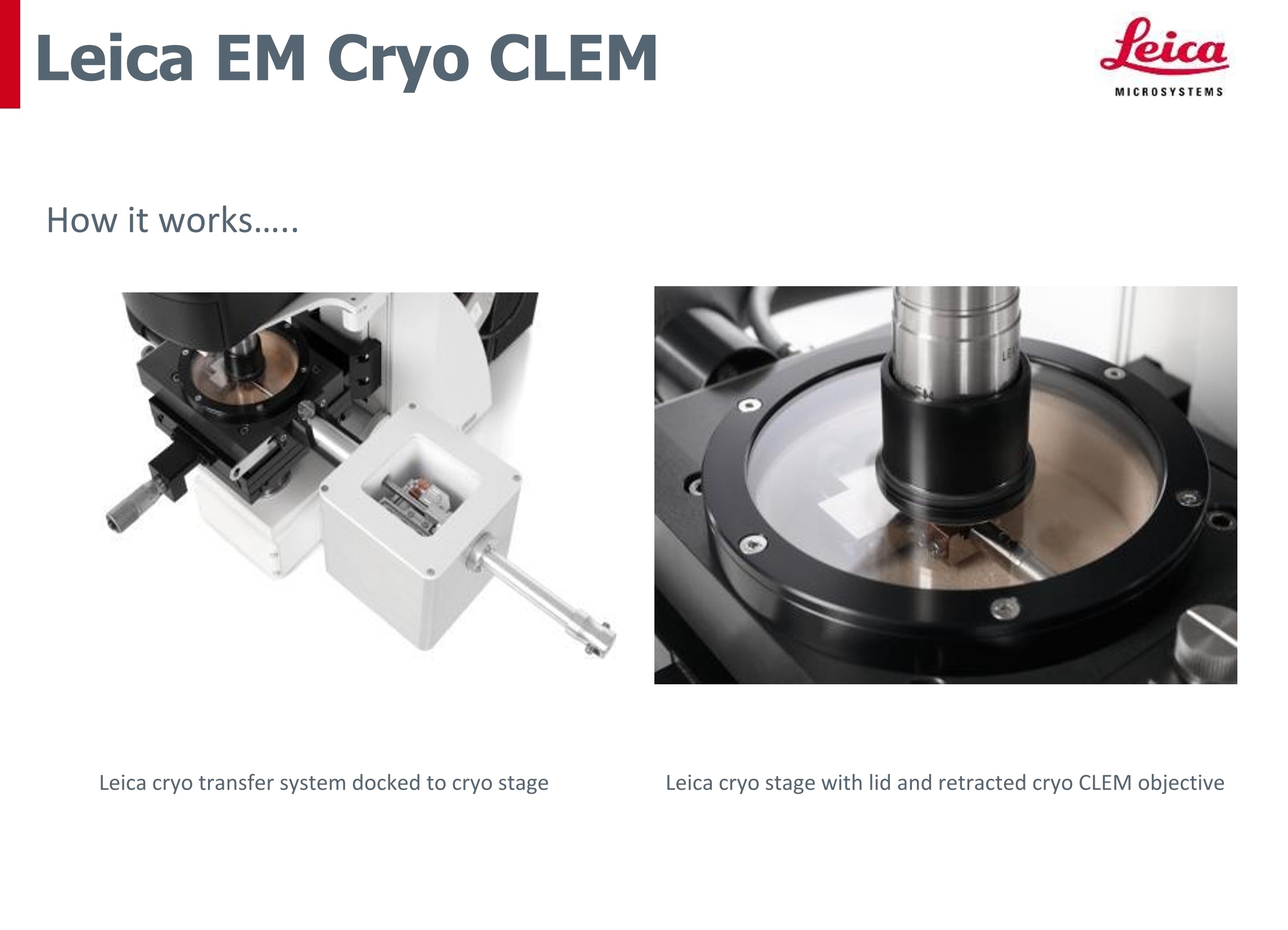
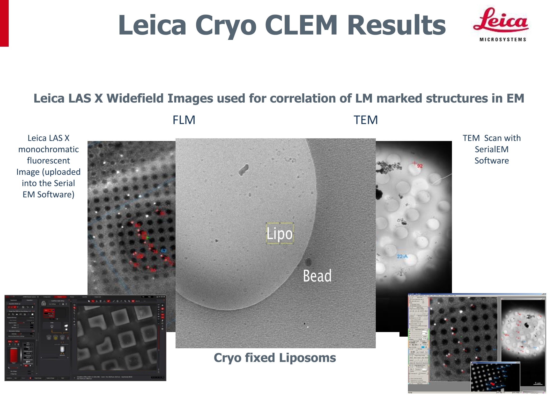
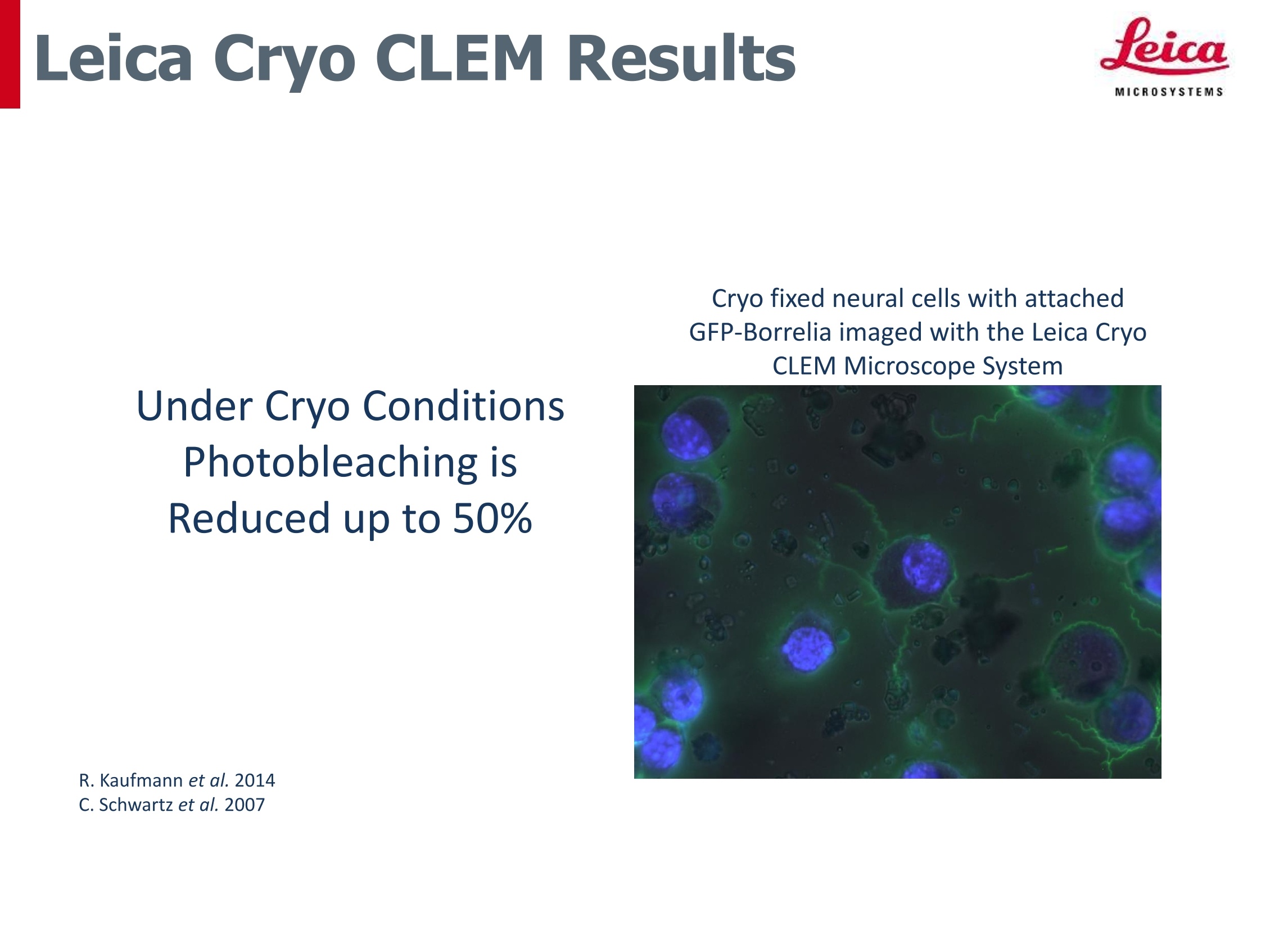
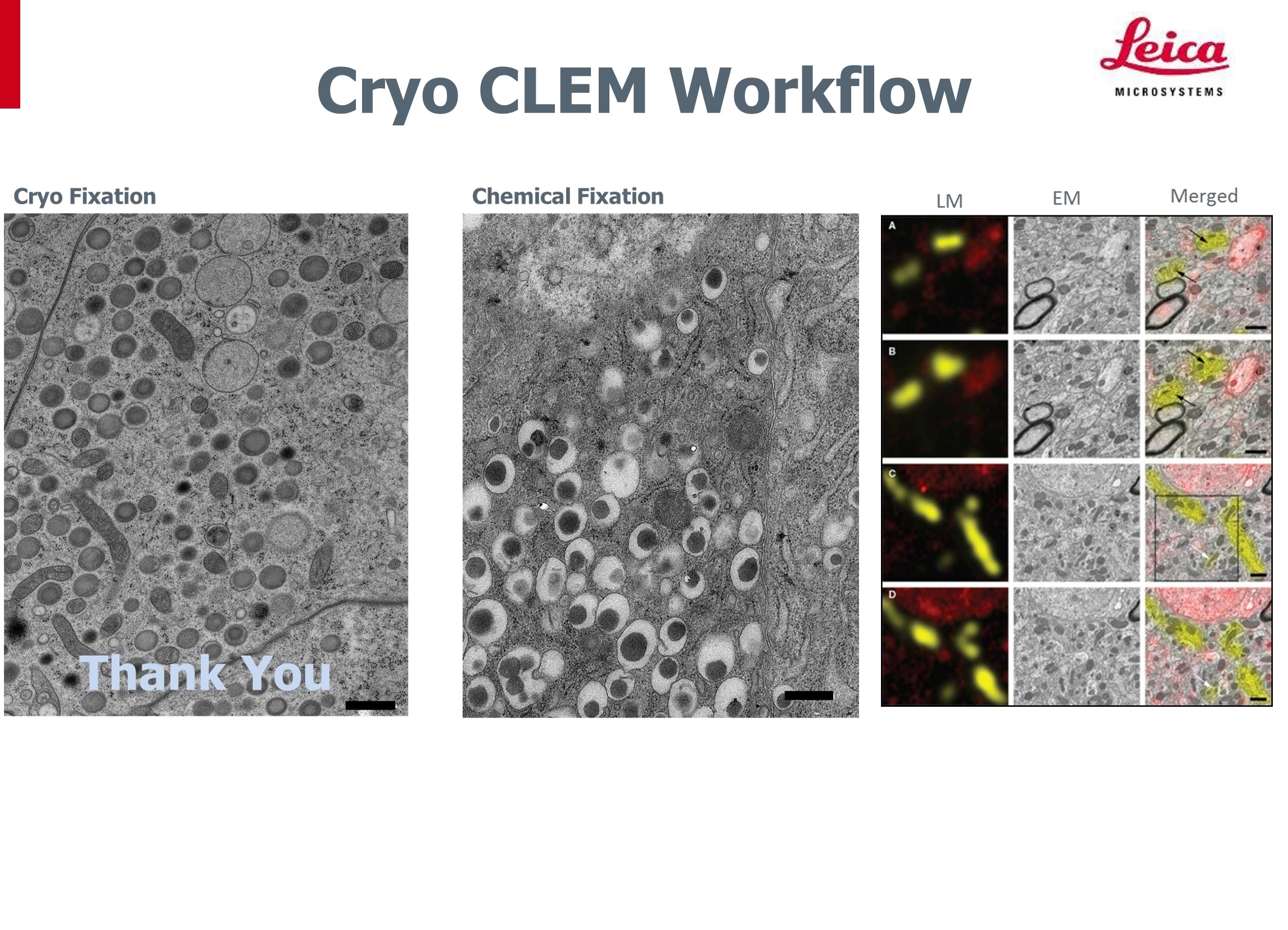
MICROSYSTEMS 介绍MICROSYSTEMS Living up to Life MICROSYSTEM S CLEM 介绍 Leica NanotechnologyLeica Microsystems Light Microscopy-Electron Microscopy MICROSYSTEM S SEM Image FLM Image TEM Image Fibroblast cell from mouse embryo tissue从小鼠胚胎组织中的纤维原细胞 HeLa cells, triplelabeled Adenovirus withattached goldnanoparticles腺病毒 Light Microscopy-Electron Microscopy.etca Large virus Paramecium Human red (HIV) blood cell Ribosome Elephant Amino Hydrogen Giant sequoia Adult human Hen egg Human eggBacterium Protein acid atom 100m 10m 10cm 1cm 1mm 100pm 10pm 1pm 100nm 10nm 1nm (1A) ELECTRON MICROSCOPE LIGHT MICROSCOPE HUMAN EYE 应用领域 光镜电镜联用技术(CLEM)实现对同一样品将荧光显微学分析和电子显微镜分析的相关联。 这一方法实现光镜下对大区域的高效成像和电镜下对感兴趣区域的快速检测。 光学显微镜 电子显微镜 Fluorescence Light Microscopy Transmission Electron microscopy (FLM) (TEM) 口 Lipo gogioda Trendt ove from RT CLEM to cryo. CLEM due to significant improvement in ao g results 信 au 22 oY ao MICROSYSTEMS 光镜电镜联用技术Correlative Light Electron Microscopy 第一种: 化学样品制备/工作流程(室温流程) 优势: 简易流程(简单易用) 标准流程被广泛应用 劣势: m化学固定会导致明显假象! -化学固定本身产生的假象 化学固定时间长导致的假象 第二种: 半冷冻样品制备/工作流程 Semi Cryo Sample Preparation/Workflow 优势: 形成的假象较少=>冷冻固定步骤可降低化学固定产生的假象 也是简单易于操作的流程 MICROSYSTEM S 徕卡半冷冻CLEM工作流程图 Leica Semi Cryo CLEM Workflow -0.01mm thi7/0品ck或者0m OC000 2000o'5004301.48定位网格光学显微镜 Finder Grid高压冷冻(TCS SP8)(EM HPM100 、PACT2/RTS)利用定位网格实现手段冷冻替代位置标定和找寻(EM AFS2+FSP)(无内置软件)修块应用:(EM TRIM2)生物样品光学与电镜图像比对分析125超薄切片 (EM UC7) 透射电子显微镜 染色(EM AC20) 13.12.2016 Protein localization in electron micrographs usingfluorescence nanoscopy Shigeki Watanabel, Annedore Punge, Gunther Hollopeter, Katrin I Willig2, Robert John Hobson,M Wayne Davisl, Stefan W Hell& Erik M Jorgensenl 活细胞I !!! Fluorescence correlated with organelles imaged inelectron micrographs from the same sections. We used these methods to localize histones, amitochondrial protein and a presynaptic denseprojection protein in electron micrographs. Fluorescence nanoscopy can be used to localize proteins precisely,but the cellular context is limited in these images. The advantage of fluorescence microscopy is that allproteins can potentially be tagged with a fluorophore. Immunocytochemical electron microscopy (immuno-EM) can be usedto localize proteins to organelles. However, this method iscompromised by technical difficulties including the destruction ofantigens, inaccessibility of antigens, the lack of suitable antibodiesand nonspecific binding of antibodies. Even when this method is usedsuccessfully, the size of antibodies (~19 nm long) limits the ultimateresolution, particularly when secondary antibodies are used. The advantage of electron microscopy is its exquisitedepiction of subcellular structure. As their strengths are complementary, these two methodscan be very effective if combined12,13. CLEM: 光镜与电镜联用技术 MICROSYSTEM S活细胞成像高压冷冻固定13T;MochooLive cell imaging with the carrier mounted in the戰rapid loader (Leica EMPACT-2)定位网格,可标记细胞位置A10114011001001100Spiegelhalter C. et al.PLoS One Feb 2010 活细胞超微结构观察的整体方案 MICROSYSTEMS 高压冷冻,冷冻替代 荧光显微镜活细胞系统 一 Aism SP8 STED 3X:基于共聚焦系统的超高分辨率 rapid high pressure freezingFreeze-substitution SR GSD 3D:基于分子定位技术,宽场超高分辨率 CLEM: 光镜与电镜联用技术 MICROSYSTEMS 培养细胞 晶 侣a Spiegelhalter C. et al. PLoS One Feb 2010 微小生物(线虫、斑马鱼...) 1.5 mm <200 pmt_ 1,5 um 1um 0,5 pm Kolotueva I., Schwab Y. and Labouesse M., 2010, Biol Cell. MICROSYSTEM S Detection of the sample MICROSYSTEMS CLEM:光镜与电镜联用技术 MICROSYSTEMS Correlation between the LM of a live cell courtesy of Y. Schwab, unpublished results CLEM:光镜与电镜联用技术 MICROSYSTEMS courtesy of Y. Schwab, unpublished results MICROSYSTEMS CLEM: 光镜与电镜联用技术 MICROSYSTEMS 最近发表的文章(至2013年2月) Methods in Cell Biology· Volume 111 40CORRELATIVE LIGHT 35 ELECTRON IMICROSCOPY 3025 Thomas Muller-Reichert and Paul Verkade A MICROSYSTEMS 第三种: 全冷冻样品制备/工作流程 Cryo Sample Preparation/Workflow 优势: 细胞保持在“原始”状态=>最原生态的固定方法(可获得最好的样品保持质量) 传统的化学固定 冷冻固定 传统的化学固定 冷冻固定 一个初级内体的拓扑结构 视网膜组织 MICROSYSTE M S Cryo CLEM工作流程实例 New ! 冷冻光镜 品 冷冻传输 载网投入冷冻(Leica EM GP) 徕卡冷冻光学显微镜 (Leica DM6000 CFS or FS) 高压冷冻和冷冻超薄切片 (Leica HPF 和 EM UC7) CLEM软件 (提供位置标记与找寻功能) Cryo-LM和Cryo-TEM图像比对分析 冷冻传输 Cryo-TEM 冷冻透射电子显微镜. 3.12.2016 Correlative Light and Electron Microscopy (CLEM)光镜电镜联用技术:对同一样品位置集荧光显微图像与高分辨率电镜图像于一体 Electron Microscopy (EM)电镜:可保存样品结构信息,及生活环境,因此目标位置以极高分辨率状态保存。但是电镜图像难以保存生物功能结构的活体信息。 Fluorescence Light Microscopy (FLM)荧光显微镜:是观察和分析已固定或活体生物样品内部生理过程的有效方法。另外,荧光显微镜可以实现对大样本区域的快速扫描,以及对目标位点 (Region of interest, ROI) 的快速定位,从而为电镜中快速识别目标位点提供便利。 Cryo Fixation冷冻固定技术:在样品制备技术中使得保存样本近乎原始生活状态成为可能。 Cryo CLEM冷冻光镜电镜联用技术:集合以上所有技术优势。该技术包含了以下各技术手段的优势:冷冻固定,荧光显微观察,电镜观察,并以对无假象保存的样品实现同一位点的高效成像,以及图像信息叠加,从而实现对样品信息的充分认 知。 Leica Cryo CLEM System on Manualeicc Stage Leica DFC Cameras (DFC365FX orDFC310FX)Leica 3x Mag. Changer Leica EL6000external lightsource Leica LN2- Pump Leica 50x Leica cryo Stage 51 Cryo Obj. Dewar 5LD. Manual Stage Leica cryo Transfer System Leica Leica LN2-PumpController Leica MicroscopeController DM6000FSMicroscope PC-System +Leica LAS Widefield SoftwareManual correlation of LM markedstructures in EM Leica STP6000 SmartTouch Panel MICROSYSTEMS How it works..... Leica cryo transfer system docked to cryo stage Leica cryo stage with lid and retracted cryo CLEM objective Leica LAS X Widefield Images used for correlation of LM marked structures in EMFLM TEM Leica LAS XmonochromaticfluorescentImage (uploadedinto the SerialEM Software) TEM Scan withSerialEM Cryo fixed Liposoms 5i MICROSYSTEMS Cryo fixed neural cells with attachedGFP-Borrelia imaged with the Leica CryoCLEM Microscope System Under Cryo ConditionsPhotobleaching isReduced up to 50% ( R. Kaufmann et al. 2014 ) C. Schwartz et al. 2007 Cryo Fixation Science Lab 光镜电镜联用技术(CLEM)实现对同一样品将 荧光显微学分析和 电子显微镜分析的相关联。这一方法实现光镜下对大区域的高效成像和电镜下对感兴趣区域的快速检测。
确定
还剩24页未读,是否继续阅读?

铂瑞达(北京)科技有限公司为您提供《材料中材料类产品制样检测方案(电镜制样)》,该方案主要用于材料中材料类产品制样检测,参考标准--,《材料中材料类产品制样检测方案(电镜制样)》用到的仪器有
相关方案
更多