推荐厂家
暂无
暂无
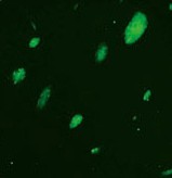
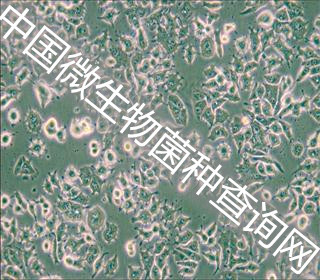
[align=center][font='times new roman'][size=16px][color=#000000]上皮细胞[/color][/size][/font][font='times new roman'][size=16px][color=#000000]-[/color][/size][/font][font='times new roman'][size=16px][color=#000000]间充质转化[/color][/size][/font][/align][font='times new roman'][size=16px][color=#000000]上皮细胞[/color][/size][/font][font='times new roman'][size=16px][color=#000000]-[/color][/size][/font][font='times new roman'][size=16px][color=#000000]间充质转化,是指上皮细胞转化为具有间质表型细胞的生物学过程,[/color][/size][/font][font='times new roman'][size=16px][color=#000000]在肿瘤的侵袭和转移等方面[/color][/size][/font][font='times new roman'][size=16px][color=#000000]具有[/color][/size][/font][font='times new roman'][size=16px][color=#000000]重要作用[/color][/size][/font][font='times new roman'][sup][size=16px][color=#000000][31][/color][/size][/sup][/font][font='times new roman'][size=16px][color=#000000]。[/color][/size][/font][font='times new roman'][size=16px][color=#000000]经历[/color][/size][/font][font='times new roman'][size=16px][color=#000000]EMT[/color][/size][/font][font='times new roman'][size=16px][color=#000000]过程的细胞具有类似干细胞的特性,耐药性也随之增强[/color][/size][/font][font='times new roman'][sup][size=16px][color=#000000][32][/color][/size][/sup][/font][font='times new roman'][size=16px][color=#000000]。研究发现,在乳腺癌的小鼠模型中,降低[/color][/size][/font][font='times new roman'][size=16px][color=#000000]snail[/color][/size][/font][font='times new roman'][size=16px][color=#000000]的表达,肺转移的发生显著减少[/color][/size][/font][font='times new roman'][sup][size=16px][color=#000000][33][/color][/size][/sup][/font][font='times new roman'][size=16px][color=#000000]。胰腺癌、肺腺癌细胞在[/color][/size][/font][font='times new roman'][size=16px][color=#000000]EMT[/color][/size][/font][font='times new roman'][size=16px][color=#000000]过程后表现出更强的迁移和侵袭能力[/color][/size][/font][font='times new roman'][sup][size=16px][color=#000000][34, 35][/color][/size][/sup][/font][font='times new roman'][size=16px][color=#000000]。[/color][/size][/font][font='times new roman'][size=16px][color=#000000]从[/color][/size][/font][font='times new roman'][size=16px][color=#000000]EMT[/color][/size][/font][font='times new roman'][size=16px][color=#000000]转录调控的角度看,[/color][/size][/font][font='times new roman'][size=16px][color=#000000]与非小细胞肺癌相比,人小细胞肺癌细胞系中[/color][/size][/font][font='times new roman'][size=16px][color=#000000]EMT[/color][/size][/font][font='times new roman'][size=16px][color=#000000]相关转录因子的表达量较高[/color][/size][/font][font='times new roman'][sup][size=16px][color=#000000][36][/color][/size][/sup][/font][font='times new roman'][size=16px][color=#000000]。[/color][/size][/font][font='times new roman'][size=16px][color=#000000]Krohn[/color][/size][/font][font='times new roman'][size=16px][color=#000000]等人研究发现,[/color][/size][/font][font='times new roman'][size=16px][color=#000000]E[/color][/size][/font][font='times new roman'][size=16px][color=#000000]-cadherin[/color][/size][/font][font='times new roman'][size=16px][color=#000000]表达下调,[/color][/size][/font][font='times new roman'][size=16px][color=#000000]Vimentin[/color][/size][/font][font='times new roman'][size=16px][color=#000000]表达上调的贴壁的小细胞肺癌亚系[/color][/size][/font][font='times new roman'][size=16px][color=#000000]NCIH69V[/color][/size][/font][font='times new roman'][size=16px][color=#000000]具有更高的侵袭性[/color][/size][/font][font='times new roman'][sup][size=16px][color=#000000][37][/color][/size][/sup][/font][font='times new roman'][size=16px][color=#000000]。在小细胞肺癌中,[/color][/size][/font][font='times new roman'][size=16px][color=#000000]EMT[/color][/size][/font][font='times new roman'][size=16px][color=#000000]与[/color][/size][/font][font='times new roman'][size=16px][color=#000000]PARP[/color][/size][/font][font='times new roman'][size=16px][color=#000000]抑制剂、顺铂、[/color][/size][/font][font='times new roman'][size=16px][color=#000000]Bcl-2[/color][/size][/font][font='times new roman'][size=16px][color=#000000]抑制剂等药物的耐药具有显著关联性。[/color][/size][/font][font='times new roman'][size=16px][color=#000000]E[/color][/size][/font][font='times new roman'][size=16px][color=#000000]-cadherin[/color][/size][/font][font='times new roman'][size=16px][color=#000000]表达量较高的小细胞肺癌对[/color][/size][/font][font='times new roman'][size=16px][color=#000000]Bcl-2[/color][/size][/font][font='times new roman'][size=16px][color=#000000]抑制剂具有更好的应答率[/color][/size][/font][font='times new roman'][sup][size=16px][color=#000000][38][/color][/size][/sup][/font][font='times new roman'][size=16px][color=#000000]。[/color][/size][/font][font='times new roman'][size=16px][color=#000000]Contactin 1[/color][/size][/font][font='times new roman'][size=16px][color=#000000]通过诱导[/color][/size][/font][font='times new roman'][size=16px][color=#000000]EMT[/color][/size][/font][font='times new roman'][size=16px][color=#000000]过程调节小细胞肺癌聚乙二醇精氨酸酶的耐药性[/color][/size][/font][font='times new roman'][sup][size=16px][color=#000000][39][/color][/size][/sup][/font][font='times new roman'][size=16px][color=#000000]。越来越多的研究证明,小细胞肺癌[/color][/size][/font][font='times new roman'][size=16px][color=#000000]EMT[/color][/size][/font][font='times new roman'][size=16px][color=#000000]过程受到多种通路的调节,促进小细胞肺癌细胞的增殖、侵袭和转移,并促进化疗的耐药性[/color][/size][/font][font='times new roman'][sup][size=16px][color=#000000][40-44][/color][/size][/sup][/font][font='times new roman'][size=16px][color=#000000]。[/color][/size][/font][font='times new roman'][size=16px][color=#000000]EMT[/color][/size][/font][font='times new roman'][size=16px][color=#000000]指的是上皮细胞变成具备间质细胞形态和特性的细胞的[/color][/size][/font][font='times new roman'][size=16px][color=#000000]过程[/color][/size][/font][font='times new roman'][size=16px][color=#000000]。[/color][/size][/font][font='times new roman'][size=16px][color=#000000]在[/color][/size][/font][font='times new roman'][size=16px][color=#000000]EMT[/color][/size][/font][font='times new roman'][size=16px][color=#000000]的发生过程中[/color][/size][/font][font='times new roman'][size=16px][color=#000000],[/color][/size][/font][font='times new roman'][size=16px][color=#000000]细胞丧失上皮表型[/color][/size][/font][font='times new roman'][size=16px][color=#000000],[/color][/size][/font][font='times new roman'][size=16px][color=#000000]同时获得间质表型。[/color][/size][/font][font='times new roman'][size=16px][color=#000000]E-cadherin[/color][/size][/font][font='times new roman'][size=16px][color=#000000]水平下降可以导致细胞的粘附力降低[/color][/size][/font][font='times new roman'][size=16px][color=#000000],[/color][/size][/font][font='times new roman'][size=16px][color=#000000]使细胞获得易于侵袭和转移的特性[/color][/size][/font][font='times new roman'][size=16px][color=#000000],这[/color][/size][/font][font='times new roman'][size=16px][color=#000000]已经被认为是[/color][/size][/font][font='times new roman'][size=16px][color=#000000]EMT[/color][/size][/font][font='times new roman'][size=16px][color=#000000]最显著的特征。[/color][/size][/font][font='times new roman'][size=16px][color=#000000]在乳腺癌及宫颈癌中发现,[/color][/size][/font][font='times new roman'][size=16px][color=#000000]MSI1[/color][/size][/font][font='times new roman'][size=16px][color=#000000]促进肿瘤[/color][/size][/font][font='times new roman'][size=16px][color=#000000]EMT[/color][/size][/font][font='times new roman'][size=16px][color=#000000]过程[/color][/size][/font][font='times new roman'][sup][size=16px][color=#000000][29][/color][/size][/sup][/font][font='times new roman'][size=16px][color=#000000]。通过[/color][/size][/font][font='times new roman'][size=16px][color=#000000]Western blot[/color][/size][/font][font='times new roman'][size=16px][color=#000000]技术检测[/color][/size][/font][font='times new roman'][size=16px][color=#000000]MSI1[/color][/size][/font][font='times new roman'][size=16px][color=#000000]低表达后[/color][/size][/font][font='times new roman'][size=16px][color=#000000]EMT[/color][/size][/font][font='times new roman'][size=16px][color=#000000]相关蛋白的表达情况。[/color][/size][/font][font='times new roman'][size=16px][color=#000000]如果[/color][/size][/font][font='times new roman'][size=16px][color=#000000]与对照相比,[/color][/size][/font][font='times new roman'][size=16px][color=#000000]H69[/color][/size][/font][font='times new roman'][size=16px][color=#000000]-sh[/color][/size][/font][font='times new roman'][size=16px][color=#000000]MSI1[/color][/size][/font][font='times new roman'][size=16px][color=#000000]组[/color][/size][/font][font='times new roman'][size=16px][color=#000000]N-cadherin[/color][/size][/font][font='times new roman'][size=16px][color=#000000]、[/color][/size][/font][font='times new roman'][size=16px][color=#000000]Vimentin[/color][/size][/font][font='times new roman'][size=16px][color=#000000]表达量下降,而[/color][/size][/font][font='times new roman'][size=16px][color=#000000]E-cadherin[/color][/size][/font][font='times new roman'][size=16px][color=#000000]表达量上升,表明[/color][/size][/font][font='times new roman'][size=16px][color=#000000]MSI1[/color][/size][/font][font='times new roman'][size=16px][color=#000000]低表达的[/color][/size][/font][font='times new roman'][size=16px][color=#000000]H69[/color][/size][/font][font='times new roman'][size=16px][color=#000000]细胞的[/color][/size][/font][font='times new roman'][size=16px][color=#000000]EMT[/color][/size][/font][font='times new roman'][size=16px][color=#000000]过程受到了抑制,减少了肿瘤转移的概率。[/color][/size][/font][font='times new roman'][size=16px][color=#000000]Wnt[/color][/size][/font][font='times new roman'][size=16px][color=#000000]信号通路能通过抑制[/color][/size][/font][font='times new roman'][size=16px][color=#000000]GSK3β[/color][/size][/font][font='times new roman'][size=16px][color=#000000](糖原合成酶激酶[/color][/size][/font][font='times new roman'][size=16px][color=#000000]3β[/color][/size][/font][font='times new roman'][size=16px][color=#000000],[/color][/size][/font][font='times new roman'][size=16px][color=#000000]glycogen synthase kinase -3β[/color][/size][/font][font='times new roman'][size=16px][color=#000000])介导的磷酸化作用以及抑制胞质中的[/color][/size][/font][font='times new roman'][size=16px][color=#000000]β-catenin[/color][/size][/font][font='times new roman'][size=16px][color=#000000]降解等作用来诱发[/color][/size][/font][font='times new roman'][size=16px][color=#000000]EMT[/color][/size][/font][font='times new roman'][size=16px][color=#000000]转换[/color][/size][/font][font='times new roman'][sup][size=16px][color=#000000][75][/color][/size][/sup][/font][font='times new roman'][size=16px][color=#000000]。[/color][/size][/font][font='times new roman'][size=16px][color=#000000]研究显示,抑制[/color][/size][/font][font='times new roman'][size=16px][color=#000000]Wnt[/color][/size][/font][font='times new roman'][size=16px][color=#000000]信号通路逆转非小细胞肺癌[/color][/size][/font][font='times new roman'][size=16px][color=#000000]EMT[/color][/size][/font][font='times new roman'][size=16px][color=#000000]过程[/color][/size][/font][font='times new roman'][sup][size=16px][color=#000000][76][/color][/size][/sup][/font][font='times new roman'][size=16px][color=#000000],而肿瘤干细胞[/color][/size][/font][font='times new roman'][size=16px][color=#000000]LGR5[/color][/size][/font][font='times new roman'][size=16px][color=#000000]通过活化[/color][/size][/font][font='times new roman'][size=16px][color=#000000]Wnt[/color][/size][/font][font='times new roman'][size=16px][color=#000000]通路促进神经胶质瘤[/color][/size][/font][font='times new roman'][size=16px][color=#000000]EMT[/color][/size][/font][font='times new roman'][size=16px][color=#000000]过程[/color][/size][/font][font='times new roman'][sup][size=16px][color=#000000][77][/color][/size][/sup][/font][font='times new roman'][size=16px][color=#000000]。参与[/color][/size][/font][font='times new roman'][size=16px][color=#000000]EMT[/color][/size][/font][font='times new roman'][size=16px][color=#000000]过程的信号通路还有:[/color][/size][/font][font='times new roman'][size=16px][color=#000000]TGF-β[/color][/size][/font][font='times new roman'][size=16px][color=#000000]、[/color][/size][/font][font='times new roman'][size=16px][color=#000000]Notch[/color][/size][/font][font='times new roman'][size=16px][color=#000000]、[/color][/size][/font][font='times new roman'][size=16px][color=#000000]SMAD[/color][/size][/font][font='times new roman'][size=16px][color=#000000]、[/color][/size][/font][font='times new roman'][size=16px][color=#000000]PI3K-AKT-MTOR[/color][/size][/font][font='times new roman'][size=16px][color=#000000]信号通路[/color][/size][/font][font='times new roman'][size=16px][color=#000000]等,其中涉及[/color][/size][/font][font='times new roman'][size=16px][color=#000000]MSI1[/color][/size][/font][font='times new roman'][size=16px][color=#000000]调节的信号通路。在人小细胞肺癌细胞系中,[/color][/size][/font][font='times new roman'][size=16px][color=#000000]MSI1[/color][/size][/font][font='times new roman'][size=16px][color=#000000]可能通过[/color][/size][/font][font='times new roman'][size=16px][color=#000000]Wnt[/color][/size][/font][font='times new roman'][size=16px][color=#000000]通路调节[/color][/size][/font][font='times new roman'][size=16px][color=#000000]EMT[/color][/size][/font][font='times new roman'][size=16px][color=#000000]过程,也可能通过其他通路协同作用,[/color][/size][/font][font='times new roman'][size=16px][color=#000000]具体机制仍需进一步关注与研究。[/color][/size][/font]
[align=center][b][color=#33383D]奶牛疾病之泌尿系统检查——尿液检查[/color][/b][/align][align=center]作者:宋娜娜[/align][color=#33383D]正常奶牛的尿液性质较为稳定,病牛尿液的性质往往发生一定变化。检查尿液的物理性状(尿量、颜色、透明度、粘稠度、气味、比重等),化学性质(酸碱性质、蛋白质、蛋白胨,葡萄糖、血液及血红蛋白、胆色素、尿兰母)及尿沉渣(红细胞、白纲胞、脓细胞、上皮细胞、管型及无机物等),对诊断肾脏疾病具有十分重要意义。[/color][color=#33383D]1[/color][color=#33383D]尿液的采集法[/color][color=#33383D]检查前采尿,最好用导尿法导尿,或趁奶牛排尿时直接接取,或将手伸入直肠内按压膀胱促其排尿。导尿时,奶牛须站立保定,用2%来苏儿洗净外阴部,将消毒过的右手伸入阴道,用中指挑起尿道外口。左手持消毒过的导尿背沿手指下插入尿道。如插入尿道忙囊时,要抽出导尿管重新插入,严禁粗暴,防止尿道损伤。[/color][color=#33383D]尿中蛋白质检查正常尿液含极微量蛋白质,用一般方法不能检出,如在尿中检出蛋白质称为蛋白尿,见于肾炎,膀胱炎和尿道炎等。检查方法常用的有以下两种。[/color][color=#33383D]血尿是指尿液中含有红细胞。奶牛排尿过程中,常用三杯法判定血尿来源。尿初有血,而后无血,表示尿道出血,尿初无血,仅最后排出血尿,表示膀胱出血,排尿自始至终都含有血液。表示肾脏出血。血尿常见于肾盂肾炎、膀胱炎、尿道炎、尿结石、某些传染病(如钩端螺旋体病、炭疽等)、中毒病及肾、膀胱、尿道损伤。[/color][color=#33383D]血红蛋白尿及肌红蛋白尿尿液呈淡红色、红色、深红色乃至黑红色,无红细胞,尿中含有血红蛋白,称血红蛋白尿,见于产后血红蛋白尿病、牛焦虫病等。尿中含有肌红蛋白,称肌红蛋白尿,多见于犊牛自肌病等。[/color][b][color=#33383D] [/color][color=#33383D]三、尿中酮体的检查可用改良骆氏法[/color][/b][color=#33383D] 1[/color][color=#33383D]、骆氏试剂[/color][color=#33383D]硫酸铵100克,无水碳酸钠50克,亚硝基铁氰化钠8克,共研成粉末储于褐色瓶内备用。[/color][color=#33383D] 2[/color][color=#33383D]、检查方法[/color][color=#33383D]在一干燥小试管内加入骆氏试剂l~2厘米高。然后小心地将被检尿重积于试剂之上,放置数分钟后,如有酮体存在。则呈现红色,如酮体量多,呈现紫色。多应用于牛醋酮血病的检查。[/color][color=#33383D]3[/color][color=#33383D]、检查尿沉渣种类[/color][color=#33383D]尿沉渣显微镜检查尿沉渣可分为两类,一类为无机沉渣,包括各种无机盐类的结晶,另一类为有饥沉渣,包括红细胞、白细胞、上皮细胞、管型和微生物等。有机沉渣的检查,对诊断肾脏和尿道疾病有重要意义。[/color][color=#33383D] [/color][color=#33383D]制作尿沉渣标本前,要采取新鲜尿液5~10毫升,用离心机按每分钟l000~1500转,离心5~10分钟,倾去上清液。播匀沉淀物,用吸管吸取一滴置载玻片上,加上盖玻片,即可镜检。如无离心机,可将尿液静置2~8小时,使之自然沉淀。[/color][b][color=#33383D]四、镜检时[/color][/b][color=#33383D]镜检时,应缩小光圈,在较暗的视野先用低倍接物镜全而观察标本,找出需要检查的区域后,再换高倍接物镜,仔细辨认细胞成分和管型等。检查结果,对细胞成分可按每个高倍视野内最低至最高数值报告,对管型和无机盐类,按偶见、少量或多量等记载。[/color][b][color=#33383D]五、红细胞[/color][/b][color=#33383D]红细胞,健康奶牛尿中无红细胞。如上面说到,尿中出现多量红细胞时,则表示肾脏、输尿管、膀胱或尿道有出血[/color][color=#33383D]奶牛排尿时直接接取,或将手伸入直肠内按压膀胱促其排尿。导尿时,奶牛须站立保定,用2%来苏儿洗净外阴部,将消毒过的右手伸入阴道,用中指挑起尿道外口。左手持消毒过的导尿背沿手指下插入尿道。如插入尿道忙囊时,要抽出导尿管重新插入,严禁粗暴,防止尿道损伤。[/color][b][color=#33383D]六、脓细胞[/color][/b][color=#33383D]在肾脏和尿道有炎症时,白细胞积聚成堆,其结构模糊,在细胞中有残存颗粒,即称为脓细胞,但应与自细胞区别。[/color][b][color=#33383D]七、上皮细胞[/color][/b][color=#33383D]是泌尿系统发生病变时脱落下来的上皮组织。当肾脏病变时,特别是肾小管病变时,可见到肾上皮细胞,比白细胞约大1/3,多呈多角形、圆形、椭圆形或圆锥形。肾盂、输尿管发生疾患时,可见到肾盂上皮细胞,其形状呈梭形或犁形,有较大的核,比脓细胞大2~4倍。膀胱炎时,可见到膀胱上皮细胞,细胞大而扁平,核小而圆,有时呈长尾状。[/color][b][color=#33383D]八、管型[/color][/b][color=#33383D]也称尿圆柱。正常尿液中没有管型,肾脏发生病变时,肾小球滤出的蛋白质在肾小管中变性凝固,形成透明管状物,故称管型。管型是直的或稍弯曲的圆柱状物,两端多钝圆,或呈折断样。长短和宽窄很不一致。根据管型的来源及构造不同,可分下列几种。[/color][b][color=#33383D]九、上皮细胞管型[/color][/b][color=#33383D]是细尿管中脱落的上皮细胞与蚤自质粘集而成,见于急性肾炎。[/color][b][color=#33383D]10[/color][color=#33383D]、玻璃样管型(即透明管型)[/color][/b][color=#33383D]玻璃样管型(即透明管型)是肾小管中蛋白质凝固而成。结构均匀,无色半透明,多见于慢性肾病。 [/color][b][color=#33383D]十、血细胞性管型[/color][/b][color=#33383D]肾小管中的血细胞互相粘集而成。红细跑管型是在透明或颗粒管型内含有多量红细胞,见于肾脏出血性炎症。白细胞或脓细胞管型为白细胞或脓细胞所构成,地予化脓性肾炎、肾盂肾炎、急性肾炎。[/color][b][color=#33383D]十一、颗粒性管型[/color][/b][color=#33383D]是由肾上皮细胞的分解产物互相粘集而形成,较粗短,内含有颗粒,见于慢性肾炎或肾脏变性。[/color][b][color=#33383D]十二、蜡样管型[/color][/b][color=#33383D]一般较粗,有蜡样光泽,末端往往折断呈正方形。边缘常有缺口,近似玻璃样管型,但色较灰暗。尿中出现蜡样管型,表明肾小管有严重变性和坏死,表示预后不良,见于重症慢性肾炎。[/color][color=#33383D] [/color][color=#33383D]尿中无机沉渣检查在尿沉渣中有很多无机盐类结晶和非结品形物,各种结晶有其特有形状,但在奶牛疾病诊断上意义不大。[/color]
我们体内所有的正常细胞都配备一种自动的自我摧毁机制:在经过大约60次分裂之后,它们都死亡。这种内在时钟引起癌症研究人员的极大兴趣,这是因为大多数类型的癌症在这种天生的定时机制上存在缺陷。癌细胞的分裂发生差错而不受控制,因而它们能够继续无限分裂下去而导致肿瘤快速生长。在一项新研究中,瑞士研究人员发现一种蛋白复合物参与这种不受控制的过程。2012年7月4日,相关研究成果发表在《自然》杂志上。http://www.bioon.com/biology/UploadFiles/201207/2012070615373433.jpg


 400-860-5168转3826
 留言咨询
留言咨询
 400-860-5168转4480
留言咨询
400-860-5168转4480
留言咨询
 400-860-5168转4480
留言咨询
400-860-5168转4480
留言咨询


 我要推广仪器
 下载APP
下载APP