
 关注
关注
已关注
![]() 已认证
已认证
粉丝量 0
趋化因子受体
趋化因子受体1988年IL-8基因克隆成功以来,已形成了称之为趋化因子(chemokine)的一个家族。到目前为止,趋化因子家族的成员至少有19个。部分趋化因子的受体已基本搞清,它们都性属于G蛋白偶联受体(GTP-binding protein coupled receptor),由于此类受体有7个穿膜区,又称7个穿膜区受体超家族(sevenpredicated transmembrane domain receptor superfamily,STR superfamily)。G蛋白偶联受体(或STR)包括的范围很广,除了趋化因子受体外,如某些氨基酸、乙酰胆硷、单胺受体,经典的趋化剂(C5a、fMLP、PAF)受体等都属于G蛋白偶联受体/STR。图7-6注:TNF-RⅠ和TNF-RⅡ胞膜外均有4个结构域,N端一个区域均有6个Cys,TMF-RⅠ其它3个区域均含6个Cys,TNF-RⅡ第二个区域含6个Cys,第3、4区域各含4个Cys。1.趋化因子受体的种类和结构(1)趋化因子受体的种类:已发现的趋化因子受体种类有IL-8RA、IL-8RB、MIP-1α/RANTEsR、NCP-1R和细胞趋化因子受体(red bloodcell chemokine receptor,RBCCKR)。有人将能与IL-8结合的IL-8RA、IL-8RB和RBCCKR(Duffy抗原)归为IL-8受体家族。趋化因子受体的种类和分布见表4-17。表4-17 趋化因子受体的种类和分布趋化因子受体种类受体基因(a)、(c)染色体定位受体长度 (氨基酸)受体分布 (b)相应配体IL-8RA2q34-q35350中性粒细胞、内皮细胞、单核细胞、T细胞、黑素瘤细胞、嗜酸性粒细胞、嗜碱性粒细胞IL-8IL-8RB2q34-q35355中性粒细胞、髓样细胞前体、成纤维细胞、嗜碱性粒细胞IL-8GROαGROβGROγNAP-2MCP-1R3q21355单核细胞、T细胞、髓样细胞前体细胞、B细胞、嗜碱性粒细胞、嗜碱性粒细胞单核细胞、髓样细胞前体、嗜碱性粒细胞MIP-1αRANTESMCP-1RBCCKR1q21-q25339红细胞、肾脏 大脑IL-8GROαNAP-2MCP-1RANTES注 :(a)趋化因子受体有功能基因目前已克隆成功4种。MCP-1R cDNA最近已分离成功,但尚未见有基因染色体定位的报道。(b)受体分布主要根据功能试验和/或配体结合试验。(c)α亚族中γIP-10、NAP-4、PE-4和ENA-78以及β亚族中CPP-2、MCP-3、MIP-1β和I-309相应受体尚未鉴定出。(2)趋化因子受体的结构:所有趋化因子受体都属于G蛋白偶联受体/STR,N端在胞膜外,C端位于胞浆内。7个穿膜区(transmembranedomain,TMD)为α螺旋,在TMDⅡ、Ⅳ、Ⅴ、Ⅵ和Ⅶ由α螺旋内保守的肺腑氨酸所扭结(kinked),胞膜外和胞浆内各有由亲水氨基酸所组成的三个一不,分别简称为e1~e3(e:extracellular connecting loops)和il~i3(i:intracellular connecting loops)。e1和e2之间由两个保守的Cys形成一个二硫键,有些受体在胞外N端和e3之间也形成二硫键,如IL-8Ra30Cys与277ys形成二硫键。在STR超家族中,趋化因子受体以及经典的趋化剂受体具有以下特点:(1)其长度在STR超家族中最短,约为350氨基酸,其主要原因是N端、C端较短,i3环只含16~22个氨基酸;(2)在氨基酸水平上同源性大于20%;(3)i3富含碱性氨基酸,带正电;(4)N端仿酸,带负是电;(5)胞浆区含有多个丝氨酸和苏氨酸,可能是磷酸化位点;(6)mRNAs多表达于白细胞。
厂商
2020.06.11

神经生长因子受体超家族
神经生长因子受体超家族1.NGFR超家族的成员属于该家族成员除神经生长因子受体(nerve growth factor receptor,NGFR)外,不有 TNF-RⅠ(CD120a)、TNF-RⅡ(CD120b)、CD40、CD27、T细胞cDNA-41BB编码产物、大鼠T细胞抗原OX40和人髓样细胞表面活化抗原Fas(CD95)。各成员的主要功能参见表4-16。表4-16NGFR超家族成员的功能成 员结构域数目主 要 功 能NGFR4介导神经细胞生长TNF-RⅠ(CD120a)4介导溶解靶细胞活性TNF-RⅡ(CD120b)4信号传递和T细胞增殖CD274CD70的配体CD404B细胞生长和记忆细胞产生4-1BB4表达在小鼠活化T细胞表面OX-404大鼠T细胞标记Fas(CD95)3参与程序性细胞死亡CD306Hodgkin氏肿瘤特征性抗原,与淋巴细胞存活、增殖有关2.NGFR超家族的结构特点NGFR超家族成员其胞膜外由3~6个约40个氨基酸组成的富含Cys区域,如NGFR、TNF-RⅠ、TNF-RⅡ有4个结构域,CD95有3个结构域,CD30有6个结构域。所有成员N端一个区域中均含6个保守的Cys以及Tyr、Gly、Thr残基各一个,其它区域亦含4~6个Cys。TNF-RⅠ、CD95、CD40分子之间胞浆区约有40~50%同源性。
厂商
2020.06.11

造血细胞因子受体超家族
造血细胞因子受体超家族造血细胞因子受体超家族(haemopoieticcytokine receptor superfamily)又称细胞因子受体家族(cytokinereceptor family),可分为红细胞生成素受人本超家族(erythropoietinreceptor superfamily,ERS)和干扰素受体家族(interferon receptorfamily。1.ERSERS所有成员胞膜外区与红细胞生成素(erythropoietin,EPO)受体胞膜外区体在氨基酸序列上有较高的同源性,分子结构上也有较大的相似性,故得名。(1)ERS的成员:属于ERS的成员有EPOR、血小板生成素R、IL-2β链(CD122)、IL-2Rγ链、IL-3Rα链(CD123)、IL-3Rβ、IIL-4R(CDw124)、IL-5Rα链、IL-5βα链、IL-5Rβ链、IL-6Rα链(CD126)、gp130(CDw123)、IL-7R、IL-9R、IL-11R、IL-1240kDa亚单位、G-CSFR、GM-CSFRα链、GM=CSFRβ链、LIFR、CNTFR等,此外,某些激素如生长激素受体(GRGR)和促乳素受体(PRLR)亦属于ERS。(2)ERS的结构特征:红细胞生成素受体超家族成员在胞膜外与配体结合部位有一个约含210氨基酸残基的牲性同源区域,主要特点①同源区靠近N端有4个高并能保守的半胱氨酸残基Cysl、Cys2、Cys3、Cys4和1个保守的钯氨酸,Cys1与Cys2之间、Cys3与Cys4之间形成两个二硫键。②同源区靠近细胞膜处,约在细胞膜外18~22氨基酸基处有一个色氨酸一丝氨酸-X-色氨酸-丝氨酸基序,所谓Trp-Ser-Xaa-Trp-Ser即WSXWS基序,其生物学功能尚不明了。IL-3α链、IL-3Rβ链、GM-CSFRβ链、LIFR办有两个ERS结构域,其中GM-CSFRβ链一个ERS结构中有一个类似WSXWS基序,即为脯氨酸一丝氨酸-赖氨酸-色氨酸-丝氨酸(PSKWS)基序。1994年Hilton等合成WSXWS基序相应的寡核苷酸为探针,从成鼠肝cDNA文库中克隆小鼠IL-11受体α链cDNA获得成功。IL-6Rα链和gp130以及G-CSFrN端有一个IGSF结构。IL-7R靠近N端侧的部位只有Cys1和Cys3,与其它成员相比,缺乏Cys2和Cys4以及色氨酸残基。IL-1240kDa亚单位有ERS的同源结构,但为非膜结合的,而且与IL-12另一35kDa亚单位通过二硫键开成异源双体。GM-CSFr N端在ERS中可以看作由2个Ⅲ型纤维粘连素组成,每个Ⅲ型纤维粘连素结构域由7股反平行β折叠股形成一个桶状结构,两个桶状结构之间的槽是配体膜外保守区域有明显的进化同源性,这种同源性的程度与IGSF成员间相似。EPo R似科与其它家族成员有更高的同源性,在进化上可能处于主导的地位。ERS的胞浆区长度不一,从54个氨基酸残基到568个氨基酸残基,除IL-2Rβ链与EPOR之间胞浆区有一定同源性外,其它成员在胞浆区未见明显的同源性。ERS成员胞浆区本身均不具备PTK结构,其信号传递的途径和机理也有所不同。IL-2Rβ链胞浆区的本双重性区与胞浆中酪氨酸激酶相关联,富含丝氨酸区与非激酶信赖途径有关。IL-2Rγ链胞浆区具有SH2结构,参与信号传递。细胞浆中的PTK和PKC可能参与IL-4R介导的信号传递。gp130胞浆区丝氨酸富含区以及酷氨酸磷酸化与gp130介导的信号转导有关。此外,酪氨酸磷酸化与IL-7R、GM-CSFRβ链、IL-3Rβ链、IL-5Rβ链介导的信号转导有关。注:根据ERS胞膜外结构特点,可分为以下四种类型:(a)1个ERS结构域的穿膜受体,包括IL-2Rβ、IL-2Rγ、IL-4R、IL-5R、IL-7R、IL-9R、GM-CSFR、EPOR;(b)2个ERS结构域的穿膜受体,包括LIFR、KH97、AIC2A、AIC2B;(c)N端有1个IGSF结构域和1个ERS结构域的穿膜受体,包括IL-6R、IL-11R(?)、G-CSFR和gp130;(d)N端1个IGSF结构域和1个ERS结构域GPI连接的受体,如CNTFR。2.干扰素受体家族属于这一家族的成员有IFN-α/βR、IFN-γR和组织因子(TF)(为凝固白酶因子Ⅶ的细胞膜受体),其结构与红细胞生成素受体家族相似,但N端只含有两个保守性的Cys,两个Cys之间有7个氨基酸。近膜处也有两个保守的Cys,两个Cys之间间隔有20~22个氨基酸。IFN-α/βR由两个上述的结构域所组成。
厂商
2020.06.11

2020年春节放假通知
厂商
2020.01.17

中秋放假通知
厂商
2019.09.12
Nanoprobes
nanoprobes国别:美国nanoprobes1.4nm的纳米金探针 授权签约代理nanoprobes公司专业生产免疫金标记和免疫测定试验产品。公司建立之初即研发用于检测生物分子的蕞灵敏的试剂和技术。其独特的金标技术使用化学交联的金属和纳米颗粒团簇作为标记。与胶体金颗粒静电吸附在抗体和蛋白质上的传统免疫金探针不同,本公司的金标是与生物分子在特定位点交联的不带电荷的分子。这使我们的探针具有胶体金不具备的范围和灵活性。我们的标记可以附着在任何带有反应基的分子上– 蛋白质、多肽、寡核苷酸、小分子和脂类 - 以检测和定位这些分子。其他标记可以与我们的金标共同使用,我们独特的荧光纳米金探针在一个探针中结合了纳米金(nanogold®)和荧光素,用于荧光和电镜两种技术共同进行样品成像。新探针可基于天然生物分子的一个片段设计制作,标记可定位于远离结合位点处从而不影响探针与目标位点的结合。nanoprobe生产的1.4nm的纳米金探针被80多篇文献引用。我们目前正在研发更大的可交联的标记,为客户提供更大的、可共价连接的,具有与1.4nm 纳米金探针相同优点的探针。nanoprobe研发了扩展金标应用的新技术,可用于更灵敏、更快速的医学诊断。 我们提供一系列用于化学扩增、染色和成像的辅助试剂。 同时也研发金属团簇和纳米颗粒作为新材料、传感器和数据存储介质的组分的新应用。nanoprobe提供蕞新的检测技术和试剂。我们技术精湛的研究团队由化学家和生物物理学家组成,并与在此领域领先的研究人员合作。公司获得nih和nsf小企业创新项目的几项拨款,这些款项资助了公司的多个科研活动。nanoprobe公司独家生产和销售独特的产品nanogold®及结合了荧光与免疫金探针的fluoronanogold。另外还生产一个完整的3、5、10、15、30nm胶体金探针的产品系列,和一个包括具有独特优点的银增强和负染试剂产品系列。公司产品畅销全球。主要产品:结合了抗体/荧光标记/脂类/链霉亲和素的纳米金金增强/银增强试剂enzmettm-替代hrp底物,检测灵敏度高,分辨率高,用于原位杂交/免疫组化/关联显微系统/免疫斑点/蛋白杂交等3-5nm/2-4nm/0.8nm/1.4nm, 带正电/负电/不带电荷,亲水/疏水的纳米金颗粒检测his标签重组蛋白的goldiblot(用于蛋白杂交)和ni-nta纳米金纳米磁珠:超顺磁,具生物兼容性负染试剂:nanovan(钒),nano-w(有机钨)带不同反应基的纳米金颗粒x-射线造影剂:aurovisttm, enzmettm产品:enzmettm - 新的生物标记和染色方法(nanoprobe研发的专利产品),使用酶探针选择性的将银沉积在目标位置,可替代hrp底物。应用:可替代hrp底物,用于原位杂交/免疫组化/电镜/关联显微系统/免疫斑点/蛋白杂交等优点:染色清晰度优于传统生色或荧光底物,永久不褪色,不扩散 可检测内源单拷贝基因(原位杂交中)!超高灵敏度 高分辨率 背景接近零 无需更多处理,染色即在电镜下可见 免疫组化:dab(左)enzmet(右)染her2扩增的组织品名产品描述货号规格enzmet™ ihc / ish hrp detection kit专为免疫组化和原位杂交优化的hrp检测试剂盒6001150张载玻片 30mlenzmet™ western blot hrp detection kit在蛋白杂交中获得蕞高灵敏度的enzmet6002100mlenzmet™ for general research applications普通实验用的enzmet601045mlreferences:j. f. hainfeld, et al., microsc. microanal. 8 (suppl. 2) (2002) 916 cd.r. tubbs, j. pettay, et al. j. mol. histol. 35 (2004) 589.r. tubbs, j. pettay, j., et al. appl. immunohistochem. mol. morphol. 13 (2005) 371.a. cali & p. m. takvorian, southeast asian trop. med. public health, 35 (suppl. 1) (2004) 58.a. cali, l. m. weiss, and p. m. takvorian, j. euk. microbiol., 49 (2003) 164.e.m. keohane, g.a. orr, et al., mol. biochem. parasitol., 94 (1998) 227. this work was supported by nih sbir grant 2r44 gm064257-02a1 and nih grant 2r01 ai031788. the authors thank ventana medical systems, incorporated for the i-view reagent.ni-nta纳米金 – 特异性结合his标签蛋白,用于标记his标签蛋白:应用:透射电镜/扫描透射电镜、光学显微镜、蛋白杂交、纯化his标签蛋白时,识别含目的蛋白的部分等实验。 优点:灵敏度高 探针小,分辨率高,1.8 nm ni-nta-纳米金比抗体探针小 高溶解度,稳定 无背景 永久染色 5 nm ni-nta-纳米金在电镜下可见,不需金增强/银增强 使用简单 左:1.8nm ni-nta-纳米金与his标签蛋白相互作用右:用ni-nta-纳米金标记6xhis腺病毒knob蛋白,未染色的扫描电镜照片left: structure of ni-nta-nanogold® showing interaction with interaction with a his-tagged protein; right: knob protein from adenovirus cloned with 6x-his tag, labeled with ni-nta-nanogold, column purified from excess gold, and viewed in the scanning transmission electron microscope (stem) unstained (full width approximately 245 nm).上:5nm ni-nta-纳米金的结构,显示金属螯合物与his标签蛋白结合下:5nm ni-nta-纳米金的扫描电镜图,平均直径5.11±0.84nm。top: structure of nta-ni(ii)-5 nm nanogold®, showing the binding of the incorporated metal chelate to a his-tagged protein; distance from the gold particle surface to the his tag is estimated to be 1.5 nm. above: transmission electron micrograph of 5 nm nta nanogold: average diameter 5.11±0.84nm.品名产品描述货号规格1.8 nm ni-nta-nanogold®比抗体小,更易渗透入组织,定位和检测细胞、组织或蛋白复合物中的his标签蛋白。208010um 30ml5 nm ni-nta-nanogold®目标量达一定程度时,无需银增强/金增强可直接用电镜观察20820.5um 3ml相关产品:goldenhance lm/blot (gelm)金增强试剂,用于光镜样品2112initiator/moderator/activator/buffer各15 ml(共60 ml,足够做600张载玻片)goldenhance em(geem)金增强试剂,用于电镜样品2113initiator/moderator/activator/buffer各2 ml(共8 ml,足够做200个网格)hq silver用于纳米金的质量蕞佳的增强试剂,均匀显影,极好的保持结构,特别适合电镜,光敏感2012initiator/moderator/activator各15 ml(共45 ml)li silver纳米金的银增强,用于电镜、光镜、凝胶、杂交,光不敏感2013initiator/enhancer125 ml (共250 ml)references: 1. kollman, j. m.; zelter, a.; muller, e. g.; fox, b.; rice, l. m.; davis, t. n., and agard, d. a.: the structure of the gamma-tubulin small complex: implications of its architecture and flexibility for microtubule nucleation. mol. biol. cell, 19, 207-215 (2008).2. adami, a.; garcia-alvarez, b.; arias-palomo, e.; barford, d., and llorca, o.: structure of tor and its complex with kog1. mol. cell., 27509-516 (2007).3. balasingham, s. v.; collins, r. f.; assalkhou, r.; homberset, h.; frye, s. a.; derrick, j. p., and tonjum, t.: interactions between the lipoprotein pilp and the secretin pilq in neisseria meningitidis. j. bacteriol., 189, 5716-5727 (2007).4. jiang, z. g.; simon, m. n.; wall, j. s., and mcknight, c. j.: structural analysis of reconstituted lipoproteins containing the n-terminal domain of apolipoprotein b. biophys. j., 92, 4097-4108 (2007).5. pye, v. e, beuron, f, keetch, c. a, mckeown, c, robinson, c. v, meyer, h. h, zhang, x, and freemont, p. s.: structural insights into the p97-ufd1-npl4 complex. proc. natl. acad. sci. usa, 104, 467-472 (2007).6. promnares, k.; komenda, j.; bumba, l.; nebesarova, j.; vacha, f., and tichy, m.: cyanobacterial small chlorophyll-binding protein scpd (hlib) is located on the periphery of photosystem ii in the vicinity of psbh and cp47 subunits. j. biol. chem., 281, 32705-32713 (2006).7. collins, r. f.; beis, k.; clarke, b. r.; ford, r. c.; hulley, m.; naismith, j. h.; and whitfield, c.: periplasmic protein-protein contacts in the inner membrane protein wzc form a tetrameric complex required for the assembly of escherichia coli group 1 capsules. j. biol. chem.,281, 2144-2150 (2006).8. wolfe, c. l.; warrington, j. a.; treadwell, l., and norcum, m. t.: a three-dimensional working model of the multienzyme complex of aminoacyl-trna synthetases based on electron microscopic placements of trna and proteins. j. biol. chem., 280, 38870-38878 (2005).9. bumba, l.; tichy, m.; dobakova, m.; komenda, j., and vacha, f.: localization of the psbh subunit in photosystem ii from the synechocystis 6803 using the his-tagged ninta nanogold labeling. j. struct. biol., 152, 28-35 (2005)10. collins, r. f.; frye, s. a.; balasingham, s.; ford, r. c.; tonjum, t., and derrick, j. p.: interaction with type iv pili induces structural changes in the bacterial outer membrane secretin pilq. j. biol. chem., 280, 18923-18930 (2005).11. chatterji, a.; ochoa, w. f.; ueno, t.; lin t., and johnson, j. e.: a virus-based nanoblock with tunable electrostatic properties. nano lett.,5, 597-602 (2005).12. buchel, c.; morris, e.; orlova, e., and barber, j.: localisation of the psbh subunit in photosystem ii: a new approach using labelling of his-tags with a ni(2+)-nta gold cluster and single particle analysis. j. mol. biol., 312, 371-379 (2001).13. hainfeld, j. f.; liu, w.; halsey, c. m. r.; freimuth, p., and powell, r. d.: ni-nta-gold clusters target his-tagged proteins. j. struct. biol., 127, 185-198 (1999).14. hainfeld, j. f.; liu, w.; joshi, v., and powell r. d.: nickel-nta-nanogold binds his-tagged proteins. microsc. microanal., 8, (suppl. 2: proceedings) (proceedings of microscopy and microanalysis 2002); voekl, e.; piston, d.; gauvin, r.; lockley, a. j.; bailey, g. w., and mckernan, s., eds.; cambridge university press, new york, ny, 2002, p. 832cd.goldiblot™ his western blot kit:ni-nta-纳米金颗粒应用:1.蛋白杂交检测带his标签的重组蛋白(染色时间1小时) 2.识别细胞裂解液或提取物中的his标签蛋白 3.证实转染细胞中his标签蛋白的表达 优点:更快更灵敏低背景 永久信号 品名产品描述货号规格goldiblot™ his western blot kit用于在蛋白杂交中检测his重组蛋白的209015个杂交references:dubendorff, j.; cruz, m.; gonzalez, c.; hainfeld, j.; liu, w.: rapid detection of his-tagged proteins on western blots proc. 47th ann. mtg., amer. soc. cell biol., 47; pres. # 1918., poster # b265 (2007).金纳米颗粒:3-5nm/2-4nm/0.8nm/1.4nm, 带正电/负电/不带电荷,溶于两性/亲水/疏水溶剂的化学功能化纳米金颗粒,用带化学基团的不同配基包被和稳定,溶解性不同 应用:在多种系统和环境中的应用广泛品名产品描述货号规格1-mercapto-(triethylene glycol) methyl ether functionalized gold nanoparticles两性3 - 5 nm纳米金颗粒,溶于甲苯、氯仿、乙酸乙酯、丙酮、水、乙醇等溶剂301280 mg(1-mercaptoundec-11-yl) tetraethyleneglycol functionalized gold nanoparticles亲水3 - 5 nm纳米金颗粒,溶于乙醇、水301380 mgdodecanethiol functionalized gold nanoparticles疏水3 - 5 nm纳米金,用于甲苯等有机溶剂301480 mgoctanethiol functionalized gold nanoparticles疏水2 - 4 nm纳米金,用于甲苯等有机溶剂301580 mgaurovisttm :x-射线造影剂 优点:用于活体,不破坏组织低毒性高对比度,比碘造影剂高3倍显微ct可成像直径为20um的血管1.9nm纳米金在血液中存留时间比碘造影剂长使用浓度可比碘造影剂高4倍多高浓度时渗透压低低粘度,可注射肾脏将其清除 live mouse 1 hour after injection with aurovist™, showing kidney contrast and fine structure (bar = 1 mm).(upper): live mouse, 2 minutes after injection showing vascular fine structure; (lower) microct of mouse inferior vena cava (bar = 1mm).品名产品描述货号规格aurovisttm-1.9nm在水、pbs或其他缓冲液中即溶110240 mg auaurovisttm-15nm产品已溶于pbs,并经0.22 um 膜过滤111540 mg aureferences:microct imaging hainfeld, j. f.; slatkin, d. n.; focella, t. m, and smilowitz, h. m.: gold nanoparticles: a new x-ray contrast agent. br. j. radiol., 79, 248-253 (2006).in vivo vascular casting hainfeld, j. f.; slatkin, d. n.; focella, t. m., and smilowitz, h. m.: in vivo vascular casting. microsc. microanal., 11, (suppl. 2: proceedings); price, r.; kotula, p.; marko, m.; scott, j. h.; vander voort, g. f.; nanilova, e.; mah lee ng, m.; smith, k.; griffin, p.; smith, p., and mckernan, s., eds.; cambridge university press, new york, ny, p. 1216cd (2005).radiation therapy enhancement hainfeld, j. f., slatkin, d. n., and smilowitz, h. m.: the use of gold nanoparticles to enhance radiotherapy in mice. phys. med. biol.,49, n309-n315 (2004).纳米金结合物:共价结合了fab’/igg/链酶亲和素的纳米金,是蕞小的免疫金标探针,优于胶体金 应用:市场上蕞小的免疫金标探针优点:金颗粒与fab’或igg比例接近1粒径小(1.4nm),且均匀低背景灵敏度高稳定品名产品描述货号规格nanogold-链酶亲和素20160.5ml1mlnanogold-山羊抗生物素igg20150.5ml1mlnanogold-山羊抗小鼠iggigg20010.5ml1mlfab’20020.5ml1mlnanogold-山羊抗兔iggigg20030.5ml1mlfab’20040.5ml1mlnanogold-兔抗山羊iggigg20050.5ml1mlfab’20060.5ml1mlnanogold-山羊抗大鼠iggigg20070.5ml1mlfab’20080.5ml1mlnanogold-兔抗绵羊iggigg20500.5ml1mlfab’20510.5ml1mlnanogold-山羊抗人iggigg20520.5ml1mlfab’20530.5ml1mlnanogold-山羊抗豚鼠iggigg20540.5ml1mlfab’20550.5ml1ml用card扩增的纳米金+银增强检测hela细胞中单个拷贝hpv16的原位杂交 扫描透射电镜图片 (左)羊抗小鼠胶体金 (右)羊fab’抗小鼠纳米金 references: nanogold® antibody conjugatesbendayan, m.: worth its weight in gold. science, 291, 1363-5 (2001).bergles, d. e.; roberts, j. d. b.; somogyi, p., and jahr, c. e.: glutamatergic synapses on oligodendrocyte precursor cells in the hippocampus. nature, 405, 187-190 (2000).d’este, l.; kulaksiz, h.; rausch, u.; vaccaro, r.; wenger, t.; tokunaga, y.; renda, t. g.; cetin, y.: expression of guanylin in "pars tuberalis-specific cells" and gonadotrophs of rat adenohypophysis proc. natl. acad. sci. usa, 97, 1131-1136 (2000).feng, d.; nagy, j. a.; brekken, r. a.; pettersson, a.; manseau, e. j.; pyne, l.; mulligan, r.; thorpe, p. e.; dvorak, h. f., and dvorak, a. m.: ultrastructural localization of the vascular permeability factor/vascular endothelial growth factor (vpf/vegf) receptor-2 (flk-1, kdr) in normal mouse kidney and in the hyperpermeable vessels induced by vpf/vegf-expressing tumors and adenoviral vectors j. histochem. cytochem., 48, 545-555 (2000).grondin, g., and beaudoin, a. r.: a new pre-embedding immunogold method that permits to obtain a very high signal with a very good ultrastructure. microsc. microanal., 7, (suppl. 2: proceedings) (proceedings of the fifty-ninth annual meeting, microscopy society of america); bailey, g. w.; price, r. l.; voelkl, e., and musselman, i. h., eds.; springer-verlag, new york, ny, 2001, pp. 1044-1045.hainfeld, j. f.: labeling with nanogold and undecagold: techniques and results. scanning microsc. suppl. (proc. 14th pfefferkorn conf.); malecki, m., and roomans, g. m. (eds.). scanning microscopy international, chicago, il, 10, 309-322 (1996).ikeda, y.; martone, m.; gu, y.; hoshijima, m.; thor, a.; oh, s. s.; peterson, k. l., and ross, j., jr.: altered membrane proteins and permeability correlate with cardiac dysfunction in cardiomyopathic hamsters am. j. physiol. heart circ. physiol., 278, h1362-h1370 (2000).kohler, a.; lauritzen, b., and van noorden, j. f.: signal amplification in immunohistochemistry at the light microscopic level using biotinylated tyramide and nanogold-silver staining j. histochem. cytochem., 48, 933-941 (2000).malecki, m.: preparation of plasmid dna in transfection complexes for fluorescence and spectroscopic imaging. scanning microsc. suppl. (proc. 14th pfefferkorn conf.); malecki, m., and roomans, g. m. (eds.). scanning microscopy international, chicago, il, 10, 1-16 (1996).robinson, j. m.; takizawa, t., and vandré, d. d.: applications of gold cluster compounds in immunocytochemistry and correlative microscopy: comparison with colloidal gold. j. microsc., 199, 163-79 (2000).robinson, j. m.; takizawa, t., and vandré: enhanced immunolabeling efficiency using ultrasmall immunogold probes: immunocytochemistry j. histochem. cytochem., 48, 487-492 (2000).sawada, h., and esaki, m.: a practical technique to postfix nanogold-immunolabeled specimens with osmium and to embed them in epon for electron microscopy j. histochem. cytochem., 48, 493-498 (2000). tolstonog, g. v.; sabasch, m., and traub, p.: cytoplasmic intermediate filaments are stably associated with nuclear matrices and potentially modulate their dna-binding function. dna cell biol., 21, 213-39 (2002).yang, r.; tabata, s.; crowley, h. h.; margolskee, r. f., and kinnamon, j. c.: ultrastructural localization of gustducin immunoreactivity in microvilli of type ii taste cells in the rat. j. comp. neurol., 11, 139-151 (2000).yoshimori, t.; yamagata, f.; yamamoto, a.; mizushima, n.; kabeya, y.; nara, a.; miwako, i.; ohashi, m.; ohsumi, m., and ohsumi, y.: the mouse skd1,a homologue of yeast vps4p, is required for normal endosomal trafficking and morphology in mammalian cells mol. biol. cell, 11, 747-763 (2000).zirwes, r. f.; eilbracht, j.; kneissel, s., and schmidt-zachmann, m. s.: a novel helicase-type protein in the nucleolus: protein noh61mol. biol. cell, 11, 1153-1167 (2000).ackerly, c. a., becker, l. e., tilups, a., rutlka, j. t., and mancuso, j. f.: ccd cameras facilitate the imaging of small gold particles in immunogold-labelled ultrathin cryosections. in proc 54th ann. mtg. micros. soc. amer., g. w. bailey, j. m. corbett, r. v. w. dimlich, j. r. michael and n. j., zaluzec (eds.). san francisco press, san francisco, ca, pp. 904-905 (1996).adams, i. r., and kilmartin, j. v.: localization of core spindle body (spb) components during spb duplication in saccharomyces cerevisiae. j. cell biol., 145, 809-823 (1999).aoki, t.; hagiwara, h., and fujimoto, t.: peculiar distribution of fodrin in fat-storing cells; exp. cell. res., 234, 313-320 (1997).baude, a.; nusser, z.; molnar, e.; mcilhinney, r. a. j., and somogyi, p.: high-resolution immunogold localization of ampa type glutamate receptor subunits at synaptic and non-synaptic sites in rat hippocampus. neuroscience, 69, 1031-1055 (1995).baude, a., nusser, a., roberts, j.d.b., mulvihill, e., mcilhinney, r.a.j., and somogyi, p. the metabotropic glutamate receptor (mglur1 a) is concentrated at perisynaptic membranes of neuronal subpopulations as detected by immunogold reaction. neuron, 11, 771-787 (1993).bernard, v.; levey, a. i., and bloch, b.: regulation of the subcellular distribution of m4 muscarinic acetylcholine receptors in striatal neurons in vivo by the cholinergic environment: evidence for regulation of cell surface receptors by endogenous and exogenous stimulation j. neurosci., 19, 10237-10249 (1999).bernard, v.; somogyi, p., and bolam, j. p.: cellular, subcellular and subsynaptic distribution of ampa-type glutamate receptor subunits in the neostriatum of the rat. j. neuroscience, 17, 819-833 (1997).boisset, n. et al. electron microscopy of alpha-2- macrogolobulin with a thiol ester bound ligand. j. struct. biol., 108, 221-226 (1992).braig, k., simon, m., furuya, f., hainfeld, j.f., and horwich, a.l. a polypeptide bound by the chaperonin groel is localized within a central cavity. proc. natl. acad.sci., 90, 3978-3982 (1993).burry, r.w., vandrée, d. d., and hayes, d. m.: silver enhancement of gold antibody probes in pre-embedding electron microscopic immunocytochemistry. j. histochem. cytochem., 40, 1849-1856 (1992).du, j.; tao-cheng, j.-h.; zerfas, p., and mcbain, c. j. the k+ channel, kv2.1, is apposed to astrocytic processes and is associated with inhibitory postsynaptic membranes in hippocampal and cortical principal neurons and inhibitory interneurons. neuroscience, 84, 37-48 (1998).fagotto, f.; jho, e.-h.; zeng, l.; kurth, t.; joos, t.; kaufmann, c., and constantini, f.: domains of axin involved in protein-protein interactions, wnt pathway inhibition, and intracellular localization. j. cell biol., 145, 741-756 (1999).gardiol, a., racca, c., and triller, a.: dendritic and postsynaptic protein synthetic machinery. j. neuroscience, 19, 168-179 (1999).gregori, l., hainfeld, j. f., simon, m. n., and goldgaber, d. binding of amyloid beta protein to the 20s proteasome. j. biol. chem., 272, 58-62 (1997).gilerovitch, h.g., bishop, g.a., king, j.s., and burry, r.w. demonstration of gad in purkinje-cell terminals with silver enhanced gold immunocytochemistry. in g. bailey and c.l. rieder (eds.), proc. 51st ann. mtg. micros. soc. amer., san francisco press, pp. 288-289 (1993).gilerovitch, h.g., bishop, g.a., king, j.s., and burry, r.w. the use of electron microscopic immunocytochemistry with silver-enhanced 1.4nm gold particles to localize gad in the cerebellar nuclei. j. histochem. cytochem., 43, 337-43 (1995).griffing, l. r., villanueva, m. a., taylor, j., and moon, s.: confocal epipolorization microscopy of gold probes in plant cells and protoplasts; methods in cell biology, 49, 109-121 (1995).gutekunst, c.-a.; li, s.-h.; yi, h.;mulroy, j. s.; kuemmerle, s.; jones, r.; rye, d.; ferrante, r. j.; hersch, s. m., and li, x.-j.: nuclear and neuropil aggregates in huntington’s disease: relationship to neuropathology. j. neuroscience, 19, 2522-2534 (1999).hainfeld, j.f. site specific cluster labels. ultramicroscopy, 46, 135-144 (1992).hainfeld, j. f., and furuya, f. r.: silver-enhancement of nanogold and undecagold: in immunogold-silver staining: principles, methods and applications," m. a. hayat (ed.); crc press, boca raton, fl, 1995, pp. 71-96.hainfeld, j.f. and furuya, f.r. a 1.4nm gold cluster covalently attached to antibodies improves immunolabeling, j. histochem. cytochem., 40, 177-184 (1992).hainfeld, j. f., and powell, r. d.: nanogold technology: new frontiers in gold labeling. cell vision, 4, 408-432 (1997).halasy, k.; buhl, e. h.; l?rinczi, z.; tamás, g., and somogyi, p: synaptic target selectivity and input of gabaergic basket and bistratified interneurons in the ca1 area of the rat hippocampus. hippocampus, 6, 306-329 (1996).hanson, j. e., and smith, y.: group i metabotropic glutamate receptors at gabaergic synapses in monkeys; j. neurosci., 19, 6488-6496 (1999).hearne, c. e.; johnson, d. l., and van campen, h.: immunogold-silver staining (igss) of (ncp and cp) bvdv-infected subcellular fraction bands; in proc 55th ann. mtg. micros. soc. amer., g. w. bailey, r. v. w. dimlich, k. b. alexander, j. j. mccarthy and t. p. pretlow (eds.). springer-verlag, new york, ny, pp. 155-156 (1997).hearne, c. e., and van campen, h.: immunogold-silver staining (igss) of agarose embedded (ncp) bvdv-infected cell suspensions; in proc 54th ann. mtg. micros. soc. amer., g. w. bailey, j. m. corbett, r. v. w. dimlich, j. r. michael and n. j., zaluzec (eds.). san francisco press, san francisco, ca, pp. 902-903 (1996).hermann, r., walther, p., and muller, m.: immunogold labeling in scanning electron microscopy; histochem. cell biol., 106, 31-39 (1996).humbel, b. m.; de jong, m. d. m.; müller, w. h., and verkleij, a. j.: pre-embedding immunolabeling for electron microscopy: an evaluation of permeabilization methods and markers. microsc. res. tech., 42, 43-58 (1998).humbel, b. m.; sibon, o. c. m.; stierhof, y.-d., and schwarz, h.: ultra-small gold particles and silver enhancement as a detection system in immunolabeling and in situ hybridization experiments; j. histochem. cytochem., 43, 735-737 (1995).kloboucek, a.; behrisch, a.; faix, j., and sackmann, e.: adhesion-induced receptor segregation and adhesion plaque formation: a model membrane study. biophys. j., 77, 2311-2328 (1999).krenács, t., and krenács, l.: immunogold-silver staining (igss) for single and multiple antigen detection in archived tissues following antigen retrieval. cell vision, 4, 387-393 (1997).krenács, t. and dux, l. silver-enhanced immunogold labeling of calcium-atpase in sarcoplasmic reticulum of skeletal muscle. j. histochem. cytochem., 42, 967-968 (1994).lackie, p. m.: immunogold silver staining for light microscopy; histochem. cell. biol., 106, 9-17 (1996).larrson, l.-i.: immunogold labeling of neuroendocrine peptides with special reference to antibody specificity and multiple staining techniques [review]; histochem. cell. biol., 106, 93-103 (1996).li, h.; ohishi, h.; kinoshita, a.; shigemoto, r.; nomura, s., and mizuno, n.: localization of a metabotropic glutamate receptor, mglur7, in axon terminals of presumed nociceptive, primary afferent fibers in the superficial layers of the spinal dorsal horn: an electron microscope study in the rat. neuroscience lett., 223, 153-156 (1997).li, s.-h.; cheng, a. l.; li, h., and li, x.-j.: cellular effects and altered gene expression in pc12 cells stably expressing mutant huntingtin. j. neuroscience, 19, 5159-5172 (1999).lipschitz, d. l., and michel, w. c.: physiological evidence for the discrimination of l-arginine from structural analogues by the zebrafish olfactory system j. neurophysiol., 82, 3160-3167 (1999).lujan, r.; nusser, z.; roberts, j. d. b.; shigemoto r.; ohishi, h., and somogyi, p.: differential plasma membrane distribution of metabotropic glutamate receptors mglur1-alpha, mglur1 and mglur5, relative to neurotransmitter release sites. j. chem. neuroanat., 13, 219-241 (1997).lujan, r.; nusser, z.; roberts, j. d. b.; shigemoto r., and somogyi, p.: perisynaptic location of metabotropic glutamate receptors mglur1 and mglur5 on dendrites and dendritic spines in the rat hippocampus. eur. j. neuroscience, 8, 1488-1500 (1996).matsubara, a., laake, j. h., davanger, s., usami, s.-i., and otterson, o. p.; organization of ampa receptor subunits at a glustamate synapse: a quantitative immunogold analysis of hair cell synapses in the rat organ of corti. j. neuroscience, 16, 4457-4467 (1996).matsuzaki, t.; suzuki, t.; koyama, h.; tanaka, s., and takata, k.: water channel protein aqp3 is present in epithelia exposed to the environment of possible water loss. j. histochem. cytochem., 47, 1275-1286 (1999).mizoguchi, a.; yano, y.; hamaguchi, h.; yanagida, h.; ide, c.; zahraoui, a.; shirataki, h.; sasaki, t., and takai, y.: localization of rabphilin-3a on the synaptic vesicle. biochem. biophys. res. commun, 202, 1235-1243 (1999).nixon, g. f., mignery, g. a., and somlyo, a. v.: immunogold localization of inositol 1,4,5-trisphosphate receptors and characterization of ultrastructural features of the sarcoplasmic reticulum in phasic and tonic smooth muscle; j. muscle res. cell mot., 15, 682-700 (1994).nusser, z.; cull-candy, s., and farrant, m.; differences in synaptic gabaa receptor number underlie variation in gaba mini amplitude;neuron, 19, 697-709 (1997).nusser, z.; hajos, n.; somogyi, p., and mody, i.: increased number of synaptic gabaa receptors underlies potentiation at hippocampal inhibitory synapses; nature, 395, 172-177 (1998).nusser, z.; mulvihill, e.; streit, p., and somogyi, p.: subsynaptic segregation of metabotropic and ionotropic glutamate receptors as revealed by immunogold localization. neuroscience, 61, 421-427 (1994).nusser, z.; roberts, j. d. b.; baude, a.; richards, j. g., and somogyi, p. relative densities of synaptic and extrasynaptic gaba-a receptors on cerebellar granule cells as determined by a quantitative immunogold method. j. neuroscience; 15, 2948-2960 (1995).nusser, z.; roberts, j. d. b.; baude, a.; richards, j. g.; sieghart, w., and somogyi. p. immunocytochemical localization of the alpha-1 and beta-2/3 subunits of the gaba-a receptor in relation to specific gabaergic synapses in the dentate gyrus. eur. j. neurosci., 7, 630-646 (1995).nusser, z.; sieghart, w.; benke, d.; fritschy, j.-m., and somogyi, p.: differential synaptic localization of two major gamma-aminobutyric acid type a receptor alpha subunits on hippocampal pyramidal cells. proc. natl. acad. sci. usa, 93, 11939-11944 (1996).nusser, z., sieghart, w., and somogyi, p.: segregation of different gabaa receptors to synaptic and extrasynaptic membranes of cerebellar granule cells. j. neuroscience, 18, 1693-1703 (1998).nusser, z.; sieghart, w.; stephenson, f. a., and somogyi, p.: the alpha-6 subunit of the gabaa receptor is concentrated in both inhibitory and excitatory synapses on cerebellar granule cells. j. neuroscience, 16, 103-114 (1996).nusser, z., and somogyi, p.: compartmentalised distribution of gabaa and glutamate receptors in relation to transmitter release sites on the surface of cerebellar neurones. prog. brain research, (c. i. de zeeuw, p. strata and j. voogd, eds.), elsevier, oxford, uk.; 114, 1488-1500 (1997).pohl, k., and stierhof, y.-d.: action of gold chloride ("gold toning") on silver-enhanced 1 nm gold markers. microsc. res. tech., 42, 66-79 (1998).punnonen, e.-l., fages, c., wartiovaara, j., and rauvala, h.: ultrststructural localization of beta-actin and amphoterin mrna in cultured cells: application of tyramide signal amplification and comparison of detection methods; j. histochem. cytochem., 47, 99 (1999).robinett, c. c., straight, a., li, g., willhelm, c., sudlow, g., murray, a., and belmont, a. s.: in vivo localization of dna sequences and visualization of large-scale chromatin organization using lac operator/repressor recognition. j. cell biol., 135, 1685-1700 (1996).robinson, j. m., and takizawa, t.: novel labeling methods for em analysis of ultrathin cryosections. in proc 54th ann. mtg. micros. soc. amer., g. w. bailey, j. m. corbett, r. v. w. dimlich, j. r. michael and n. j., zaluzec (eds.). san francisco press, san francisco, ca, pp. 894-895 (1996).salas, p. j. i.: insoluble gamma-tubulin-containing structures are anchored to the apical network of intermediate filaments in polarized caco-2 epithelial cells. j. cell biol., 146, 645-657 (1999).sawada, h., sugawara, i., kitami, a., and hayashi, m. vitronectin in the cytoplasm of leydig cells in the rat testis. biol. reprod., 54, 29-35 (1996).sawada, h., and esaki, h: use of nanogold followed by silver enhancement and gold toning for preembedding immunolocalization. j. elect. micro., 43, 361-66 (1994).shigemoto, r., kulik, a., roberts, j. d. b., ohishi, h., nusser, z., kaneko, t., and somogyi, p.; target-cell-specific concentration of a metabotropic glutamate receptor in the presynaptic active zone. nature, 381, 523-525 (1996).somogyi, p.; tamas, g.; lujan, r., and buhl, e. h.: salient features of synaptic organization in the cerebral cortex. brain res., brain res. rev., 26 113-135 (1999).soussan, l.; burakov, m.; daniels, m. p.; toister-achituv, m.; porat, a.; yarden, y., and elazar, z.: erg30, a vap-33-related protein, functions in protein transport mediated by copi vesicles. j. cell biol., 146, 301-311 (1999).sun, x.j., tolbert, l.p. and hildebrand, j.g. using laser scanning confocal microscopy as a guide for electron microscopic study: a simple method for correlation of light and electron microscopy. j. histochem. cytochem., 43, 329-35 (1995).suzuki, e., and hirosawa, k. immunolocalization of a drosphila phosphatidylinositol transfer protein (rdgb) in normal and rdga mutant photoreceptor cells with special reference to the subrhabdomeric cisternae. j. electron microsc., 43, 183-189 (1994).suzuki, y.; itakura, m.; kashiwagi, m.; nakamura, n.; matsuki, t.; sakuta, h.; naito, n.; takano, k.; fujita, t., and hirose, s.: identification by differential display of a hypertonicity-inducible inward rectifier potassium channel highly expressed in chloride cells. j. biol. chem,274, 11376-11382 (1999).takizawa, t.: high-resolution immunocytochemical labeling of replicas with ultrasmall gold. j. histochem. cytochem., 47, 569-573 (1999).takizawa, t. and robinson, j.m. use of 1.4-nm immunogold particles for immunocytochemistry on ultra-thin cryo- sections. j. histochem. cytochem., 42, 1615-1623 (1994).tanner, v. a., ploug, t., and tao-cheng, j.-h. subcellular localization of sv2 and other secretory vesicle components in pc12 cells by an efficient method of preembedding em immunocytochemistry for cell cultures. j. histochem. cytochem., 44, 1481-1488 (1996).tao-cheng, j.-h. and tanner, v.a. a modified method of pre-embedding em immunocytochemistry which improves specificity and simplifies the process for in vitro cells. in g. bailey and a.j. garratt-reed (eds.), proc. 52nd ann. mtg. micros. soc. amer.; (pp. 306-307). san francisco press (1994).thompson, w. f., beven, a. f., wells, b., and shaw, p. j. sites of rdna transcription genes are widely dispersed through the nucleolus in pisum sativum and can comprise single genes. plant j., 12, 571-581 (1997).vandre, d.d. and burry, r.w. immunoelectron microscopic localization of phosphoproteins associated with the mitotic spindle. j. histochem.cytochem., 40, 1837-1847 (1992).yazama, f., esaki, m., and sawada, h.: immunocytochemistry of extracellular matric components in the rat seminiferous tubule: electron microscopic localization with improved methodology. anat. rec., 248, 51-62 (1997).yoshida, m.;wakatsuki, y.; kobayashi, y.; itoh, t.; murakami, k.; mizoguchi, a.; usui, t.; chiba, t., and kita, t.; cloning and characterization of a novel membrane-associated antigenic protein of helicobacter pylori. infection and immunity, 67, 286-293 (1999).immunogold-silver staining: principles, methods and applications," m. a. hayat (ed.); crc press, boca raton, fl, 1995. see chapters by: burry, r. w.; hainfeld, j. f., and furuya, f. r. (pp. 71-96); and hacker, g.nanogold® streptavidinbendayan, m.: worth its weight in gold. science, 291, 1363-5 (2001).graf, a. h.; cheung, a. l.; hauser-kornberger, c.; dandachi, n.; tubbs, r. r.; dietze, o., and hacker, g. w.: clinical relevance of hpv 16/18 testing methods in cervical squamous cell carcinoma. appl. immunohistochem. molecul. morphol., 8, 300-9 (2000).tubbs, r.; pettay, j.; skacel, m.; powell, r.; stoler, m.; roche, p., and hainfeld, j.: gold-facilitated in situ hybridization: a bright-field autometallographic alternative to fluorescence in situ hybridization for detection of her-2/neu gene amplification. am. j. pathol., 160, 1589-1595 (2002).weipoltshammer, k.; schéfer, c.; almeder, m., and wachtler, f.: signal enhancement at the electron microscopic level using nanogold and gold-based autometallography. histochem. cell biol., 114, 489-495 (2000).cheung, a. l.; graf, a. h.; hauser-kronberger, c.; dietze, o.; tubbs, r. r., and hacker, g. w.: detection of human papillomavirus in cervical carcinoma: comparison or peroxidase, nanogold, and catalyzed reporter deposition (card)-nanogold in situ hybridization mod. pathol., 12, 689 (1999).hacker, g. w., and danscher, g.: recent advances in immunogold-silver staining-autometallography; cell vision 1, 102 (1994).hacker, g. w., hauser-kronberger, c., zehbe, i., su, h., schiechl, a., dietze, o., and tubbs, r.: in situ localization of dna and rna sequences: super-sensitive in situ hybridization using streptavidin-nanogold-silver staining: minireview, protocols and possible applications. cell vision, 4, 54-65 (1997).hacker, g. w.; hauser-kronberger, c.; zehbe, i.; su, h., and tubbs, r.: new advances in super-sensitive dna-, rna- and antigen detection: combination of labeled tyramides with nanogold-silver staining (ngss). proc. 56th ann. mtg., micros. soc. amer.; bailey, g. w.; alexander, k. b.; jerome, w. g.; bond, m. g., and mccarthy, j. j., eds.; springer, new york, ny, 1998, 996-997.hacker, g. w.; zehbe, i.; hainfeld, j.; s?llstr?m, j.; hauser-kronberger, c.; graf, a.-h.; su, h.; dietze, o., and bagasra, o; high-performance nanogold® in situ hybridization and in situ pcr. cell vision, 3, 209 (1996).hacker, g. w., zehbe, i., hainfeld, j., graf, a.-h., hauser-kronberger, c., schiechl, a., su, h., and dietze, o.: high performance nanogold®-in situ hybridization and its use in the detection of hybridized and pcr-amplified microscopical preparations; in proc 54th ann. mtg. micros. soc. amer., g. w. bailey, j. m. corbett, r. v. w. dimlich, j. r. michael and n. j., zaluzec (eds.). san francisco press, san francisco, ca, pp. 896-897 (1996).hauser-kronberger, c.: highly sensitive dna, rna and antigen detection methods. streptavidin-nanogold-silver staining. cell vision, 5, 83 (1998).sch?fer, c.; weipoltshammer, k.; hauser-kronberger, c., and wachtler, f.: high-resolution detection of nucleic acids at the electron microscope level - review of in situ hybridization technology, the use of gold, and catalyzed reporter deposition (card). cell vision, 4, 443-454 (1997).tbakhi, a.; totos, g.; hauser-kronberger, c.; pettay, j.; baunoch, d.; hacker, g. w., and tubbs, r. r.: fixation conditions for dna and rna in situ hybridization: a reassessment of molecular morphology dogma. am. j. pathol., 152, 35-41 (1998).totos, g.; tbakhi, a.; hauser-kronberger, c., and tubbs, r. r.: catalyzed reporter deposition: a new era in molecular and immunomorphology - nanogold-silver staining and colorimetric detection and protocols. cell vision, 4, 433-442 (1997).woldin, c. n.; hing, f. s.; lee, j.; pilch, p. f., and shipley, g. g.: structural studies of the detergent-solubilized and vesicle-reconstituted insulin receptor j. biol. chem, 274, 34981-34992 (1999).zehbe, i., hacker, g.w., su, h., hauser-kronberger, c., hainfeld, j.f., and tubbs, r.: sensitive in situ hybridization with catalyzed reporter deposition, streptavidin-nanogold, and silver acetate autometallography. detection of single-copy human papillomavirus. am. j. pathol. 150, 1553-1561 (1997).荧光纳米金: 连接了fab’的荧光纳米金 alexa fluor® 488 荧光纳米金结合物: 品名产品描述货号规格兔抗山羊igg alexa fluor® 488 荧光纳米金1.4nm, 连接了alexa fluor® 488的亲和纯化的fab’72060.5ml1ml山羊抗豚鼠igg alexa fluor® 488 荧光纳米金1.4nm, 连接了alexa fluor® 488的亲和纯化的fab’72550.5ml1ml山羊抗人igg alexa fluor® 488 荧光纳米金1.4nm, 连接了alexa fluor® 488的亲和纯化的fab’72530.5ml1ml山羊抗小鼠igg alexa fluor® 488 荧光纳米金1.4nm, 连接了alexa fluor® 488的亲和纯化的fab’72020.5ml1ml山羊抗兔igg alexa fluor® 488 荧光纳米金1.4nm, 连接了alexa fluor® 488的亲和纯化的fab’72040.5ml1ml山羊抗大鼠igg alexa fluor® 488 荧光纳米金1.4nm, 连接了alexa fluor® 488的亲和纯化的fab’72080.5ml1ml兔抗绵羊igg alexa fluor® 488 荧光纳米金1.4nm, 连接了alexa fluor® 488的亲和纯化的fab’72510.5ml1ml链酶亲和素alexa fluor® 488 荧光纳米金1.4nm, 连接了alexa fluor® 488的亲和纯化的fab’72160.5ml1ml alexa fluor®* 546荧光纳米金结合物:品名产品描述货号规格山羊抗豚鼠igg alexa fluor®* 546荧光纳米金1.4nm, 连接了alexa fluor® 546的亲和纯化的fab’74550.5ml1ml山羊抗小鼠igg alexa fluor®* 546荧光纳米金1.4nm, 连接了alexa fluor® 546的亲和纯化的fab’74020.5ml1ml山羊抗兔igg alexa fluor®* 546荧光纳米金1.4nm, 连接了alexa fluor® 546的亲和纯化的fab’74040.5ml1ml链酶亲和素alexa fluor®* 546荧光纳米金1.4nm, 连接了alexa fluor® 546的亲和纯化的fab’74160.5ml1mlalexa fluor® 594荧光纳米金结合物:品名产品描述货号规格山羊抗豚鼠igg alexa fluor® 594荧光纳米金1.4nm, 连接了结合alexa fluor® 594的亲和纯化的fab’73550.5ml1ml山羊抗小鼠igg alexa fluor® 594荧光纳米金1.4nm, 连接了结合alexa fluor® 594的亲和纯化的fab’73020.5ml1ml山羊抗兔igg alexa fluor® 594荧光纳米金1.4nm, 连接了结合alexa fluor® 594的亲和纯化的fab’73040.5ml1ml链酶亲和素alexa fluor®* 594荧光纳米金1.4nm, 连接了结合alexa fluor® 594的亲和纯化的fab’73160.5ml1mlreferences:fluorescein fluoronanogold conjugates powell, r. d., and hainfeld, j. f.: combined fluorescent and gold probes for microscopic and morphological investigations. ingold and silver staining: techniques in molecular morphology, (g. w. hacker and j. gu, eds.), crc press, boca raton, fl; pp. 107-118 (2002).robinson, j. m.; takizawa, t., and vandré, d. d.: applications of gold cluster compounds in immunocytochemistry and correlative microscopy: comparison with colloidal gold. j. microsc., 199, 163-79 (2000).robinson, j. m.; takizawa, t., and vandré: enhanced immunolabeling efficiency using ultrasmall immunogold probes: immunocytochemistry j. histochem. cytochem., 48, 487-492 (2000).takeuchi, s.; takagishi, y.; yasui, k.; murata, y.; toyama, j., and kodama, i.: voltage-gated k(+)channel, kv4.2, localizes predominantly to the transverse-axial tubular system of the rat myocyte j. mol. cell.cardiol., 32, 1361-1369 (2000).takizawa, t., and robinson, j. m.: analysis of antiphotobleaching reagents for use with fluoronanogold in correlative microscopy j. histochem. cytochem., 48, 433-436 (2000).takizawa, t., and robinson, j. m.: fluoronanogold is a bifunctional immunoprobe for correlative fluorescence and electron microscopyj. histochem. cytochem., 48, 481-485 (2000).fluorescein fluoronanogold conjugateshumbel, b. m.; de jong, m. d. m.; müller, w. h., and verkleij, a. j.: pre-embedding immunolabeling for electron microscopy: an evaluation of permeabilization methods and markers. microsc. res. tech., 42, 43-58 (1998).powell, r. d.; halsey, c. m. r.; gutierrez, e.; hainfeld, j. f., and furuya, f. r.: dual-labeled probes for fluorescence and electron microscopy. proc. 56th ann. mtg., micros. soc. amer.; bailey, g. w.; alexander, k. b.; jerome, w. g.; bond, m. g., and mccarthy, j. j., eds.; springer, new york, ny, 1998, 992-993.powell, r. d.; halsey, c. m. r., and hainfeld, j. f.: combined fluorescent and gold immunoprobes: reagents and methods for correlative light and electron microscopy. microsc. res. tech., 42, 2-12 (1998).powell, r. d.; halsey, c. m. r.; spector, d. l.; kaurin, s. l.; mccann, j.;, and hainfeld, j. f. a covalent fluorescent-gold immunoprobe: "simultaneous" detection of a pre-mrna splicing factor by light and electron microscopy. j. histochem. cytochem., 45, 947-956 (1997).powell, r. d.; hainfeld, j. f.; halsey, c. m. r.; spector, d. l.; kaurin, s.; mccann, j.; craig, r.; fay, f. s., and mcnamara, k. e.: large cluster and combined fluorescent and gold immunoprobes. in proc 54th ann. mtg. micros. soc. amer., g. w. bailey, j. m. corbett, r. v. w. dimlich, j. r. michael and n. j., zaluzec (eds.). san francisco press, san francisco, ca, pp. 892-893 (1996).powell, r.d., hainfeld, j.f., churchill, m.e.a., and belmont, a.s.i. combined fluorescent and gold nucleic acid probes. in g. bailey and a.j. garratt-reed (eds.). proc 52nd ann. mtg. micros. soc. amer., san francisco press, pp.176-177 (1994).robinson, j. m.: fluoronanogold: an efficient labeling reagent for immunocytochemistry. proc. 56th ann. mtg., micros. soc. amer.; bailey, g. w.; alexander, k. b.; jerome, w. g.; bond, m. g., and mccarthy, j. j., eds.; springer, new york, ny, 1998, 990-991.robinson, j. m.; takizawa, t.: biological labeling and correlative microscopy; proc. 57th ann. mtg., micros. soc. amer.; g. w. bailey, w. g. jerome, s. mckernan, j. f. mansfield, and r. l. price (eds.); springer-verlag, new york, ny; 1999, 474-475.robinson, j. m.; takizawa, t.; vandré, d. d., and burry, r. w.: ultrasmall immunogold particles: important probes for immunocytochemistry; microsc. res. tech., 42, 13-23 (1998).robinson, j. m., and vandré, d. d. efficient immunocytochemical labeling of leukocyte microtubules with fluoronanogold: an important tool for correlative microscopy. j. histochem. cytochem., 45, 631-642 (1997).takizawa, t., and robinson, j. m.: fluoronanogold as a probe for high resolution correlation between immunofluorescence and electron microscopy; proc. 57th ann. mtg., micros. soc. amer.; g. w. bailey, w. g. jerome, s. mckernan, j. f. mansfield, and r. l. price (eds.); springer-verlag, new york, ny; 1999, 476-477.takizawa, t.; suzuki, k., and robinson, j. m.: correlative microscopy using fluoronanogold on ultrathin cryosections: proof of principle;j. histochem. cytochem., 46, 1097-1102 (1998).a prototype was used in the following publication:huang, s.; deerinck, t. j.; ellisman, m. h., and spector, d. l.: in vivo analysis of the stability and transport of nuclear poly(a)+ rna; j. cell. biol., 126, 877-899 (1994).cy3®-fluoronanogold conjugateskeohane, e. m.; orr, g. a.;takvorian, p. m.;cali, a.; tanowitz, h. b.; wittner, m., and weiss, l. m.: polar tube proteins of microsporida of the family encephalitozoonidae; j. euk. microbiol., 46, 1-5 (1999). powell, r. d., and hainfeld, j. f.: combined fluorescent and gold probes for microscopic and morphological investigations. ingold and silver staining: techniques in molecular morphology, (g. w. hacker and j. gu, eds.), crc press, boca raton, fl; pp. 107-118 (2002).powell, r. d.; halsey, c. m. r., and hainfeld, j. f.: combined fluorescent and gold immunoprobes: reagents and methods for correlative light and electron microscopy. microsc. res. tech., 42, 2-12 (1998).powell, r. d.; joshi, v. n.; halsey, c. m. r.; hainfeld, j. f.; hacker, g. w.; hauser-kronberger, c.; muss, w. h., and takvorian, p. m.: combined cy3 / nanogold conjugates for immunocytochemistry and in situ hybridization; proc. 57th ann. mtg., micros. soc. amer.; g. w. bailey, w. g. jerome, s. mckernan, j. f. mansfield, and r. l. price (eds.); springer-verlag, new york, ny; 1999, 478-479.连接了脂类的纳米金:品名产品描述货号规格palmitoyl nanogold®共价结合c15棕榈酸的1.4nm纳米金颗粒402030 nmoldppe nanogold®共价结合二棕榈酰磷酯酰乙醇胺的1.4nm纳米金颗粒402130 nmolpalmitoyl undecagold共价结合c15棕榈酸的0.8nm纳米金颗粒402230 nmoldppe undecagold共价结合二棕榈酰磷酯酰乙醇胺的0.8nm纳米金颗粒402330 nmolreferences:adler-moore, j.: ambisome targeting to fungal infections. bone marrow transplantation, 14, s3-s7 (1994).hainfeld, j. f.; furuya, f. r., and powell, r. d.: metallosomes. j. struct. biol., 127, 152-160 (1999).hainfeld, j. f.: gold liposomes. in proc 54th ann. mtg. micros. soc. amer., g. w. bailey, j. m. corbett, r. v. w. dimlich, j. r. michael and n. j., zaluzec (eds.). san francisco press, san francisco, ca, pp. 898-899 (1996).hainfeld, j. f., and powell, r. d.: new frontiers in gold labeling j. histochem. cytochem., 48, 471-480 (2000).thurston, g., mclean, j. w., rizen, m., baluk, p., haskell, a., murphy, t. j., hanahan, d., and mcdonald, d. m.: cationic liposomes target endothelial cells in tumors and chronic infalmmation in mice. j. clin. invest., 101, 1401-1413 (1998).带不同反应基的纳米金颗粒:品名产品描述货号规格monomaleimido nanogold®(mmn)用于标记巯基,可标记fab’、抗体、含半胱氨酸的蛋白质或其他含氢巯基的复合物, 1.4 nm202030 nmolmonoamino nanogold®1.4nm, 标记糖蛋白的碳水化合物部分或其他应用。202130 nmolmono-sulfo-nhs-nanogold®14nm,标记伯胺202530 nmolpositively charged nanogold®1.4nm,带正电,含多个胺,用于结合带负电的位点,或其他偶联方案。202230 nmolnegatively charged nanogold®1.4nm,带负电,含多个胺,用于结合带正电的位点,或其他偶联方案。202330 nmolnanogold® particles, non- functionalized1.4nm金颗粒,冻干粉,非活性形式201030 nmolreferences:positively charged nanogold®seron, k., tieaho, v., prescianotto-baschong, c., aust, t., blondel, m. o., guillard, p., devilliers, g., rossanese, o. w., glick, b. s., riezman, h., keranen, s., and haguenauer-tapis, r.: a yeast t-snare involved in endocytosis; mol. biol. cell, 9, 2873 (1998).prescianotto-baschong, c., and riezman, h.: morphology of the yeast endocytic pathway. mol. cell biol., 9, 173-189 (1998).negatively charged nanogold® akaki, m.; nagayasu, e.; nakano, y., and aikawa, m.: surface charge of plasmodium falciparum merozoites as revealed by atomic force microscopy with surface potential spectroscopy. parasitol. res., 88, 16-20 (2002).recent references: monomaleimido-nanogold®ackerly, c. a.; tilups, a., and becker, l. e.: strategies insuring the optimal use of igg or fab’ fragments covalently bound to 1.4 nm nanogold® in immunogold labeling procedures. proc. 56th ann. mtg., micros. soc. amer.; bailey, g. w.; alexander, k. b.; jerome, w. g.; bond, m. g., and mccarthy, j. j., eds.; springer, new york, ny, 1998, 988-989.hainfeld, j. f., and powell, r. d.: new frontiers in gold labeling j. histochem. cytochem., 48, 471-480 (2000).jeon, h., and shipley, g. g. localization of the n-terminal domain of the low density lipoprotein receptor. j. biol. chem.,, 275, 30465-30470 (2000).jeon, h., and shipley, g. g. vesicle-reconstituted low density lipoprotein receptor: visualization by cryoelectron microscopy. j. biol. chem., 275, 30458-30464 (2000). mahdi, f.; madar, z. s.; figueroa, c. d., and schmaier, a. h.: factor xii interacts with the multiprotein assembly of urokinase plasminogen activator receptor, gc1qr, and cytokeratin 1 on endothelial cell membranes. blood, 99, 3585-3596 (2002).malecki, m.; hsu, a.; truong, l., and sanchez, s: molecular immunolabeling with recombinant single-chain variable fragment (scfv) antibodies designed with metal-binding domains; proc. natl. acad. sci. usa, 99, 213-218 (2002).medalia, o.; heim, m.; guckenberger, r.; sperling, r., and sperling, j.: gold-tagged rna-a probe for macromolecular assemblies. j. struct. biol., 127, 113-119 (1999).montesano-roditis, l.; glitz, d. g.; traut, r. r., and stewart, p. l.: cryo-electron microscopic localization of protein l7/l12 within the escherichia coli 70s ribosome by difference mapping and nanogold labeling. j. biol. chem., e-publication ahead of print.qualmann, b.; kessels, m. m.; thole, h. h., and sierralta, w. d.; a hormone pulse induces transient changes in the subcellular distribution and leads to a lysosomal accumulation of the estradiol receptor alpha in target tissues. eur. j. cell biol., 79, 383-93 (2000).robinson, j. m.; takizawa, t.; vandré, d. d., and burry, r. w.: ultrasmall immunogold particles: important probes for immunocytochemistry; microsc. res. tech., 42, 13-23 (1998).scheibel, t.; kowal, a. s.; bloom, j. d., and lindquist, s.l.: bidirectional amyloid fiber growth for a yeast prion determinant. current biology, 11, 366-369 (2001).schwartz, m. p., and matouschek, a.: the dimensions of the protein import channels in the outer and inner mitochondrial membranes.proc. natl. acad. sci. usa, 96, 13086-13090 (1999). tolstonog, g. v.; sabasch, m., and traub, p.: cytoplasmic intermediate filaments are stably associated with nuclear matrices and potentially modulate their dna-binding function. dna cell biol., 21, 213-39 (2002).traxler, k. w.; norcum, m. t.; hainfeld, j. f., and carlson, g. m.: direct visualization of the calmodulin subunit of phosphorylase kinase via electron microscopy following subunit exchange. j. struct. biol., 135, 231-8 (2001).woldin, c. n.; hing, f. s.; lee, j.; pilch, p. f., and shipley, g. g.: structural studies of the detergent-solubilized and vesicle-reconstituted insulin receptor j. biol. chem, 274, 34981-34992 (1999).monomaleimido-nanogold®alivisatos, a. p., johnsson, k. p., peng, x., wilson, t. e., loweth, c. j., bruchez, m. p., jr., and schultz, p. g.: organization of ’nanocrystal molecules’ using dna. nature, 382, 609 (1996).boisset, n., penczek, p., pochon, f., frank, j., and lamy, j. three-dimensional reconstruction of human alpha 2-macroglbulin and refinement of the localization of thiol ester bonds with monomaleimido nanogold;. ann. ny acad. sci., 737, 229-44 (1994).boisset, n., grassucci, r., penczek, p., delain, e., pochon, f., frank, j., and lamy, j.n. three-dimensional reconstruction of a complex of human alpha-2-macroglobulin with monomaleimido nanogold; (au1.4nm) embedded in ice. j. struct. biol., 109;39-45 (1992).gregori, l., hainfeld, j. f., simon, m. n., and goldgaber, d. binding of amyloid beta protein to the 20s proteasome. j. biol. chem., 272, 58-62 (1997).hainfeld, j. f.: labeling with nanogold and undecagold: techniques and results. scanning microsc. suppl. (proc. 14th pfefferkorn conf.); malecki, m., and roomans, g. m. (eds.). scanning microscopy international, chicago, il, 10, 309-322 (1996).hainfeld, j. f., and powell, r. d.: nanogold technology: new frontiers in gold labeling. cell vision, 4, 408-432 (1997).lin, m., sistina, y., and rodger, j. c.: electron-microscopic localisation of thiol and disulphide groups by direct monomaleimido-nanogold labeling in the spermatozoa of a marsupial, the tammar wallaby (macropus eugenii); cell tisue res., 282, 291-296 (1995).malecki, m.: energy filtering transmission electron microscopy of transfected dna; in proc 54th ann. mtg. micros. soc. amer., g. w. bailey, j. m. corbett, r. v. w. dimlich, j. r. michael and n. j., zaluzec (eds.). san francisco press, san francisco, ca, pp. 924-925 (1996).rayner, s. l., and stephenson, f. a. labelling and characterization of gamma-aminobutyric acida receptor subunit-specific antibodies with monomaleimido-nanogold. biochem. soc. trans., 25, 546s (1997).spin, j. m., and atkinson, d.: cryoelectron microscopy of low density lipoprotein in vitreous ice. biophys. j., 68, 2115-2123 (1995).wagenknecht, t,; berkowitz, j.; grassucci, r.; timerman, a. p., and fleischer, s.: localization of calmodulin binding sites on the ryanodine receptor from skeletal muscle by electron microscopy. biophys. j., 67, 2286-2295 (1994).wenzel, t., and baumeister, w.: conformational restraints in protein degradation by the 20s proteasome. nature struct. biol., 2, 199-204 (1995).wilkens, s. and capaldi, r.a. monomaleimidogold labeling of the g subunit of the e. coli f1 atpase examined by cryoelectron microscopy. arch biochem. biophys., 229, 105-109 (1992).yanase, k.; smith, r. m.; cizman, b.; foster, m. h.; peachey, l. d.; jarrett, l., and madaio, m. p.: a subgroup of murine monoclonal anti-deoxyribonucleic acid antibodies traverse the cytoplasm and enter the nucleus in a time- and temperature- dependent manner;laboratory investigation, 71, 52-60 (1994).yang, y. s.; datta, a.; hainfeld, j. f.; furuya, f. r.; wall, j. s., and frey, p. a.: mapping the lipoyl groups of the pyruvate dehydrogenase complex by use of gold cluster labels and scanning transmission electron microscopy. biochemistry, 16;33(32), 9428-9437 (1994).mono-sulfo-nhs-nanogold®hainfeld, j. f.: labeling with nanogold and undecagold: techniques and results. scanning microsc. suppl. (proc. 14th pfefferkorn conf.); malecki, m., and roomans, g. m. (eds.). scanning microscopy international, chicago, il, 10, 309-322 (1996).hainfeld, j. f., and powell, r. d.: new frontiers in gold labeling j. histochem. cytochem., 48, 471-480 (2000).hainfeld, j. f., and powell, r. d.: nanogold technology: new frontiers in gold labeling. cell vision, 4, 408-432 (1997).hamad-schifferli, k.; schwartz, j. j.; santos, a. t.; zhang, s., and jacobson, j. m.: remote electronic control of dna hybridization through inductive coupling to an attached metal nanocrystal antenna. nature, 2002, 415, 152-155.luo, r. z.-t.; beniac, d. r.; fernandes, a.; yip, c. c., and ottensmeyer, f. p.: quaternary structure of the insulin-insulin receptor complex. science, 285, 1077-1080 (1999).ottensmeyer, f. p.; luo, r. z.-t.; fernandes, a. b.; beniac, d., and yip, c. c.: insulin receptor: 3d reconstruction from darkfield stem images, structural interpretation and functional model; proc. 57th ann. mtg., micros. soc. amer.; g. w. bailey, w. g. jerome, s. mckernan, j. f. mansfield, and r. l. price (eds.); springer-verlag, new york, ny; 1999, 408-409.segond von banchet, g., schindler, m., hervieu, g. j.; beckmann, b., emson, p. c., and heppelmann, b.: distribution of somatostain receptor subtypes in rat lumbar spinal cord examined with gold-labelled somatostatin and anti-receptor antibodies; brain res., 816, 254 (1999).ribrioux, s., kleymann, g., haase, w., heitmann, k., ostermeier, c., and michel, h. use of nanogold- and fluorescent-labeled antibody fv fragments in immunocytochemistry. j. histochem. cytochem., 44, 207-213 (1996).segond von banchet, g., and heppelman, b.: non-radioactive localization of substance p binding sites in rat brain and spinal cord using peptides labeled with 1.4 nm gold particles; j. histochem. cytochem., 43, 821 (1995).monoamino-nanogold®wille, h.; michelitsch, m. d.; guenebaut, v.; supattapone, s.; serban, a.; cohen, f. e.; agard, d. a, and prusiner, s. b.: structural studies of the scrapie prion protein by electron crystallography. proc. natl. acad. sci. usa, 99, 3563-8 (2002).shah, n., zhang, s., harada, s., smith, r. m., and jarrett, l.: electron microscopic visualization of insulin translocation into the cytoplasm and nuclei of intact h35 hepatoma cells using covalently linked nanogold-insulin. endocrinology, 136, 2825-2835 (1995).other nanogold® reagents nanogold® nitrilotriacetic acid-ni(ii)hainfeld, j. f.; liu, w.; halsey, c. m. r.; freimuth, p., and powell, r. d.: ni-nta-gold clusters target his-tagged proteins. j. struct. biol., 127, 185-198 (1999).hainfeld, j. f.; powell, r. d.; halsey, c. m. r., and freimuth, p.: ni-nta-nanogold for binding his tags. proc. xiv int. congress on electron microscopy, calderon benevides, h. a., and jose yacaman, m. (eds.); institute of physics publishing, bristol, uk, 1998, p. 859.nanogold® enzyme substratesmayer, g.; leone. r. d.; hainfeld, j. f., and bendayan, m.: introduction of a novel hrp substrate-nanogold probe for signal amplification in immunocytochemistry j. histochem. cytochem., 48, 461-469 (2000).undecagold (au11):比纳米金小,直径仅0.8nm。适用于超高分辨率电镜(如扫描透射电镜)或透射电镜与图像处理结合的技术。单个聚合物在tem下通常不能被直接观测到,染色更慢,与纳米金相比,银沉积的量更少。因此在很多应用中,建议选用纳米金而非undecagold。品名货号规格monomaleimido undecagold203050 nmolmonoamino undecagold203150 nmolmono-sulfo-nhs-undecagold204550 nmolpositively charged undecagold204350 nmolnegatively charged undecagold204450 nmolundecagold particles, non- functionalized206050 nmolesreferences: undecagold: recent new referencesother publicationsoliver, r. m.: negative stain electron microscopy of protein macromolecules. meth. enzym., 27, 616-672 (1973).shayakhmetov, d. m.; papayannopoulou, t.; stamatoyannopoulos, g., and lieber, a.: efficient gene transfer intohuman cd34+ cells by a retargeted adenovirus vector j. virol., 74, 2567-2583 (2000). franzetti, b.; schoehn, g.; hernandez, j. f.; jaquinod, m.; ruigrok, r. w.; and zaccai, g.: tetrahedral aminopeptidase: a novel large protease complex from archaea. embo j., 21, 2132-2138 (2002).gregori, l., hainfeld, j. f., simon, m. n., and goldgaber, d. binding of amyloid beta protein to the 20s proteasome. j. biol. chem., 272, 58-62 (1997).hainfeld, j.f., safer, d., wall, j.s., simon, m., lin, b., and powell, r. d. methylamine vanadate (nanovan) negative stain. in proc. 52nd ann. mtg. micros. soc. amer.; bailey, g.w. and garratt-reed, a.j. (eds.), san francisco press, san francisco. 132-133, (1994).tracz, e., dickson, d. w., hainfeld, j. f., and ksiezak-reding, h. paired helical filaments in corticobasal degeneration: the fine fibrillary structure with nanovan. brain res., 773, 33-44 (1997).tracz, e., dickson, d.w., hainfeld, j.f., and ksiezak-reding, h. the ultrastructure of paired helical filaments with nanovan, a novel negative stain reagent. proc. xiiith int. cong. for elec. micros., paris (1994) pp. 675-676.zagursky, r. j.; ooi, p.; jones, k. f.; fiske, m. j.; smith, r. p., and green, b. a. identification of a haemophilus influenzae 5’-nucleotidase protein: cloning of the nuca gene and immunogenicity and characterization of the nuca protein. infect. immun., 68, 2525-34 (2000).负染试剂:hainfeld, j. f., and furuya, f. r.: silver-enhancement of nanogold and undecagold: in immunogold-silver staining: principles, methods and applications," m. a. hayat (ed.); crc press, boca raton, fl, 1995, pp. 71-96.rayner, s. l., and stephenson, f. a. labelling and characterization of gamma-amminobutyric acida receptor subunit-specific antibodies with monomaleimido-nanogold. biochem. soc. trans., 25, 546s (1997).baude, a.; nusser, z.; molnar, e.; mcilhinney, r. a. j., and somogyi, p.: high-resolution immunogold localization of ampa type glutamate receptor subunits at synaptic and non-synaptic sites in rat hippocampus. neuroscience, 69, 1031-1055 (1997).baude, a.; nusser, z.; molnar, e.; mcilhinney, r. a. j., and somogyi, p.: high-resolution immunogold localization of ampa type glutamate receptor subunits at synaptic and non-synaptic sites in rat hippocampus. neuroscience, 69, 1031-1055 (1995).baude, a., nusser, a., roberts, j.d.b., mulvihill, e., mcilhinney, r.a.j., and somogyi, p. the metabotropic glutamate receptor (mglur1 a) is concentrated at perisynaptic membranes of neuronal subpopulations as detected by immunogold reaction. neuron, 11, 771-787 (1993).bernard, v.; levey, a. i., and bloch, b.: regulation of the subcellular distribution of m4 muscarinic acetylcholine receptors in striatal neurons in vivo by the cholinergic environment: evidence for regulation of cell surface receptors by endogenous and exogenous stimulation j. neurosci., 19, 10237-10249 (1999).bernard, v.; somogyi, p., and bolam, j. p.: cellular, subcellular and subsynaptic distribution of ampa-type glutamate receptor subunits in the neostriatum of the rat. j. neuroscience, 17, 819-833 (1997).du, j.; tao-cheng, j.-h.; zerfas, p., and mcbain, c. j. the k+ channel, kv2.1, is apposed to astrocytic processes and is associated with inhibitory postsynaptic membranes in hippocampal and cortical principal neurons and inhibitory interneurons. neuroscience, 84, 37-48 (1998).gardiol, a., racca, c., and triller, a.: dendritic and postsynaptic protein synthetic machinery. j. neuroscience, 19, 168-179 (1999).hainfeld, j. f., and furuya, f. r.: silver-enhancement of nanogold and undecagold: in immunogold-silver staining: principles, methods and applications," m. a. hayat (ed.); crc press, boca raton, fl, 1995, pp. 71-96.halasy, k.; buhl, e. h.; l?rinczi, z.; tamás, g., and somogyi, p: synaptic target selectivity and input of gabaergic basket and bistratified interneurons in the ca1 area of the rat hippocampus. hippocampus, 6, 306-329 (1996).hanson, j. e., and smith, y.: group i metabotropic glutamate receptors at gabaergic synapses in monkeys; j. neurosci., 19, 6488-6496 (1999).huang, s.; deerinck, t. j.; ellisman, m. h., and spector, d. l.: in vivo analysis of the stability and transport of nuclear poly(a)+ rna; j. cell. biol., 126, 877-899 (1994).humbel, b. m.; sibon, o. c. m.; stierhof, y.-d., and schwarz, h.: ultra-small gold particles and silver enhancement as a detection system in immunolabeling and in situ hybridization experiments; j. histochem. cytochem., 43, 735-737 (1995).lin, m., sistina, y., and rodger, j. c.: electron-microscopic localisation of thiol and disulphide groups by direct moomaleimido-nanogold labeling in the spermatozoa of a marsupial, the tammar wallaby (macropus eugenii); cell tisue res., 282, 291-296 (1995).lujan, r.; nusser, z.; roberts, j. d. b.; shigemoto r.; ohishi, h., and somogyi, p.: differential plasma membrane distribution of metabotropic glutamate receptors mglur1-alpha, mglur1 and mglur5, relative to neurotransmitter release sites. j. chem. neuroanat., 13, 219-241 (1997).lujan, r.; nusser, z.; roberts, j. d. b.; shigemoto r., and somogyi, p.: perisynaptic location of metabotropic glutamate receptors mglur1 and mglur5 on dendrites and dendritic spines in the rat hippocampus. eur. j. neuroscience, 8, 1488-1500 (1996).matsubara, a., laake, j. h., davanger, s., usami, s.-i., and otterson, o. p.; organization of ampa receptor subunits at a glustamate synapse: a quantitative immunogold analysis of hair cell synapses in the rat organ of corti. j. neuroscience, 16, 4457-4467 (1996).nixon, g. f., mignery, g. a., and somlyo, a. v.: immunogold localization of inositol 1,4,5-trisphosphate receptors and characterization of ultrastructural features of the sarcoplasmic reticulum in phasic and tonic smooth muscle; j. muscle res. cell mot., 15, 682-700 (1994).nusser, z.; cull-candy, s., and farrant, m.; differences in synaptic gabaa receptor number underlie variation in gaba mini amplitude;neuron, 19, 697-709 (1997).nusser, z., sieghart, w., and somogyi, p.: segregation of different gabaa receptors to synaptic and extrasynaptic membranes of cerebellar granule cells. j. neuroscience, 18, 1693-1703 (1998).nusser, z.; sieghart, w.; benke, d.; fritschy, j.-m., and somogyi, p.: differential synaptic localization of two major gamma-aminobutyric acid type a receptor alpha subunits on hippocampal pyramidal cells. proc. natl. acad. sci. usa, 93, 11939-11944 (1996).nusser, z.; sieghart, w.; stephenson, f. a., and somogyi, p.: the alpha-6 subunit of the gabaa receptor is concentrated in both inhibitory and excitatory synapses on cerebellar granule cells. j. neuroscience, 16, 103-114 (1996).nusser, z., and somogyi, p.: compartmentalised distribution of gabaa and glutamate receptors in relation to transmitter release sites on the surface of cerebellar neurones. prog. brain research, (c. i. de zeeuw, p. strata and j. voogd, eds.), elsevier, oxford, uk.; 114, 1488-1500 (1997).nusser, z.; roberts, j. d. b.; baude, a.; richards, j. g., and somogyi, p. relative densities of synaptic and extrasynaptic gaba-a receptors on cerebellar granule cells as determined by a quantitative immunogold method. j. neuroscience; 15, 2948-2960 (1995).nusser, z.; mulvihill, e.; streit, p., and somogyi, p.: subsynaptic segregation of metabotropic and ionotropic glutamate receptors as revealed by immunogold localization. neuroscience, 61, 421-427 (1994).nusser, z.; roberts, j. d. b.; baude, a.; richards, j. g.; sieghart, w., and somogyi. p. immunocytochemical localization of the alpha-1 and beta-2/3 subunits of the gaba-a receptor in relation to specific gabaergic synapses in the dentate gyrus. eur. j. neurosci., 7, 630-646 (1995).powell, r. d.; halsey, carol m. r.; spector, d. l.; kaurin, s. l.; mccann, j.;, and hainfeld, j. f. a covalent fluorescent-gold immunoprobe: "simultaneous" detection of a pre-mrna splicing factor by light and electron microscopy. j. histochem. cytochem., 45, 947-956 (1997).punnonen, e.-l., fages, c., wartiovaara, j., and rauvala, h.: ultrststructural localization of beta-actin and amphoterin mrna in cultured cells: application of tyramide signal amplification and comparison of detection methods; j. histochem. cytochem., 47, 99 (1999).shigemoto, r., kulik, a., roberts, j. d. b., ohishi, h., nusser, z., kaneko, t., and somogyi, p.; target-cell-specific concentration of a metabotropic glutamate receptor in the presynaptic active zone. nature, 381, 523-525 (1996).soussan, l.; burakov, m.; daniels, m. p.; toister-achituv, m.; porat, a.; yarden, y., and elazar, z.: erg30, a vap-33-related protein, functions in protein transport mediated by copi vesicles. j. cell biol., 146, 301-311 (1999).suzuki, y.; itakura, m.; kashiwagi, m.; nakamura, n.; matsuki, t.; sakuta, h.; naito, n.; takano, k.; fujita, t., and hirose, s.: identification by differential display of a hypertonicity-inducible inward rectifier potassium channel highly expressed in chloride cells. j. biol. chem,274, 11376-11382 (1999).thompson, w. f., beven, a. f., wells, b., and shaw, p. j. sites of rdna transcription genes are widely dispersed through the nucleolus in pisum sativum and can comprise single genes. plant j., 12, 571-581 (1997).yanase, k.; smith, r. m.; cizman, b.; foster, m. h.; peachey, l. d.; jarrett, l., and madaio, m. p.: a subgroup of murine monoclonal anti-deoxyribonucleic acid antibodies traverse the cytoplasm and enter the nucleus in a time- and temperature- dependent manner;laboratory investigation, 71, 52-60 (1994).bergles, d. e.; roberts, j. d. b.; somogyi, p., and jahr, c. e.: glutamatergic synapses on oligodendrocyte precursor cells in the hippocampus. nature, 405, 187-190 (2000).feng, d.; nagy, j. a.; brekken, r. a.; pettersson, a.; manseau, e. j.; pyne, l.; mulligan, r.; thorpe, p. e.; dvorak, h. f., and dvorak, a. m.: ultrastructural localization of the vascular permeability factor/vascular endothelial growth factor (vpf/vegf) receptor-2 (flk-1, kdr) in normal mouse kidney and in the hyperpermeable vessels induced by vpf/vegf-expressing tumors and adenoviral vectors j. histochem. cytochem., 48, 545-555 (2000).yoshimori, t.; yamagata, f.; yamamoto, a.; mizushima, n.; kabeya, y.; nara, a.; miwako, i.; ohashi, m.; ohsumi, m., and ohsumi, y.: the mouse skd1,a homologue of yeast vps4p, is required for normal endosomal trafficking and morphology in mammalian cells mol. biol. cell, 11, 747-763 (2000).ackerley, c. a.; tilups, a.; bear, c. e., and becker, l. e.: correlative lm/tem studies are essential in evaluating the effectiveness of liposome mediated delivery of the cystic fibrosis transmembrane regulator (cftr) as a corrective therapy in a cftr knockout mouse that develops lung disease; proc. 57th ann. mtg., micros. soc. amer.; g. w. bailey, w. g. jerome, s. mckernan, j. f. mansfield, and r. l. price (eds.); springer-verlag, new york, ny; 1999, 484-485.cheung, a. l.; graf, a. h.; hauser-kronberger, c.; dietze, o.; tubbs, r. r., and hacker, g. w.: detection of human papillomavirus in cervical carcinoma: comparison or peroxidase, nanogold, and catalyzed reporter deposition (card)-nanogold in situ hybridization mod. pathol., 12, 689 (1999).graf, a. h.; cheung, a. l.; hauser-kornberger, c.; dandachi, n.; tubbs, r. r.; dietze, o., and hacker, g. w.: clinical relevance of hpv 16/18 testing methods in cervical squamous cell carcinoma. appl. immunohistochem. molecul. morphol., 8, 300-9 (2000).grondin, g., and beaudoin, a. r.: a new pre-embedding immunogold method that permits to obtain a very high signal with a very good ultrastructure. microsc. microanal., 7, (suppl. 2: proceedings) (proceedings of the fifty-ninth annual meeting, microscopy society of america); bailey, g. w.; price, r. l.; voelkl, e., and musselman, i. h., eds.; springer-verlag, new york, ny, 2001, pp. 1044-1045.hainfeld, j. f.; powell, r. d.; stein, j. k.; hacker, g. w.; hauser-kronberger, c.; cheung, a. l. m., and schofer, c.: gold-based autometallography; proc. 57th ann. mtg., micros. soc. amer.; g. w. bailey, w. g. jerome, s. mckernan, j. f. mansfield, and r. l. price (eds.); springer-verlag, new york, ny; 1999, 486-487.owen, g. r.; meredith, d. o.; ap gwynn, i., and richards, r., g.: enhancement of immunogold-labelled focal adhesion sites in fibroblasts cultured on metal substrates: problems and solutions. cell biol. int., 25, 1251-1259 (2001)powell, r. d.; joshi, v. n.; halsey, c. m. r.; hainfeld, j. f.; hacker, g. w.; hauser-kronberger, c.; muss, w. h., and takvorian, p. m.: combined cy3 / nanogold conjugates for immunocytochemistry and in situ hybridization; proc. 57th ann. mtg., micros. soc. amer.; g. w. bailey, w. g. jerome, s. mckernan, j. f. mansfield, and r. l. price (eds.); springer-verlag, new york, ny; 1999, 478-479.scheibel, t.; kowal, a. s.; bloom, j. d., and lindquist, s.l.: bidirectional amyloid fiber growth for a yeast prion determinant. current biology, 11, 366-369 (2001). tubbs, r.; pettay, j.; skacel, m.; powell, r.; stoler, m.; roche, p., and hainfeld, j.: gold-facilitated in situ hybridization: a bright-field autometallographic alternative to fluorescence in situ hybridization for detection of her-2/neu gene amplification. am. j. pathol.,160, 1589-1595 (2002).weipoltshammer, k.; schéfer, c.; almeder, m., and wachtler, f.: signal enhancement at the electron microscopic level using nanogold and gold-based autometallography. histochem. cell biol., 114, 489-495 (2000).bartels, h., bennett, w. s., hansen, h. a., eisenstein, m., and weinstein s. mussig, j., volkmann, n., schlunzen, f., agmon, i., and franceschi, f., et al: the suitability of a monofunctional reagent of an undecagold cluster for phasing data collected from the large ribosomal subunits from bacillus stearothermophilus [review]; biopolymers, 37, 411-419 (1995).bartlett, p.a., bauer, b., and singer, s.j. synthesis of water-soluble undecagold cluster compounds of potential importance in electron microscopic and other studies of biological systems; j. am. chem. soc., 100, 5085 (1978).blechschmidt, b., jahn, w. hainfeld, j.f., sprinzl, m., and boublik, m. visualization of a ternary complex of the escherichia coli phe-trna(phe) andtu.-gtp from thermus thermophilus by scanning transmission electron microscopy. j. struct. biol., 110, 84-89 (1993).blechschmidt, b., shirokov, v., and sprinzl m. undecagold cluster modified trna (phe) from escherichia coli and its activity in the protein elongation cycle. j. biochem., 219, 65-71 (1994).crum, j., gruys, k.j., and frey, t.g. undecagold labeling of cytochrom cooxidase dimer crystals. in bailey, g.w. and hall, e.l., eds. proc. 49th ann. meeting elec. micros. soc. amer., san francisco press, 278-279 (1991).crum, j., gruys, k.j., and frey, t.g. electron microscopy of cytochrome c oxidase crystals: labeling of subunit iii with a monomaleimide undecagold cluster compound. biochemistry, 33, 13719-26 (1994).hainfeld, j.f. gold, electron microscopy, and cancer therapy. scanning micros., in press (1995).hainfeld, j.f., sprinzl, m., mandiyan, v., tumminia, s.j. and boublik, m. localization of a specific nucleotide in yeast trna by scanning transmission electron microscopy using an undecagold cluster. j. struct. biol., 107, 1-5, (1991) (cover picture).hainfeld, j.f., foley, c.f., srivastava, s.c., mausner, l.f. feng, n.i., meinken, g.e., and steplewski, z. radioactive gold cluster immunoconjugates: potential agents for cancer therapy. nucl. med. biol., 17, 287-294 (1990).hainfeld, j.f. undecagold-antibody method. in colloidal gold: principles methods, and applications., m.a. hayat (ed.), san diego, academic press; vol. 2, pp. 413-429 (1989).hainfeld, j.f. gold cluster-labelled antibodies. nature, 333, 281-282 (1988).hainfeld, j.f. a small gold-conjugated antibody label: improved resolution for electron microscopy. science, 236, 450 (1987).kessler, p., kotzyba-hilbert, f., leonetti, m., bouet, f., ringler, p., brisson, a., mendez, a., goeldner, m. p.and hirth, c. synthesis of an acetylcoline receptor-specific toxin derivative regioselectively labeled with an undecagold cluster. bioconj. chem., 5, 199-204 (1994).lipka, j.j., hainfeld, j.f., and wall, j.s. undecagold labeling of a glycoprotein: stem visualization of an undecagoldphosphine cluster labeling the carbohydrate sites of human haptoglobin-hemoglobin complex. j. ultrastruct. res., 84, 120 (1983).milligan, r.a., whittaker, m., and safer, d. molecular structure of f-actin and location of surface binding sites. nature,, 348, 217-221 (1990).reardon, j. e., and frey, p. a.: synthesis of undecagold cluster molecules as biochemical labeling reagents. 1. monoacyl and mono [n-(succinimidoxy) succinyl clusters; biochemistry, 23, 3849-3856 (1984).safer, d., bolinger, l., and leigh, j.s. undecagold clusters for site-specific labeling of biological macromolecules: simplified preparation and model applications. j. inorg. biochem., 26, 77 (1986).safer, d., hainfeld, j. f., wall, j. s., and reardon j. e. biospecific labeling with undecagold: visualization of the biotin-binding site on avidin. ,science 218, 290 (1982).sagi, i., weinrich, v., levin, i., glotz, c., laschever, m., melamud, m., franceschi, f., weinstein, s., and yonath, a.: crystallography of ribosomes: attempts at decorating the ribosomal surface; biophys. chem., 55, 31-42 (1995).schnyder, t., tittmann, p., winkler, h., gross, h., and wallimann, t.: localization of reactive cysteine residues by maleidoyl undecagold in the mitochondrial creatine kinase octamer; j. struct. biol., 14, 209-217 (1995).servent, d., menez, a., and kessler, p. site-directed disulfide reduction using an affinity reagent: application on the nicotinic acetylchloine receptor. febs lett., 360, 261-5 (1995).skripkin, e., yusupova, g., yusupov, m., kessler, p. kessler, p., ehresmann, c., and ehresmann, b. syntheses and ribosome binding properties of model mrnas modified with undecagold cluster. bioconj. chem., 4, 549-553 (1993).thygesen, j., weinstein, s., franceshi, f., and yonath, a.: the suitability of multi-metal clusters for phasing in crystallography of large macromolecular assemblies [review]; structure, 4, 513-518 (1996).valdivia, e., gabel, c., reardon, j.e., cajacob, c.a., yang, h., wehbi, r.s., scott, g.l., frey, p.a., and fahien, l.a., functional and morphological studies of mitochondria exposed to undecagold clusters: biologic surfaces labeling with gold clusters. scanning microsc., 6, 799-814 (1992).wall, j.s., hainfeld, j.f., bartlett, p.a., and singer, s.j. observation of an undecagold cluster compound in the scanning transmission electron microscope. ultramicroscopy, 8, 397 (1982).watts, n. r. m.; hainfeld, j. f., and coombs, d. h.: localization of the proteins gp7, gp8 and gp10 in the bacteriophage t4 baseplate with colloidal gold: f(ab’)2 and undecagold: fab’ conjugates. j. mol. biol., 216, 315-325 (1990).weinstein s., jahn, w., hansen, h., wittmann, h.g., and yonath, a. j. novel procedures for derivatization of ribosomes for crystallographic studies. biol. chem., 264, 19138-19142 (1989).wilkinson, d.a., marion, t.n., tillman, d.m., norcum, m.t., hainfeld, j.f., seyer, j. m., and carlson, g.m. an epitope proximal to the carobxyl terminus of the a - subunit is located near the tips of the phosphorylase kinase hexadecamer. j. mol. biol., 235, 974-982 (1994).yang, y.s., datta, a., hainfeld, j.f., furuya, f.r., wall, j.s. and frey, p.a. mapping the lipoyl groups of the pyruvate dehydrogenase complex by use of gold cluster-labels and scanning transmission electron microscopy. biochemistry, 33, 9428-37 (1994).zlotnick, a., cheng, n., stahl, s. j., conway, j. f., steven, a. c., and wingfield, p. t.: localization of the c terminus of the assembly domain of hepatitis b virus capsid protein: implicatins for morphogenesis and organization of encapsidated rna; proc. natl. acad. sci. usa, 94, 9556-9561 (1997).金增强/银增强试剂品名产品描述货号规格goldenhance lm/blot (gelm)金增强试剂,用于光镜样品,4种溶液,使用前混合2112initiator/moderator/activator/buffer各15 ml(共60 ml,足够做600张载玻片)goldenhance em(geem)金增强试剂,用于电镜样品,4种溶液,使用前混合2113initiator/moderator/activator/buffer各2 ml(共8 ml,足够做200个网格)hq silver用于纳米金的质量蕞佳的银增强试剂,均匀显影,极好的保持结构,特别适合电镜,光敏感2012initiator/moderator/activator各15 ml(共45 ml)li silver纳米金的银增强,用于电镜、光镜、凝胶、杂交,光不敏感2013initiator/enhancer125 ml (共250 ml)references: goldenhancehq and li silverhq silverli silvernanovan(钒)可用于标准负染,建议用于纳米金标记的样品,使1.4 nm极小的纳米金清晰可见,而高原子数的染料使其模糊。nano-w(有机钨)扩展性优良,密度高,对比度高,可与nanovan混合得到中等密度负染试剂。品名产品描述货号规格nanovan® (methylamine vanadate)2%钒负染染料20115 mlnano-w® (methylamine tungstate)2%基于有机钨复合物的负染染料20185 mlreferences: nanovan negative stainnano-w negative stainfrey, p. a., and frey, t. g.: synthesis of undecagold labeling compounds and their applications in electron microscopic analysis of multiprotein complexes. j. struct. biol., 127, 94-100 (1999).hainfeld, j. f.; liu, w., and barcena, m.: gold-atp. j. struct. biol., 127, 120-134 (1999).jahn, w.: chemical aspects of the use of gold clusters in structural biology. j. struct. biol., 127, 106-112 (1999).mosseson, m. w., siebenlist, k. r., meh, d. a., wall, j. s., and hainfeld, j. f. the location of the carboxy-terminal region of gamma chains in fibrinogen and fibrin d domains. proc. natl. acad.sci. usa, 95, 10511-10516 (1998).safer, d.: undecagold cluster labeling of proteins at reactive cysteine residues. j. struct. biol., 127, 101-105 (1999). schwartz, m. p., and matouschek, a.: the dimensions of the protein import channels in the outer and inner mitochondrial membranes. proc. natl. acad. sci. usa, 96, 13086-13090 (1999).steinmetz, m. o., stoffler, d., muller, s. a., jahn, w., wolpensinger, b., goldie, k. n., engel, a., faulstich, h., and aebi, u.: evaluating atomic models of f-actin with an undecagold-tagged phalloidin derivative; j. mol. biol., 276, 1 (1998).
厂商
2018.12.06
Euro Diagnostica 高品质ELISA
Euro Diagnostica 国别:瑞典Euro DiagnosticaEuro Diagnostica,全方位服务 测定解决方案公司Euro Diagnostica通过提供尖端化验,有助于开发安全有效的药物和蕞佳治疗患者。通过为制药,生物科技和CRO客户提供多样化的检测解决方案Euro Diagnostica是从探索阶段到临床试验之外的药物开发的蕞佳合作伙伴。我们还为临床实验室和医师提供帮助诊断,预后,监测和治疗炎症及相关疾病的工具。高品质ELISA和RIA试剂盒:我们的放射免疫测定和酶联免疫吸附测定组合中的 主要产品是ANCA,抗CCP2,嗜铬粒蛋白和补体测定试剂盒,是与世界一流科学家和主要意见领袖合作开发的。我们的大多数ELISA试剂盒在Dynex系统上实现了自动化。独特的基于细胞的分析:我们 ILITE ® 平台是基于报告基因技术,可为我们的客户,主要在制药和生物技术行业,在试验准备冷冻细胞的形式(现成的,货架),并为自定义细胞系发展。可以使用相同的细胞系来评估药物效力和用于检测针对特定药物靶标的中和抗体(NAb)。定制解决方案和OEM:拥有20年的经验,我们提供优化的开发和生产流程。由于我们将瑞典马尔默的研发,制造和管理结合在一起,我们能够为客户提供可靠和经济的解决方案,以满足其测定开发和制造需求。专用实验室服务: 我们的内部实验室Wieslab提供临床诊断测试,以及广泛的生物分析服务,用于非临床以及药物,生物技术和CRO客户的临床研究。所有服务均遵循符合ISO 17025,GCP和GLP标准的环境,确保满足法规要求。Euro Diagnostica的产品列表:AAlpha-MSH EURIA (RB 303 RUO)Alport's Syndrome kit WIESLAB® - Discontinued (ALP 105 RUO)ANA DIASTAT® (FANA 200)ANA Profile 8 IgG EULISA (213096)ANA Screen 6 IgG EULISA (214096)ANA Screen 8 IgG EULISA (213896)ANCA panel kit WIESLAB® (PAN 106)Angiotensin II EURIA (RB 320)Anti-CCP EDIA™ (FCCP 100)Anti-dsDNA DIASTAT® (FDNA 100)Anti-GBM semi quantitative kit WIESLAB® (GP 104X)Anti-GBM, ANCA screen kit WIESLAB® (GCP 100)Anti-Jo-1 DIASTAT® - Discontinued (FAJO 200)Anti-La (SS-B) DIASTAT® - Discontinued (FALA 200)Anti-Mitochondria DIASTAT® (FMIT 200)Anti-Ro (SS-A) DIASTAT® - Discontinued (FARO 300)Anti-Scl-70 DIASTAT® - Discontinued (FSCL 200)Anti-Sm DIASTAT® - Discontinued (FASM 200)Anti-Sm/RNP DIASTAT® - Discontinued (FRNP 200)Anti-Tg DIASTAT® (FATG 200)Anti-TPO DIASTAT® (FTPO 300)Antigens ()ASCA IgA semi quantitative kit WIESLAB® (ASCA 150)ASCA IgG semi quantitative kit WIESLAB® (ASCA 151)BBeta2-Glycoprotein-1 IgG EULISA (212496)Beta2-Glycoprotein-1 IgM EULISA (212596)CCapture MPO-ANCA WIESLAB® (Cap MPO IU)Capture PR3-ANCA WIESLAB® (Cap PR3 IU)Cardiolipin IgG EULISA (212796)Cardiolipin IgM EULISA (212896)CCK (Cholecystokinin) EURIA - Discontinued (RB 302)CCPlus® IMMUNOSCAN (RA-96PLUS)CCPoint® (CCPoint 20)CCPoint® US (CCPoint 20 US)Chromogranin A EURIA (RB 321)Chromogranin A NEOLISA™ (CGA)Complement system Alternative Pathway WIESLAB® (COMPL AP330)Complement system Classical Pathway WIESLAB® (COMPL CP310)Complement system MBL pathway WIESLAB® (COMPL MP320)Complement system Screen WIESLAB® (COMPL 300)Dds DNA IgG EULISA (212196)EENA profile DIASTAT® - Discontinued (FAID 200)ENA single well screen DIASTAT® - Discontinued (FSWS 200)GGastrin EURIA (MD 302)Glucagon EURIA (RB 310)HHelicobacter pylori IgA EULISA (211296)Helicobacter pylori IgG EULISA (211396)IiLite® Adalimumab NAb positive control (BM3159)iLite® ADCC Effector (F) Assay Ready Cells (BM4040)iLite® ADCC Effector (V) Assay Ready Cells (BM4001)iLite® ADCC Target CD20 (+) Assay Ready Cells (BM4010)iLite® ADCC Target CD20 (-) Assay Ready Cells (BM4015)iLite® ADCC Target EGFR (+) Assay Ready Cells (BM4035)iLite® ADCC Target EGFR (-) Assay Ready Cells (BM4036)iLite® ADCC Target HER2 (+) Assay Ready Cells (BM4011)iLite® ADCC Target HER2 (-) Assay Ready Cells (BM4016)iLite® ADCC Target mTNF-alpha (+) Assay Ready Cells (BM4013)iLite® ADCC Target mTNF-alpha (-) Assay Ready Cells (BM4014)iLite® anti-CD20 ADCC Activity Set (BM4070)iLite® anti-HER2 ADCC Activity Set (BM4090)iLite® Diluent A (BM3132)iLite® Diluent B (BM3134)iLite® Diluent C (BM3139)iLite® Diluent D (BM3250)iLite® Etanercept NAb positive control (BM3177)iLite® FGF21 Assay Ready Cells (BM3071)iLite® GM-CSF Assay Ready Cells (BM4050)iLite® IFN beta 1a (950 IU/mL) (BM3249)iLite® IFN beta 1a NAb positive control (BM3251)iLite® IL-12 Assay Ready Cells (BM4012)iLite® IL-2 Assay Ready Cells (BM4002)iLite® IL-23 Assay Ready Cells (BM4023)iLite® IL-6 Assay Ready Cells (BM4060)iLite® Infliximab NAb positive control (BM3136)iLite® Insulin Assay Ready Cells (BM3060)iLite® Reagent BLANK (BM3135)iLite® TLR4 Assay Ready Cells (BM4024)iLite® TNF-alpha (16 ng/mL) (BM3133)iLite® TNF-alpha Assay Ready Cells (BM3044)iLite® Type I IFN Assay Ready Cells (BM3049)iLite® VEGF Assay Ready Cells (BM4020)JJo-1 IgG EULISA (213796)MModified Gliadin Peptide IgA EULISA (215196)Modified Gliadin Peptide IgG EULISA (215096)MPO ANCA IgG EULISA (211596)MPO-ANCA WIESLAB® (MPO IU)NNPY (Neuropeptide Y) EURIA - Discontinued (RB 317)PPP (Pancreatic Polypeptide) EURIA (RB 316)PR3 ANCA IgG EULISA (211696)PR3-ANCA WIESLAB® (PR3 IU)SScl-70 IgG EULISA (213196)Somatostatin EURIA (RB 306 RUO)VVasculitis screen WIESLAB® (GCP-CAP)Vasopressin EURIA (RB 319)VIP (Vasoactive intestinal polypeptide) EURIA (RB 311)
厂商
2018.12.06

Biomatik货期短,价格优
Biomatik 国别:加拿大BiomatikBiomatik公司于2002年在加拿大成立,是生物试剂一站式购物中心,提供7000多种ELISA试剂盒,5300多种重组蛋白,350多种生物试剂。 特色产品包括定制基因和多肽、以及PCR产品和酶。特色生化试剂包括三羟甲基氨基甲烷(Tris)、琼脂糖、白蛋白、核糖核酸酶A(RNase A0)\蛋白酶 A 和溶菌酶、半乳糖苷等。 Biomatik部分产品品牌货号品名biomatikE91046GeELISA Kit for 1,5-Anhydroglucitol (1,5-AG), GeneralbiomatikE92268HuELISA Kit for 11-Beta-Hydroxysteroid Dehydrogenase Type 1 (HSD11b1), Homo sapiens (Human)biomatikE92268RaELISA Kit for 11-Beta-Hydroxysteroid Dehydrogenase Type 1 (HSD11b1), Rattus norvegicus (Rat)biomatikE95183HuELISA Kit for 11-Beta-Hydroxysteroid Dehydrogenase Type 2 (HSD11b2), Homo sapiens (Human)biomatikE89001HuELISA Kit for 11-Beta-Hydroxysteroid Dehydrogenase Type 3 (HSD11b3), Homo sapiens (Human)biomatikE95180MuELISA Kit for 17-Beta-Hydroxysteroid Dehydrogenase Type 10 (HSD17b10), Mus musculus (Mouse)biomatikE95175HuELISA Kit for 17-Beta-Hydroxysteroid Dehydrogenase Type 14 (HSD17b14), Homo sapiens (Human)biomatikE95173HuELISA Kit for 17-Beta-Hydroxysteroid Dehydrogenase Type 3 (HSD17b3), Homo sapiens (Human)biomatikE90454GeELISA Kit for 17-Hydroxyprogesterone (17-OHP), GeneralbiomatikE91841GeELISA Kit for 1-Desamino 8D Arginine Vasopressin (DDAVP), GeneralbiomatikE92684HuELISA Kit for 2',5'-Oligoadenylate Synthetase 1 (OAS1), Homo sapiens (Human)biomatikE92684MuELISA Kit for 2',5'-Oligoadenylate Synthetase 1 (OAS1), Mus musculus (Mouse)biomatikE92684RaELISA Kit for 2',5'-Oligoadenylate Synthetase 1 (OAS1), Rattus norvegicus (Rat)biomatikE95689HuELISA Kit for 2',5'-Oligoadenylate Synthetase 2 (OAS2), Homo sapiens (Human)biomatikE95690HuELISA Kit for 2',5'-Oligoadenylate Synthetase 3 (OAS3), Homo sapiens (Human)biomatikE95691HuELISA Kit for 2',5'-Oligoadenylate Synthetase Like Protein (OASL), Homo sapiens (Human)biomatikE93321HuELISA Kit for 20S-Proteasome (20S-PSM), Homo sapiens (Human)biomatikE93321MuELISA Kit for 20S-Proteasome (20S-PSM), Mus musculus (Mouse)biomatikE90915GeELISA Kit for 25-Hydroxyvitamin D3 (HVD3), GeneralbiomatikE93686RaELISA Kit for 3-Hydroxy-3-Methylglutaryl Coenzyme A Reductase (HMGCR), Rattus norvegicus (Rat)biomatikE96356HuELISA Kit for 3-Hydroxybutyrate Dehydrogenase 1 (BDH1), Homo sapiens (Human)biomatikE97480HuELISA Kit for 3-Oxoacid Coenzyme A Transferase 1 (OXCT1), Homo sapiens (Human)biomatikE94251HuELISA Kit for 4-Hydroxyphenylpyruvate Dioxygenase (HPD), Homo sapiens (Human)biomatikE91005GeELISA Kit for 5-Hydroxyindoleacetic Acid (5-HIAA), GeneralbiomatikE90808GeELISA Kit for 5-Hydroxytryptamine (5-HT), GeneralbiomatikE92271HuELISA Kit for 5-Hydroxytryptamine Receptor 1A (HTR1A), Homo sapiens (Human)biomatikE93486HuELISA Kit for 5-Hydroxytryptamine Receptor 2A (HTR2A), Homo sapiens (Human)biomatikE93488RaELISA Kit for 5-Hydroxytryptamine Receptor 2C (HTR2C), Rattus norvegicus (Rat)biomatikE93493HuELISA Kit for 5-Hydroxytryptamine Receptor 4 (HTR4), Homo sapiens (Human)biomatikE96462HuELISA Kit for 5-Methyltetrahydrofolate Homocysteine Methyltransferase (MTR), Homo sapiens (Human)biomatikE91250HuELISA Kit for 5'-Nucleotidase, Ecto (NT5E), Homo sapiens (Human)biomatikE91250RaELISA Kit for 5'-Nucleotidase, Ecto (NT5E), Rattus norvegicus (Rat)biomatikE92270HuELISA Kit for 7-Dehydrocholesterol Reductase (DHCR7), Homo sapiens (Human)biomatikE90701GeELISA Kit for 8-Epi Prostaglandin F2 Alpha (8-epi-PGF2a), GeneralbiomatikE90660GeELISA Kit for 8-Hydroxydeoxyguanosine (8-OHdG), GeneralbiomatikE90766HuELISA Kit for A Disintegrin And Metalloprotease 10 (ADAM10), Homo sapiens (Human)biomatikE90766RaELISA Kit for A Disintegrin And Metalloprotease 10 (ADAM10), Rattus norvegicus (Rat)biomatikE99242HuELISA Kit for A Disintegrin And Metalloprotease 12 (ADAM12), Homo sapiens (Human)biomatikE98800HuELISA Kit for A Disintegrin And Metalloprotease 15 (ADAM15), Homo sapiens (Human)biomatikE91555HuELISA Kit for A Disintegrin And Metalloprotease 17 (ADAM17), Homo sapiens (Human)Biomatik简介加拿大Biomatik成立于2002年,公司创建伊始就以为全世界的生命科学和药物研发工作者提供高质量的科研用品和服务为奋斗目标。产品涵盖生化试剂、重组蛋白、ELISA试剂盒、基因定制和多肽合成及蛋白及抗体的定制生产等。公司之所以能成功,因为我们始终把您的科研需要摆在第一位,为您独一无二的研究项目提供完美的产品和服务。我们的长远目标是成为您科研道路上中可靠的合作伙伴。Biomatik为您提供工业领先的创新产品和高效的客户服务。公司在为您提供高质量的产品,满足您科研需要的同时,也为您节省研究经费,努力创造双赢的局面。这为我们在业内带来持续不断的竞争力,成长和品牌认可,也是我们所推崇的高质量产品服务带来的回报。常规产品生化试剂酶DNA,RNA&PCR试剂标签抗体重组蛋白ELISA和CLIA试剂盒客户定制产品寡链核苷酸合成-DNA Oligos,DNA 探针和RNA 探针基因合成-Gene合成,DHA 片段合成,大规模质粒制备多肽合成-蛋白表达和纯化,大规模蛋白生产抗体定制-多抗和单抗定制 Biomatik provides one stop source for bio-reagents, ELISA kits, custom gene synthesis, peptide synthesis, custom antibody and protein production services. Established in year 2002, Biomatik provides high quality research products and services to worldwide life science researchers in their innovation and discovery. Your one stop for Bio-Reagents, Custom Gene, Peptide, Protein and Antibody Services. We offer industry-leading innovation, quality and customer-satisfaction. Featured Custom Services · Custom Oligo Synthesis · Custom Gene Synthesis · Custom Peptide Synthesis · Custom Protein Production · Custom Antibody Production Bio-Reagents · BioChemicals & Enzymes · PCR, qPCR & RT-PCR products · Bio-Kits · DNA/Protein Markers · Culture Media · Tag Antibodies Catalog Products · ELISA Kits · Kinase Inhibitors · Catalog Peptides · Recombinant Proteins · Catalog Antibodies
厂商
2018.12.06
通心络胶囊联合厄贝沙坦对早期糖尿病肾病的疗效及血清CysC IL-18 ET-1 NO水平的影响
通心络胶囊联合厄贝沙坦对早期糖尿病肾病的疗效及血清CysC IL-18 ET-1 NO水平的影响通心络胶囊联合厄贝沙坦(Irb)治疗早期糖尿病肾病(DKD)的临床效果及对患者血清胱抑素C(Cys C)、白细胞介素(IL)-18、内皮素(ET)-1、一氧化氮(NO)水平的影响。方法:选取我院2015年1月至2017年1月收治的138例早期DKD患者,采取随机数字表法均分为两组。对照组给予Irb治疗,观察组采取通心络胶囊联合Irb治疗。记录比较两组临床疗效,治疗前后血清Cys C、IL-18、ET-1、NO水平的变化及不良反应的发生情况。结果:治疗12周后,观察组总有效率较对照组明显升高(P0.05)。结论:通心络胶囊联合厄贝沙坦治疗早期DKD能有效提高其临床疗效,改善肾功能,且安全性高,可能与其显著降低患者血清Cys C、IL-18、ET-1水平,提高血清NO水平,进而改善机体微炎症状态及血管内皮功能有关。
厂商
2018.11.08

改良消化道重建术联合替吉奥治疗胃癌对患者血清G17、CCL11水平的影响
改良消化道重建术联合替吉奥治疗胃癌对患者血清G17、CCL11水平的影响改良消化道重建术联合替吉奥治疗胃癌对患者血清胃泌素17(G17)、人嗜酸粒细胞趋化蛋白(CCL11)水平的影响。方法:选取我院2015年1月—2016年3月收治的63例胃癌患者,按随机数字表法分为研究组32例与对照组31例,两组均手术切除病灶。对照组行食管(胃)空肠吻合术;研究组行改良食管(胃)空肠吻合术。术后两组均给予替吉奥胶囊(80 mg/m2)治疗。比较两组治疗前后血清CCL11、G17水平及术中出血量、重建消化道的时间、血清白蛋白、血清总蛋白浓度情况。结果:研究组血清G17、CCL11水平低于对照组,术中出血量、重建消化道时间少于对照组,血清白蛋白及血清总蛋白浓度高于对照组,差异有统计学意义(P
厂商
2018.11.08
ESM-1和β-arrestin-2蛋白在子宫内膜癌组织中的表达及相关性
ESM-1和β-arrestin-2蛋白在子宫内膜癌组织中的表达及相关性ESM-1和β-arrestin-2蛋白对子宫内膜癌发生发展的影响。方法:本研究标本资料选自我院病理科存档的石蜡标本,子宫内膜腺癌、子宫内膜不典型增生、正常子宫内膜组织各30例,采用免疫组织化学方法检测ESM-1和β-arrestin-2蛋白的表达情况。结果:ESM-1和β-arrestin-2蛋白的阳性表达在子宫不典型增生组及子宫内膜腺癌组均较正常子宫内膜组显著升高,差异具有统计学意义(P
厂商
2018.11.08
前列腺炎患者前列腺液NF-kB与IL-1β、TNF-α、IL-8的相关性
前列腺炎患者前列腺液NF-kB与IL-1β、TNF-α、IL-8的相关性探讨前列腺炎患者前列腺液NF-k B与IL-1β、TNF-α、IL-8的相关性。方法 2016年1月~2018年1月我院及深圳市第一人民医院泌尿外科收治的210例前列腺炎患者,根据NIH分类标准分为:II型组65例、III-a型组70例、III-b型组75例。选取同期进行健康体检或婚前体检50例作为对照组。比较各组间前列腺液NF-k B、IL-1β、TNF-α、IL-8水平及美国国立卫生研究院前列腺炎症状评分(NIH-CPSI),并进行pearson相关性分析和logistic回归分析。结果前列腺炎各组前列腺液中NF-k B、IL-1β、TNF-α、IL-8水平及NIH-CPSI评分均高于对照组(P
厂商
2018.10.17

金水宝胶囊联合丙酸倍氯米松治疗支气管哮喘的临床研究
金水宝胶囊联合丙酸倍氯米松治疗支气管哮喘的临床研究金水宝胶囊联合丙酸倍氯米松治疗支气管哮喘的有效性和安全性。方法选取2016年4月—2017年4月哈尔滨医科大学附属第二医院收治的支气管哮喘患者149例,随机分成对照组(74例)和治疗组(75例)两组。对照组患者吸入丙酸倍氯米松气雾剂,50μg/次,1520 min内吸入完毕,3次/d。治疗组患者在对照组基础上口服金水宝胶囊,3次/粒,3次/d。两组患者均治疗2周。观察两组患者临床疗效,同时比较治疗前后两组患者肺功能、炎症因子水平和哮喘控制测试评分(ACT)评分。结果治疗后,对照组和治疗组临床总有效率分别为85.14%和96.00%,两组比较差异具有统计学意义(P1)、最大呼气流量(PEF)、肺活量(VC)指标水平均显著升高(P
厂商
2018.10.17
不同途径移植脂肪间充质干细胞修复肝纤维化:客观标准评价的比较
不同途径移植脂肪间充质干细胞修复肝纤维化:客观标准评价的比较在干细胞移植治疗肝脏损伤的过程中,移植途径的选择可能会直接影响治疗效果。目的:对比分析经尾静脉、门静脉及肝动脉途径移植脂肪间充质干细胞修复肝纤维化大鼠的差异。方法:75只雄性大鼠中取15只作为正常对照,不进行任何干预;取剩余60只大鼠,采用皮下注射四氯化碳的方法建立肝纤维化模型,建模成功后随机分4组干预,其中3组分别经肝动脉、门静脉、尾静脉移植同种异体脂肪间充质干细胞悬液2 mL(细胞浓度为1×10⁹ L-1),模型组尾静脉注射等量的细胞培养液。细胞移植4周后,进行肝功能、肝纤维化指标检测及肝脏组织病理学观察。结果与结论:(1)与模型组相比,尾静脉移植组、肝动脉移植组和门静脉移植组大鼠谷丙转氨酶、谷草转氨酶及总胆红素水平显著降低(P
厂商
2018.09.28

抑制Toll样受体4/核因子κB(TLR4/NF-κB)通路加重鲍曼不动杆菌感染大鼠的炎症
抑制Toll样受体4/核因子κB(TLR4/NF-κB)通路加重鲍曼不动杆菌感染大鼠的炎症利用Toll样受体4(TLR4)抑制剂TAK-242处理大鼠,检测鲍曼不动杆菌(A.baumannii)感染过程中TLR4的作用。方法将健康雄性SD大鼠分为正常对照组、TAK-242处理组、A.baumannii接种组以及TAK-242和A.baumannii联合处理组。TAK-242处理组大鼠通过尾静脉注射TAK-242(1 mg/kg),感染大鼠通过气道接种方法接种A.baumannii。于接种后72 h,取肺组织匀浆后接种至LB培养基,进行肺组织细菌计数;HE染色观察肺组织炎症变化。收集支气管肺泡灌洗液(BALF),ELISA检测BALF中肿瘤坏死因子α(TNF-α)和白细胞介素6(IL-6)水平。分离外周血单个核细胞(PBMC),Western blot法检测PBMC中磷酸化核因子κBp65(p-NF-κBp65)的蛋白水平。结果免疫功能正常的大鼠感染A.baumannii 72 h后,肺内细菌基本被清除,而TAK-242处理的大鼠接种A.baumannii后,肺中细菌计数显著增加;正常大鼠感染后,肺部有轻微炎症,TAK-242处理的大鼠接种A.baumannii后肺部炎症较为明显;TAK-242处理的大鼠接种A.baumannii后,TNF-α和IL-6增加幅度小于正常感染组大鼠;正常大鼠感染A.baumannii 72 h后,PBMC中p-NF-κBp65蛋白水平升高,而经过TAK-242处理的大鼠感染后,PBMC中p-NF-κBp65的水平升高不明显。结论抑制TLR4/NF-κB通路引起大鼠A.baumannii感染加重。
厂商
2018.09.28
2018年国庆节放假安排的通知
2018年国庆节放假安排的通知 各客户、各部门: 根据《国务院办公厅关于2018年部分节假日安排的通知》精神,结合我司实际,现就2018年国庆节放假的有关事宜通知如下: 一、放假安排 2018年国庆节放假7天。 2018年10月1日至10月7日放假,共7天,10月8日正常上班。 9月26日-30日仍可正常发货,为确保不耽误贵司的正常运作,请贵司提前做好所需库存计划安排,因放假给您带来的不便,敬请谅解!在此,上海沪震实业有限公司全体员工恭祝您国庆愉快,身体健康,财源广进。希望您能继续支持我们的工作,愿我们的合作更加紧密,愿我们的事业更加辉煌! 上海沪震实业有限公司2018年9月28日
厂商
2018.09.28
三碘甲腺原氨酸对大鼠成骨细胞表达碱性磷酸酶、骨钙素的影响
三碘甲腺原氨酸对大鼠成骨细胞表达碱性磷酸酶、骨钙素的影响探讨三碘甲腺原氨酸(T3)对大鼠成骨细胞表达碱性磷酸酶(ALP)、骨钙素(OC)的影响。方法采用骨组织块法原代分离培养新生SD大鼠颅骨成骨细胞,然后配置含不同浓度(10-9、10-8、10-7 mol/L)T3的培养基,与大鼠成骨细胞共培养4 d,采用比色法测定细胞ALP,采用放射免疫法测定成骨细胞OC表达变化。结果T3可促进大鼠成骨表达ALP,随T3浓度的增高,成骨细胞表达ALP活性增强(F=17.60,P=0.039);10-8、10-7 mol/L T3促进成骨细胞表达OC(F=19.25,P=0.032)。结论 T3可以刺激成骨细胞表达ALP、OC。
厂商
2018.07.19
前列腺素E1联合血必净注射液对Ⅳ期糖尿病肾病患者尿蛋白及炎性因子的影响
前列腺素E1联合血必净注射液对Ⅳ期糖尿病肾病患者尿蛋白及炎性因子的影响观察并探讨前列腺素E1(PGE1)注射液联合血必净注射液对Ⅳ期糖尿病肾病(DN)患者尿蛋白及炎性因子的影响。方法将2013年1月至2015年6月入选的132例Ⅳ期DN患者分为观察组(67例)和对照组(65例),两组均予基础治疗,对照组加用血必净注射液,观察组在对照组基础上加用PGE1注射液,疗程均为14d,比较治疗前后两组肾功能、尿蛋白及炎性因子变化情况。结果治疗后,两组血清尿素氮(BUN)、血清肌酐(Scr)、24h尿微量总蛋白(24hUpro)、24h尿微量清蛋白(24hmAlb)、微量尿蛋白排泄率(UAER)均较治疗前显著下降(P0.05)。结论 PGE1联合血必净注射液治疗Ⅳ期DN可有效减轻肾脏炎症反应、减少尿蛋白、改善肾功能。
厂商
2018.07.19

藏药十八味欧曲膏对慢性湿疹大鼠模型血清TLR2及LTB4表达的影响
藏药十八味欧曲膏对慢性湿疹大鼠模型血清TLR2及LTB4表达的影响研究藏药十八味欧曲膏对慢性湿疹大鼠模型血清TLR2及LTB4的影响。方法:将72只大鼠随机分为空白组、模型对照组、阳性对照组、藏药十八味欧曲膏高(0.35 g生药/kg)、中(0.21 g生药/kg)、低(0.14 g生药/kg)剂量组,模型对照组、阳性对照组和藏药十八味欧曲膏高、中、低剂量组大鼠制备慢性湿疹模型,空白对照组正常饲养。模型对照组给予凡士林,阳性对照组给予冰黄肤乐,藏药十八味欧曲膏高、中、低剂量组予藏药十八味欧曲膏经皮给药,给药10 d后采用ELISA法检测大鼠血清中的TLR2及LTB4的含量。结果:模型对照组血清中TLR2及LTB4含量的表达水平显著高于空白组,差异均有统计学意义(P2、LTB4含量的表达水平均显著低于模型对照组,差异均有统计学意义(P2及LTB4含量,其治疗湿疹的作用机制可能与免疫调节及减轻炎症反应相关。
厂商
2018.07.19
舌静脉注射没食子酸的慢性低灌注大鼠学习记忆能力及脑氧化应激损伤相关指标观察
舌静脉注射没食子酸的慢性低灌注大鼠学习记忆能力及脑氧化应激损伤相关指标观察观察舌静脉注射没食子酸(GA)的慢性低灌注大鼠学习记忆能力及脑氧化应激损伤相关指标变化。方法 48只大鼠随机分为4组各12只。A、B组大鼠制备慢性低灌注模型,A组在制模前后各5天舌静脉注射100 mg/kg GA,B组给予等量生理盐水;C、D组参照A组手术方法处理大鼠但不结扎双侧颈总动脉,C组在手术前后各5天舌静脉注射100 mg/kg GA,D组给予等量生理盐水。比较各组大鼠的学习记忆能力(逃避潜伏期、目标象限所用时间百分比、游泳速度)及脑氧化应激损伤相关指标[超氧化物歧化酶(SOD)、谷胱甘肽过氧化物酶(GSH-Px)、丙二醛(MDA)、总硫醇(TT)]。结果与D组比较,B组第1~4天逃避潜伏期增加(P均
厂商
2018.07.02

安络化纤丸辅助治疗对乙型肝炎肝硬化患者血清炎性因子、肝纤维化指标及免疫功能的影响
安络化纤丸辅助治疗对乙型肝炎肝硬化患者血清炎性因子、肝纤维化指标及免疫功能的影响探讨安络化纤丸辅助治疗乙型肝炎肝硬化,对患者血清炎性因子、肝纤维化指标及免疫功能的影响。方法:112例乙型肝炎肝硬化患者依据随机数据表法分为对照组(n=55)和观察组(n=57),两组患者均给予常规护肝治疗,在此基础上,对照组患者接受阿德福韦酯片治疗,观察组患者给予阿德福韦酯片联合安络化纤丸治疗,两组均治疗48周,比较治疗前后血清炎性因子、免疫功能及肝纤维化指标变化水平。结果:治疗前,两组肿瘤坏死因子α(TNF-α)、白细胞介素6(IL-6)、超敏C反应蛋白(hsCRP)、层黏连蛋白(ⅣC)、透明质酸(HA)、Ⅲ型前胶原肽(PⅢP)、Ⅳ型胶原(LN)、CD3+、CD4+、CD8+和CD4+/CD8+水平比较,差异无统计学意义(P>0.05)。治疗后,观察组与对照组TNF-α、IL-6、hsCRP、ⅣC、HA、PⅢP、LN及CD8+水平均较组内治疗前显著降低(P
厂商
2018.07.02
糖尿病肾病患者血清炎性因子、vWf、VEGF及黏附分子水平变化及意义
糖尿病肾病患者血清炎性因子、vWf、VEGF及黏附分子水平变化及意义探讨糖尿病肾病(DN)患者血清炎性因子、血管性血友病因子(vWf)、血管内皮生长因子(VEGF)及黏附分子水平变化及其检测意义。方法:158例T2DM患者依据尿白蛋白排泄率(UAER)分为T2DM组(单纯糖尿病组,n=52)与DN组(糖尿病肾病组,n=106),后者又可分为微量白蛋白尿组(n=54)和大量白蛋白尿组(n=52);同时选取同期的健康体检者50例作为对照组,比较4组间的血清炎性因子超敏C-反应蛋白(hs-CRP)、白介素-6(IL-6)、肿瘤坏死因子-α(TNF-α),血管性血友病因子(vWf)、血管内皮生长因子(VEGF)与黏附分子(sVCAM-1、sICAM-1和E-选择素)水平。结果:T2DM组与DN组患者hs-CRP、IL-6、TNF-α、vWf、VEGF、sVCAM-1、sICAM-1和E-选择素水平均显著高于对照组(P
厂商
2018.07.02
吸烟史对持续血液透析患者炎症因子变化的影响
吸烟史对持续血液透析患者炎症因子变化的影响目的探讨吸烟对持续血液透析患者部分炎症因子水平变化的影响。方法选本院病情平稳的血液透析患者40例,分为无烟组18例和吸烟组22例,选取同期健康体检人员作为无烟和吸烟对照组,各20例,对照组晨起空腹采血,透析组检测透析前后指标。结果与无烟对照组各项指标相比较,吸烟对照组IL-10和TNF-α升高,差异有统计学意义(P
厂商
2018.06.19
西洋参茎叶皂苷对糖尿病大鼠氧化损伤和血管内皮功能的影响
西洋参茎叶皂苷对糖尿病大鼠氧化损伤和血管内皮功能的影响目的研究西洋参茎叶皂苷(Panax quinquefolium saponins,PQS)对实验性糖尿病大鼠氧化损伤及其对血管内皮功能的保护作用。方法♂Wistar大鼠适应性饲养3 d后,随机抽取56只作为正常对照组,其余均采用链脲佐菌素(STZ,30 mg·kg-1)诱导糖尿病大鼠模型。糖尿病大鼠随机分为模型组、PQS组(100 mg·kg-1)和Vitamin E组(100μmol·kg-1),正常组注射等量柠檬酸盐缓冲液,各组按方案给药4、8、16周后,检测大鼠血糖和血清、心、肾组织中LPO含量及SOD的活性,并记录各组大鼠胸主动脉离体血管环内皮依赖性舒张反应情况。结果与模型组比较,随着PQS给药时间的延长,PQS组大鼠的血糖及血清、心、肾组织中的LPO含量明显下降,血清、心、肾组织中SOD活性明显升高(P
厂商
2018.06.19

补肾化痰活血法联合针灸对多囊卵巢综合征子宫内膜容受性的影响研究
补肾化痰活血法联合针灸对多囊卵巢综合征子宫内膜容受性的影响研究目的:研究补肾化痰活血法配合针灸治疗多囊卵巢综合征的临床疗效及对患者子宫内膜容受性的影响。方法:将90例多囊卵巢综合征患者随机分为对照组和观察组各45例,对照组患者采用克罗米芬治疗,观察组患者在此基础上联合补肾化痰活血法及针灸治疗,3个月经周期为1个疗程,比较两组患者的临床疗效。结果:经过治疗,两组患者雌二醇(E2)、促黄体生成素(LH)水平均显著升高,子宫内膜螺旋动脉血流阻力指数(RI)及搏动指数(PI)显著降低,且观察组患者改善情况显著优于对照组,差异具有统计学意义(P
厂商
2018.06.19
长期摄入α-亚麻酸对自然衰老大鼠学习记忆能力和阿尔茨海默病样改变的保护作用及机制-沪震生物文献
长期摄入α-亚麻酸对自然衰老大鼠学习记忆能力和阿尔茨海默病样改变的保护作用及机制 高慧华中科技大学摘要:第一部分长期α-亚麻酸干预对自然衰老大鼠学习记忆能力的影响目的:衰老是机体不可避免的一种正常而复杂的过程,认知功能减退是其重要标志之一。α-亚麻酸(α-linolenic acid,ala)作为人体必需的一种不饱和脂肪酸,具有良好的神经保护和抗衰老作用。本部分旨在探讨长期ala摄入对自然衰老模型大鼠学习记忆功能的影响。方法:将160只specific-pathogen free(spf)级6月龄雌性sprague-dewley大鼠(体重250-300g)随机分为4组,即老年对照组(aged),高脂对照组(high fat,hf),低剂量亚麻油干预组(l-ala)和高剂量亚麻油干预组 (h-ala).aged组给予ain-93m饲料,hf组给予10%(w/w)猪油高脂饲料;l-ala组给予5%(w/w)猪油高脂饲料和5%(w/w)亚麻油;h-ala组给予10%(w/w)亚麻油,持续喂养12个月。另外选择30只相同来源和相同遗传背景的3月龄雌性sd大鼠作为年轻对照组(young),给予ain-93m饲料喂养1个月。监测各组大鼠每天的摄食量和每周体重,计算食物功效比(food effciency ratio,fer);采用morris水迷宫测试评估老年大鼠(18月龄)和年轻大鼠(4月龄)的空间学习记忆能力;大鼠处死后,称量主要脏器重量、he和尼氏染色检查脑组织病理学改变、气相色谱法分析海马和皮质脂质成分的改变。结果:对4组老年大鼠为期12个月的动态监测显示,hf大鼠体重增量和摄食量均最多,并具有最高的fer(均为p0.05)。对活化creb的上游调节因子分析显示,海马erk和akt在各组的磷酸化状态与creb-p一致;而camkiv-p和pka的表达未受影响。结论:衰老和高脂会降低海马creb信号通路的表达,损害衰老大鼠的学习记忆功能;长期膳食ala干预可呈海马依赖性地活化erk和akt介导的信号通路,调节creb活性,增强突触可塑性,从而改善自然衰老模型大鼠的学习记忆能力。第三部分长期补充ala调节perk/eif2a信号转导通路对自然衰老大鼠阿尔茨海默病样改变的保护作用目的:细胞外p-淀粉样蛋白(amyloid-β,aβ聚集沉淀形成的老年斑和细胞内过度磷酸化的tau蛋白形成的神经原纤维缠结作为阿尔茨海默病(alzheimer’s disease,ad)的典型病理特征,也发生于生理性老化的进程中。近来研究发现内质网应激在ad病变早期发生特定的保护作用。本研究旨在验证衰老进程中是否出现ad样病理改变,并在此基础上进一步探讨内质网应激在ala改善ad样病理中的作用机制。方法:将160只spf级6月龄雌性sprague-dewley大鼠(体重250-300g)随机分为4组,即老年对照组(aged),高脂对照组(hf),低剂量亚麻油干预组(l-ala)和高剂量亚麻油干预组(h-ala)。aged组给予ain-93m饲料,hf组给予10%(w/w)猪油高脂饲料;l-ala组给予5%(w/w)猪油高脂饲料和5%(w/w)亚麻油;h-ala组给予10%(w/w)亚麻油,持续喂养12个月。另外选择30只相同来源和相同遗传背景的3月龄雌性sd大鼠作为年轻对照组(young),给予ain-93m饲料喂养1个月。处死大鼠后,制作脑组织切片进行刚果红和硫黄素t染色,观察aβ在脑实质和血管的沉积:酶联免疫吸附试验检测皮质和海马区aβ40和aβ42水平;免疫组织化学法染色检测脑内磷酸化tau蛋白(ps396-tau)水平,观察是否出现了ad样病理改变;western blot方法p-分泌酶(β-site app-cleaving enzyme 1, bace1)对p-淀粉样前体蛋白(amyloid-β precursor protein, app)的剪切,以及tau蛋白的上游调节分子糖原合成酶激酶(glycogen synthase kinase 3β,gsk-3β)的活化;同时,检测er应激和未折叠蛋白反应的蛋白激酶rna样内质网激酶/真核起始因子2α(protein kinase rna-like endoplasmic reticulum kinase/eukaryotic initiation factor 2α,perk/eif2α)信号分支(调控bace1和gsk-3β活化的关键信号转导通路)分子mrna和蛋白的表达水平。结果:与young组大鼠相比,aged和hf组大鼠脑实质和血管内淀粉样斑块阳性染色明显增多(均为p还原关键词:衰老; ala; 学习记忆; 脑形态; 脑脂肪酸组成; creb-bdnf-trkb; 突触可塑性; aβ; tau;perk/eif2α信号通路;基金:国家自然科学;导师:刘烈刚;分类号:r151
厂商
2018.06.06

转SHH基因的骨髓间充质干细胞修复大鼠脊髓损伤的实验研究-沪震生物文献
转SHH基因的骨髓间充质干细胞修复大鼠脊髓损伤的实验研究贾祎佳山西医科大学摘要:由于交通事故、外伤、塌方、自然灾害等原因导致的脊髓损伤(Spinal cordinjury, SCI)一直是医学界的难题之一。脊髓损伤可以导致神经细胞的缺失,破坏神经传导,进而引起许多组织、器官功能障碍,是最常见的一种致残、致死率性疾病。脊髓损伤不仅会给患者本人带来身体和心理的严重伤害,还会对整个社会造成巨大的经济负担,严重影响人类生活质量。目前脊髓损伤的主要治疗方法包括手术解除压迫、类固醇、蛋白激酶、金属蛋白酶抑制和再生技术等,但是治疗效果有限。近年来,关于脊髓损伤后的修复治疗研究取得了很大进展,其中干细胞基因疗法移植治疗是公认的有希望的治疗方法,是目前治疗脊髓损伤的研究焦点。研究表明骨髓间充质干细胞具有来源广泛、具有良好的增殖和分化能力、易于获得和培养、免疫排斥反应低,而且不涉及伦理问题,是理想的基因治疗的载体。在治疗脊髓损伤方面,骨髓间充质干细胞可以分泌神经营养因子,改善脊髓损伤的微环境;具有桥接作用,可以为轴突再生提供一个很好的连接通道;可以促进新生血管的形成并对损伤部位进行靶点归巢。然而由于脊髓损伤后局部缺血、低氧等微环境的改变,骨髓间充质干细胞移植后不能很好的存活,这极大程度上限制了它的应用。生长因子对于脊髓损伤的修复必不可少。音猬因子(Sonic hedgehog,Shh)是胚胎发育过程中由脊索产生的一种因子,作为一种重要的调控因子Shh参与了调控了神经系统的发育和分化过程。当前研究表明Shh信号途径在胚胎干细胞分化为神经系统的过程中是必须的。它在以下方面对脊髓再生产生作用:音猬因子可以诱导干细胞生成运动神经元和少突胶质细胞,提高骨髓间充质干细胞分泌神经营养因子,促进神经元的存活和促进轴突的生长,阻碍星形胶质细胞的生成。然而因为Shh的快速清除性,Shh蛋白的注射并不能对脊髓损伤功能恢复产生很好的效果。在我们的实验中,我们使用转染音猬因子基因的骨髓间充质干细胞来治疗脊髓损伤,它可以持续稳定的分泌和表达Shh,尝试着解决音猬因子快速清除性和骨髓间充质干细胞的生存问题,进而可以很好的促进脊髓损伤后功能的恢复。本实验从细胞研究及动物实验两个方面进行了相关工作。研究内容主要包括以下三个部分:1提取骨髓间充质干细胞并进行培养和鉴定。2构建Shh载体,采用慢病毒技术转染骨髓间充质干细胞并进行鉴定。3制作大鼠脊髓损伤模型,通过蛛网膜下腔注射转Shh的骨髓间充质干细胞,观察脊髓损伤后的修复效果。第一部分大鼠骨髓间充质干细胞的分离、培养和鉴定研究目的建立大鼠骨髓间充质干细胞的分离、培养,并进行细胞形态学、细胞增殖、细胞表面标记物、成骨成脂诱导分化的鉴定。研究方法1.实验动物健康雌性Sprague Dawley(SD)大鼠,体重160-200g,由北京军事医学科学院动物实验中心提供。2.大鼠骨髓间充质干细胞(bone mesenchymal stem cells,BMSCs)的原代分离培养10%水合氯醛腹腔麻醉SD大鼠,无菌条件下快速取大鼠的股骨及胫骨,PBS清洗干净。用剪刀剪掉长骨的骨骺端,露出骨髓腔,用含有100U/ml青、链霉素的L-DMEM培养基将骨髓冲出,直至骨髓腔发白;将冲出的骨髓内容物制成单细胞悬液,1200r/min离心5分钟,弃去上清,重悬细胞置于SD MSC完全培养基中生长,接种浓度为6-8×l06/ml,置于37°C、5%CO2的细胞培养箱中。3.BMSCs的纯化及传代在原代培养过程中,48小时后半量更换培养基,以后每3d全量更换含有10%胎牛血清的L-DMEM培养基,当细胞铺满培养皿约80%时,使用胰酶-EDTA溶液消化,1:3的比例进行传代培养。4.BMSCs的形态学检查培养后使用倒置显微镜观察贴壁细胞的形态变化以及生长情况,并进行拍照。5.MTT法检测细胞增殖能力取生长状态良好的SD大鼠BMSCs传至3代并接种于96孔培养板中,接种密度为5×103/孔,将细胞培养板置入浓度为5%的CO2培养箱中培养。每日取出一板采用MTT法进行检测。选择490nm波长,在酶联免疫检测仪上连续7日测定各孔光吸收值,并记录结果。以观测时间为横轴,光吸收值为纵轴绘制细胞生长曲线。6.流式细胞术测定BMSCs的细胞表面标记物表达取第3代生长状态良好细胞,胰酶-EDTA溶液消化,4°C离心,1200r/min,5min,计数细胞,各管依次加入单克隆抗体CD29, CD34, CD44, and CD45。室温孵育30min,PBS液洗涤细胞3次,除去未结合抗体,与FITC标记和PE标记的二抗避光作用30min, PBS液重悬细胞并置于冰上,流式细胞仪进行检测分析。7.BMSCs的成骨成脂诱导分化能力取第3代BMSCs,接种于6孔的细胞培养板中,待贴壁细胞密度达到80%时,在各诱导孔的完全培养液中分别加入成骨细胞诱导剂和成脂细胞诱导剂,各诱导孔每3天换液1次,同时设置未加诱导培养液的细胞培养孔作为空白对照。成骨细胞诱导剂诱导28天后,室温下40g/L多聚甲醛固定10分钟,然后对诱导分化的成骨细胞进行茜素红染色;成脂细胞诱导剂诱导23天后,40g/L多聚甲醛固定,然后对诱导分化的脂肪细胞进行油红O染色。倒置显微镜观察并拍照。结果1.BMSCs的形态学观测SD大鼠骨髓间充质干细胞形态以梭形细胞为主,呈放射状细胞集落单位排列,细胞生长良好,可持续稳定传代10代以上。2.BMSCs的细胞生长曲线细胞生长曲线显示BMSCs符合正常细胞生长特征并且生长活跃。3.BMSCs表面标记物的表达第3代骨髓间充质干细胞CD29,CD44均呈阳性表达,而CD34,CD45呈阴性表达。4.BMSCs成骨成脂诱导分化鉴定成骨、成脂诱导分化后,茜素红染色和油红O染色均呈阳性。小结全骨髓贴壁培养法操作步骤简单,能够大量分离、纯化、扩增骨髓间充质干细胞,获得的细胞具有骨髓间充质干细胞的生物学特性,经诱导分化培养后具有成骨成脂分化潜能。实验所用的全骨髓贴壁法法为基因工程提供充足的载体细胞来源具有重要的现实意义。第二部分构建转音猬因子基因的骨髓间充质干细胞和鉴定研究目的以慢病毒为载体,体外构建音猬因子基因转染的骨髓间充质干细胞,并对转染细胞的转染效率进行鉴定,为干细胞移植治疗脊髓损伤提供稳定可靠的细胞来源。研究方法1.构建大鼠音猬因子(Sonic hedgehog,Shh)的载体首先利用重叠PCR扩增attB1-Shh-attB2;然后利用Gateway Technology构建pDown-Shh;最后使用Gateway Technology构建pLV.EX3d.P/puro-EF1α>Shh>IRES/DsRed(red fluorescent protein,红色荧光蛋白)Express2。2.构建Shh慢病毒并利用其感染BMSCs在脂质体Lipofectamine2000作用下转染293T细胞,逐孔滴度稀释法检测慢病毒滴度;以Shh慢病毒感染BMSCs,并通过荧光表达法判定Shh转染情况。3.Real-time PCR测定转染细胞Shh的mRNA表达细胞组织总RNA提取后,按照说明书将提取的RNA在一定的化学反应体系以及反转录条件下合成cDNA,在Real-time PCR仪上进行扩增,记录Ct值、扩增曲线以及溶解曲线。并相对定量计算实验结果(2-ΔΔCT)。4.Western blot测定转染细胞Shh的蛋白含量细胞组织提取蛋白质后,用BCA法测定蛋白浓度。配置12%凝胶,上样、电泳、转膜、封闭、一抗反应、二抗反应,化学发光检测。结果1.构建转音猬因子基因的骨髓间充质干细胞构建音猬因子的重组慢病毒载体,经酶切及测序方法鉴定完全正确,可以转染293T细胞并表达,滴度为2-4×10⁸TU/ml,转染BMSCs5日后红色荧光蛋白表达显著增强。2.转Shh基因的骨髓间充质干细胞Shh表达增高同未转染Shh的骨髓间充质干细胞相比,Shh慢病毒感染BMSCs能持续稳定高水平表达Shh mRNA和蛋白。小结成功构建Shh的慢病毒,然后将Shh基因成功转染至BMSCs基因组中,导致Shh的持续稳定高水平表达。为接下来的体内实验提供了基础。第三部分音猬因子转染的骨髓间充质干细胞修复脊髓损伤的效果研究目的建立统一的大鼠脊髓损伤模型,然后在大鼠脊髓损伤后的7天将转音猬因子(Sonic hedgehog,Shh)的骨髓间充质干细胞移植入大鼠脊髓损伤模型,观察骨髓间充质干细胞的存活情况和脊髓损伤的运动功能的恢复。研究方法1.实验动物健康雌性Sprague Dawley(SD)大鼠,体重200-220g,由北京军事医学科学院动物实验中心提供。2.建立大鼠脊髓损伤实验动物模型实验动物10%水合氯醛麻醉(0.3ml/100g,腹腔注射);然后采用改良的ALLEN打击法。以T10为标志,行椎板切除术,暴露脊髓硬膜囊,垫一塑料垫片,将10g中的打击杆自25mm高度下落25mm(打击杆底部直径2.5mm)撞击脊髓。3.蛛网膜下腔注射转音猬因子的大鼠骨髓间充质干细胞(Shh-BMSCs)治疗脊髓损伤在脊髓损伤造模成功后7天,在大鼠L4节段咬除椎板,通过蛛网膜下腔将Shh-BMSCs移植入大鼠脊髓损伤模型,注射BMSCs和PBS作为对照。4.Shh-BMSCs治疗脊髓损伤后Shh的蛋白测定大鼠脊髓损伤治疗后28日,通过Western blot法检测脊髓组织Shh蛋白水平的变化。5.Shh-BMSCs对脊髓损伤大鼠神经保护作用的测定5.1神经营养因子的表达大鼠脊髓损伤治疗后28日,通过Western blot法检测脊髓组织成纤维细胞生长因子和血管内皮生长因子蛋白水平的变化。5.2BMSCs的存活情况大鼠脊髓损伤治疗后28日,损伤中心冰冻切片使用荧光显微镜观察,ImageJ软件进行BMSCs数量的分析5.3神经丝蛋白200(NF200)和胶质纤维酸性蛋白(GFAP)免疫荧光染色的测定大鼠脊髓损伤治疗后28日,损伤中心周围2mm的冰冻切片进行免疫荧光染色鉴定,经过洗片、破膜、封闭、一抗孵育(神经丝蛋白200和胶质纤维酸性蛋白)、二抗孵育和DAPI染色,最后使用荧光显微镜观察。使用Image J软件进行荧光表达量的分析。5.4尼氏染色对脊髓前角运动神经元进行检测大鼠脊髓损伤治疗后28日,损伤中心冰冻切片进行尼氏染色,经过预热60°C1%的甲苯胺蓝染液染色40min、蒸馏水洗3次、95%酒精快速分化脱色、无水乙醇快速脱水,二甲苯透明,通风橱中风干后中性树胶封片。倒置显微镜下照相并对脊髓前角运动神经元进行计数,使用Image J软件进行人工定量。5.5HE染色对损伤空洞进行评估大鼠脊髓损伤后28日,损伤中心冰冻切片进行HE染色,Harris苏木素染液染色7分钟,0.5%伊红(水溶性)染液染色2-4分钟,倒置显微镜下照相并对损伤空洞体积进行评估,使用Image J软件进行定量分析。6.Shh-BMSCs对脊髓损伤大鼠行为学影响的评定脊髓损伤术后每一周对大鼠的运动情况进行Basso–Beattie–Bresnahan(BBB)评分,总共6周。由3位实验人员了解评分标准后,采用单盲法进行评分,取测量评分的平均值。7.统计处理实验数据以均数±标准差(Mean±SD)表示,多组间均数比较采用单因素方差分析,BBB评分采用重复测量的方差分析。采用SPSS15.0统计软件对数据进行统计分析。P还原关键词:骨髓间充质干细胞; 原代培养; 分化; 鉴定; 音猬因子; 慢病毒转染; 脊髓损伤; 蛛网膜下腔注射; BBB评分; 神经保护; BMSCs存活;导师:刘强;分类号:R651.2
厂商
2018.06.06
miRNA-21在伊马替尼敏感型胃肠间质瘤中的表达及通过B淋巴细胞瘤-2基位点作用机制研究-沪震生物文献
miRNA-21在伊马替尼敏感型胃肠间质瘤中的表达及通过B淋巴细胞瘤-2基位点作用机制研究曹春莉南方医科大学摘要:背景胃肠道间质瘤(gastrointestinal stromal tumors, GISTs)是较常见的胃肠道间叶细胞瘤,其发病率占整个胃肠道肿瘤的2%,最初在上个世纪60年代,病理学层定义为平滑肌肿瘤或神经源性肿瘤,直到1983年Mazur研究确定以胃肠道间质瘤命名,其命名主要是依据肿瘤的分化特征而提出。胃肠道间质瘤多见于中老年人,40岁以下病人极少见,发病中位年龄为50岁到60岁。一般60-70%多发于胃,20%到30%发生在小肠,只有10%以内发生在食管、结肠和直肠。胃肠道间质瘤在CT上主要表现为T1上肿块实质部分呈低信号,与肌肉信号相当,在T2上呈高信号,肿瘤中央可以发现含气的坏死腔和肿瘤钙化的无信号区域,如果肿瘤中央内出现出血,会出现时间不同的混杂信号。胃部的胃肠道间质瘤占胃部肿瘤的2%-3%,发病部位一般位于胃体,胃窦发病率较低,形态大小不一,大者还可以超过30cm,大多数病变肿瘤瘤体可以向胃外生长,或者向腹膜腔蔓延。生长在小肠的胃肠道间质瘤占整个胃肠道间质瘤的20%,全段小肠均可发生,其中30%的病变多发生于十二指肠,空肠病变多于回肠,通常发生在小肠的胃肠道间质瘤,其肿瘤体积较大,恶性程度较高,其病变一般位于肠腔外,病变多为偏心性,有些小肠壁可见像动脉瘤样扩张,其主要机制是肿瘤引起肠管皱缩及神经丛受损,影像学口服造影剂检查多可以发现肠瘘管,向管腔内生长者,表现为肠梗阻;发生于肛门和直肠的胃肠道间质瘤发病率不足十分之一,一旦发生绝大部分为恶性,其中2/3以上恶性程度高,具有侵袭的可能,肿瘤组织多为向肠腔内生长,境界清晰,中心可见坏死病变和囊腔。结肠的胃肠道间质瘤的发病率最低,通常是小肠或胃部的恶性间质瘤瘤体转移到结肠,一般结肠的间质瘤同上,损伤结肠壁,向腹腔蔓延。食管的胃肠道间质瘤占整个胃肠道间质瘤的1/4,患病年龄中位数是63岁,通常恶性程度高,食管的胃肠道间质瘤要与食管的其他病变鉴别,小型的食管肿物,多为食管平滑肌瘤,如果肿瘤体积较大,要与晚期食管癌和淋巴瘤或者纵膈肿瘤侵犯食管鉴别。胃肠道间质瘤转移以肝脏转移最常见,转移灶CT扫描,可呈低密度,也可呈等密度,转移灶的坏死病变较多,但是坏死后很少出血,如果胃肠道间质瘤出现转移,多表现为全腹部多发性肿块,病变一般较小,境界清楚,一般不出现肠系膜的侵润和牵拉。胃肠道间质瘤淋巴结转移很少见,因此如果在肿瘤组织周围间淋巴结肿大,则胃肠道间质瘤的可能性不大,胃肠道间质瘤肺转移很少见,因此,胃肠道间质瘤一般是沿着静脉转移,这个特征也是胃肠道间质瘤与平滑肌肉瘤的主要不同之处。从病理组织学上看,瘤细胞狭长,呈波浪状,胞浆略嗜酸性,细胞界限不清,核梭形,两端细。胃肠道间质细胞瘤最具特征的标记物是CD117,主要应用免疫组织化学的方法即可检测,还有部分胃肠道间质细胞瘤还可以表达CD34,正常胃肠道肌层内CAJAL细胞核肥大细胞CD117阳性,而平滑肌细胞、血管平滑肌细胞核神经纤维不表达CD117。胃肠道间质瘤细胞起源于胃肠壁肌层的卡哈尔间质细胞,其主要作用是胃肠道神经丛节律起搏细胞。电镜下,瘤细胞表面有较多树突状突起,胞浆内有少量细丝和神经内分泌颗粒,与CAJAL细胞相似。通常胃肠道间质细胞瘤在胃部直径大于5.5cm,在肠部大于4cm,核分裂现象在胃部大于5/50HPE,在肠道大于1/50 HPE,肿瘤表现为坏死样外观,核异型性明显,肿瘤细胞丰富,生长活跃,上皮样细胞呈细胞巢或腺泡样排列一般恶性化程度高,但现今尚无有效的指标能有预测胃肠道间质细胞瘤的转移和恶化行为,有些专家推荐使用肿瘤的大小和核分裂作为转移的危险性评估指标,但是方法尚不可靠。胃肠道间质细胞瘤长期以来一直被诊断为平滑肌肉瘤或恶性神经鞘瘤,随着免疫组织化学、电子显微镜和分子生物学技术的发展,发现该肿瘤存在C-kit基因突变及蛋白表达,才有突破新进展,组织学上多有未分化或多功的梭形细胞或上皮样细胞组成。间质瘤在性质上与其他肿瘤不甚相同,从其生物学性质上可分为良性、交界性和恶性肿瘤。胃肠道间质瘤恶性的表现主要是肿瘤具有浸润侵蚀性,常常在基层侵润或粘膜层侵润,或者发现肿瘤有远处脏器和淋巴结转移。潜在的恶性的指标有肿瘤直径大于5cm,核分裂相大于5/50hpf,肿瘤组织内出现坏死,肿瘤细胞有明显的核异型性。我们通常应用以下依据来判定胃肠道间质瘤的良恶性,具备恶性指标1项或2项以上潜在恶性指标诊断为恶性胃肠道间质瘤,如果仅仅有一项潜在的恶性指标为交界性胃肠道间质瘤,如果没有上述指标即可诊断为良性。胃肠道平滑肌瘤与间质瘤的区别主要有:胃肠道平滑肌瘤一般发病年龄较轻,瘤细胞较稀少,形态单一,A-Mat(+),CD 117(-),CD34(-),无C-KIT基因突变,而胃肠道间质瘤一般发病年龄大,瘤细胞形态多变,排列结构多样,A-Mat(-),CD 117(+),CD34(+),有C-KIT基因突变。胃肠道间质瘤目前主要临床治疗方法,以外科治疗为主,由于微创技术的发展,内镜下ESD的剥离越来越让到广大患者接受。完整切除是手术疗效提高的表现,在手术中无瘤操作,并在术中要防止肿瘤破溃,术后复发或不能手术的病人一般可以选择肿瘤分子靶向药物的治疗。胃肠道间质瘤的大小其直径可以从1-2cm大到20cm,一般呈局限性生长,多数肿瘤没有完整的包膜,有时具有假包膜,恶性化程度高,有时伴有坏死局灶性出血和肿瘤组织破裂,有时可以穿破粘膜层导致消化道溃疡。消化道间质瘤一般见于粘膜下层,通常占整个间质瘤的百分之六十,比例最少的是局限于肌层,仅仅占整个胃肠道间质瘤的十分之一。一般胃肠道间质瘤外形呈息肉状,息肉常伴有溃疡,多向胃肠道腔内生长,向浆膜层生长一般形成浆膜下肿瘤。位于腹腔内的间质细胞瘤肿块一般体积较大,粘膜面一般有溃疡形成,可有出血、坏死粘液和囊性变等等,切开肿瘤呈结节状或分叶状,切面呈红色或灰白色,内部坚硬。手术切除是唯一可以治愈早期胃肠道间质瘤的方式,一般可以做局部切除和楔形切除,一般切除的范围要大于肿瘤包膜外2厘米,胃肠道间质瘤高危患者手术后复发率较高,据相关报道,复发率可以到达80-91%,约85%的患者在手术后2-3年可以复发,通常复发的过程还伴有肝脏的转移,一旦复发,其再治疗的可能性就很低,据相关研究,即便再次手术切除,也很难提高患者的生存率,如果胃肠道间质瘤发现较早,包膜完整没有转移,其手术切除后5年生存率达到一半,如果切除时包膜不完整,向粘膜层侵袭,或者切除不彻底,其手术切除后5年生存率小于35%,不能切除者总生存时间为一年。伊马替尼为蛋白络氨酸激酶小分子抑制剂,而络氨酸激酶能催化底物磷酸化,在细胞内传递信号,激发细胞增殖,络氨酸激酶的活性部位均有ATP结合口袋,络氨酸激酶的活性即是通过催化ATP转移磷酸基团到底物的络氨酸残基上,也就是通常所说的激酶的磷酸化。伊马替尼第一个适应症是慢性粒细胞性白血病,而第二个适应症是胃肠道间质瘤。伊马替尼能够选择性的使PDGERA受体络氨酸激酶抑制,对于胃肠道间质瘤中晚期或者手术不能治疗,或存在恶性转移危险的患者的预防性治疗,对于提高患者的预后和延长生存时间具有重要的意义。伊马替尼耐药主要是由于胃肠道间质细胞瘤表达野生型的KIT或者是外显子9突变,其主要机制是突变导致KIT结构的稳定,阻止了其与伊马替尼的结合。伊马替尼作为选择性KITPDGFRA受体络氨酸激酶抑制剂,主要应用于不能手术切除,或者切除后转移的病例,或者是切除后复发的病患,或者是完整切除后预防性的治疗,以防止胃肠道间质瘤的复发。通常,如果手术治疗后发现以下现象则高度怀疑复发,首先是在切除部位或者是原发灶又发现新的肿瘤组织,或者是原发灶的周围出现远处转移,或者是患者正在实施伊马替尼治疗的过程中发现肿瘤灶还在逐渐增大,影像学诊断发现,原发灶密度增高。如果能够直接进行手术治疗的,要尽早手术切除,如果不能进行手术治疗的,要先行伊马替尼治疗后如果可能再行手术治疗。有研究表明,手术联合伊马替尼及化疗药物联合治疗可以显著的提高胃肠道间质细胞瘤的预后。但是在伊马替尼治疗过程中,胃肠道间质瘤的耐药率较高,甚至高到70%,有些患者表现为治疗之初就耐药,一般发生率为20%,通常伊马替尼在连续性用药治疗胃肠道间质瘤半年后发现肿瘤继续生长,而且呈多病灶生长,一般患者耐药其基因型多表现为GIST野生型KIT的外显子9突变;继发性耐药现在学术界主要的观点是突变导致KIT的稳定,KIT的稳定阻滞了KIT结构域与伊马替尼的结合,还有其他突变的部位是外显子12、14和17。miRNA最早是在1993年由LEE在线虫中发现了长约22nt的非编码单链小RNA-lin-4,该实验首次发现由不编码蛋白质的单链小分子RNA能够调控生物体发育的过程,后来将该小分子RNA命名为miRNA,2000年有学者又在线虫中发现了另外一个miRNA基因称异时开关基因let-7,发现其主要功能是控制线虫向成虫生长,2002年,CALIN在临床慢性淋巴细胞白血病患者血液中发现miRNA部分缺失或者表达下降,2005年,有研究发现miRNA与某些肿瘤的发生相关,也有发现miRNA与特定的靶基因例如MiR15/16h和let-7作用对于癌症的发生发展发挥重要的作用,研制新的癌症药物基因靶点具有重要的意义和价值。miRNA分子链较小,一般为19-25个核苷酸分子的单链小RNA分子,成熟的miRNA的5’端有磷酸基团,3’端为羟基,miRNA主要在DNA的修复过程中抑制或组织正常细胞DNA修复产生致癌作用,同时抑制或阻止癌细胞DNA修复达到治疗的效果。miRNA基因序列和位置高度保守,表达和结构多样性,可以作为mRNA的调节因子,主要对细胞发育、细胞凋亡、细胞分化及细胞增殖起到调节作用,同时与应激反应、病毒感染、脂肪代谢及癌症和心血管疾病均有相关性,人类基因组中有一半以上的已知miRNA成簇表达,其表达彼此相关。miRNA分子序列短小,其基因组中存在较多的互补序列,在不同的生物体内与靶基因结合的方式也不一样,一般与多种蛋白结合,至今,miRBase公布miRNA的总数大约为3200余种,一般现阶段miRNA的检测主要通过基因克隆或者生物学筛选所得。miRNA对于癌症相关的作用主要表现在,影响癌基因的表达,主要是通过let-7miRNA调节RAS蛋白的表达水平,其次,通过研究慢性淋巴细胞性白血病发现,miR-15/16靶向调节BCL2而诱导肿瘤细胞的凋亡,再次,通过C-myc在激活E2F1介导的转录同时,通过miR-17/20也减少E2F1的表达,最后,有研究报道可以通过miRNA表达谱用于人类肿瘤的分类,miRNA作为肿瘤的分类依据,不但能够显示区分正常和肿瘤,还可以进一步区分不同的肿瘤组织类型和正常组织类型。miRNA的检测方法是对miRNA在生物细胞调控作用研究的关键和基础,如何快速、准确的定量检测miRNA是揭示细胞生长、凋亡、死亡或者异常生长的关键。通常的方法是Northern blotting法,全部的生物信息学分析都是需要Northern blotting法来验证和确认。但该方法灵敏度低,检测效率低。RT-PCR或者称为反转录PCR法也可以检测miRNA前体的表达含量,但前体的表达与成熟miRNA的表达往往水平不等,实时荧光定量PCR能够较为精确的定量分析miRNA的表达,通常可以通过延生引物的方法,在5’端进行标记,可以定量的测量丰度较低的RNA的含量。原位杂交技术可以确定miRNA的空间结构及表达的差异。但近期的研究我们也发现,新的miRNA发现频率越来越高,但是miRNA的功能研究相对缓慢,主要是由于miRNA作用靶点难以确认。大量的研究发现癌症组织存在异常的miRNA, miRNA在癌症的发生发展过程中起到特定的调节作用,尤其是在抑制某类蛋白质表达方面起到重要的作用。研究miRNA对于癌症的治疗具有重大意义。尤其是近年来发现,癌症的发生与某些蛋白关系密切,这类蛋白能激活癌基因,因此miRNA与蛋白质的结构域相关,目前RNAi技术可以在体外培养细胞中沉默内源性基因,利用RNAi技术降低恶性癌症细胞中癌基因成为肿瘤药物治疗的新的思路。因此,未来如果能够进一步深入的研究miRNA在生物发育中的作用,揭示其基因表达调控机制和生命发展过程,并通过对肿瘤发生发展中作用机制的研究,为进一步认识肿瘤的起因,为肿瘤的诊断和靶向治疗奠定基础。近年来对于miRNA-21与癌症的研究较多,大量的实时荧光PCR实验或者免疫印迹实验发现,miRNA-21在多种癌症组织或细胞中阳性表达,主要包括神经胶质细胞瘤,消化系统肿瘤(胃癌,食管癌,胆管癌,肝癌、口腔癌)生殖系统癌症(卵巢癌、宫颈癌)等等。如上所说miRNA-21的致癌机制尚不清楚,主要是由于其作用靶点难以确认,但近年来随着利用Northern blotting法,全部的生物信息学分析技术的深入研究,现主要发现miRNA-21靶基因主要有33个的位点,其主要的位点有PTEN,主要的作用是抑制细胞迁移。增殖等,NFTB,主要发挥转录抑制因子作用,STAT3,调控转录,细胞运动等。APAF-1,调控细胞凋亡等。以上因子都是确认由miRNA-21直接的靶基因,以上因子的动态变化都受miRNA-21的直接调控。但是也有一些不是miRNA-21的直接靶基因,但也受miRNA-21的调控,例如,BCL-2和TIMP3等,他们往往通过别的介导因子从而受miRNA-21的调控。以上因子的调控虽然都与miRNA-21相关,但是许多过程也不仅仅和miRNA-21相关,是1miRNA-21协同其他miRNA共同作用调控,因此,miRNA-21的调控过程是较为复杂的网络结构,有学者研究对酒精敏感的靶基因,发现JAG-1是miRNA-153, miRNA-335和miRNA-21共同的基因靶点,单独敲除其中的一项,都不会影响神经细胞对酒精的敏感性。miRNA-21的表达过程通常是在细胞核内由RNA聚合酶生成转录前体,通过加工成发卡结构转运到细胞质,最后剪切成熟。上个世纪八十年代末,由日本人十本等首先从非霍其金淋巴瘤细胞染色体中分离出Bcl-2基因,而该基因的存在与Bcl-2蛋白的表达相关。Bcl-2蛋白大小为26KD,其中含有BH4同源结构,该结构与抗凋亡蛋白的结构类似,BH3结构与其他促凋亡的蛋白结构类似,因此Bcl-2蛋白的作用与其两个同源结构的比例相关,抗凋亡结构的比例高,Bcl-2蛋白就呈现抗凋亡的作用,反之亦然。Bcl-2主要存在于线粒体外膜,核膜和内质网膜上。Bcl-2蛋白家族分为两类,一类是抗凋亡蛋白,例如Bcl-XL, Bcl-W,A1等,另一类促凋亡的蛋白有BAX,BAK等。通常Bcl-2家族分为三大类,抗凋亡的Bcl-2蛋白主要结构中包含BH1/BH2/BH3/BH4等,直接或间接抑制细胞死亡,促凋亡蛋白BAX,BAK,其结构中主要含有BH1/BH2/BH3,具有促进细胞凋亡,抑制癌细胞生成的作用。BH3-only,其主要的功能为增敏剂和诱导剂的作用,既可以促进凋亡作用,又可以促进抑制凋亡的作用。Bcl-2的抑制凋亡作用主要有五个通路,分别为维持细胞钙的稳定状态,阻止线粒体膜电位下降;抑制促凋亡BAX/BAK的细胞毒性作用,维持二者的稳定性,阻止其二聚体的形成。保护线粒体膜的功能抑制线粒体释放细胞色素C、AIF等促凋亡蛋白的释放,同时可以减少细胞氧化和氧化物的形成。而以上的分析说明,对于miRNA-21在胃肠道间质细胞瘤样本的表达情况尚未有相关报道,探索miRNA-21的表达与伊马替尼敏感性胃肠道间质细胞瘤临床病理特征的相关性尚未有相关研究,探索研究的miRNA-21与Bcl-2在胃肠道间质细胞瘤的相互关系及作用机制,为进一步研究miRNA-21在胃肠道间质细胞瘤中的作用基因靶点提供新的实验证据,而胃肠道间质细胞瘤的发生、发展均较为隐匿,药物治疗效果不很理想,因此,临床急需对于胃肠道间质细胞瘤相关发生发展机制进行深入研究,为开发靶向治疗药物,提高其治疗效果,减少病患痛苦提供新的思路和方法。目的本文预通过研究miRNA-21在胃肠道间质细胞瘤样本的表达情况,探索miRNA-21的表达与伊马替尼敏感性胃肠道间质细胞瘤临床病理特征的相关性,为寻找伊马替尼敏感性胃肠道间质细胞瘤治疗新的药物靶点提供有效的基础实验研究,我们又进一步研究的miRNA-21与Bcl-2在胃肠道间质细胞瘤的相互关系及作用机制,为进一步研究miRNA-21在胃肠道间质细胞瘤中的作用基因靶点提供新的实验证据,其研究成果可以直接应用到胃肠道间质细胞瘤药物开发和临床治疗,无论是对于提高胃肠道间质细胞瘤的治疗效果,降低其患者医疗花费和社会家庭的负担均具有重大意义。为其相关miRNA-21与Bcl-2靶点的抗癌药物的开发在临床应用的进一步推广提供科学依据。方法1.胃肠道间质瘤组织样本获取,胃肠道间质瘤T1细胞培养我们收集了2013年10月至2015年10月期间,在内蒙古医科大学附属医院及广州南方医科大学南方医院就诊并诊断为胃肠道间质瘤的患者31例,整个实验过程均经内蒙古医科大学附属医院及广州南方医科大学南方医院伦理委员会审核通过,全部患者在进行标本获取前均签署知情同意书,收集的病例标本我们统计了病患的性别、年龄、临床表现、肿瘤位置,大小及其病理分型。于Cosmo Bio Co. Ltd (Tokyo, Japan)公司购买胃肠道间质瘤T1细胞,细胞用DMEM配方其主要组成为细胞溶液中添加10%的胎牛血清,1% v/v的青霉素和1%v/v的链霉素(Ameresco, Framingham, MA, USA).将细胞种植在培养瓶中,在5% CO2、370C的加湿培养箱中进行培养。将浓度为85%的胃肠道间质瘤T1细胞在加有2%小牛血清和5uM的伊马替尼(Sigma-Aldrich, St. Louis, MO, USA)的DMEM培养液中共培养,培养的细胞作为伊马替尼药物干扰细胞组。我们利用Lipofectamine RNAiMax (Invitrogen, Carlsbad, CA, USA)试剂盒转染生成含有非特异靶标miRNA和含有miRNA-21的85%胃肠道间质瘤细胞,并在DMEM培养液中共培养。2.RNA的提取与定量测量利用TRIzol试剂盒(Life Technologies, Grand Island, NY, USA)从胃肠道间质瘤细胞,正常胃组织作为照组组标本中提取总mRNA,并滴加核糖核酸酶抑制剂SUPERase·InTM(Thermo Scientific, Rockford, IL, USA)lul,通过RT-qPCR对目标基因进行扩增,42℃C,5 min,接着95℃,10秒钟逆转录,95℃,5秒,60 ℃,20秒,共40个循环。用Takara One Step RT-PCT kit (Takara, Tokyo, Japan)试剂盒定量分析Bcl-2 mRNA和miRNA-21的含量。胃肠道间质瘤细胞的miRNA用miRNA Isolation Kit (Ambion, Austin, TX,USA)试剂盒进行提取,利用1nirVanaTM qRT-PCR miRNA Detection Kit (Thermo Scientific, Rockford, IL, USA)试剂盒和Applied Biosystems 7300 Real-Time PCR仪对miRNA-21的表达进行定量测量,Bcl-2 or miRNA-21倍数变化以β-actin orU6为内参采用△△Ct= ACt(stimulus)-ACt(solvent), ACt= Ct (target gene)-Ct(contol gene)公式进行计算,基础表达水平利用2-(△Ct)公式进行计算3.荧光素酶印迹实验我们用荧光素酶把载体插入三个相同的miRNA-21的靶位点Bcl-2基因3’UTR位点,利用脂质体2000将Bcl-2位点和Bcl-2mut位点转染到浓度为85%胃肠道间质瘤细胞,同时将30和60 nM miRNA-21和对照小RNA进行转染,转染共培养24小时后利用双荧光素试剂盒(Promega, Madison, WI, USA)进行检测实验。4.细胞增殖实验、MTT比色法和细胞凋亡实验胃肠道间质瘤T1细胞种植在12孔细胞培养板,每个孔有300个细胞,12小时后培养板分为两组,第一组加入0 μM伊马替尼,第二组加入5μM伊马替尼,并且将60 nM非特异靶标miRNA和miRNA-21对细胞进行转染,转染后将细胞培养于37摄氏度5%C02箱中96个小时,(0.005%)结晶紫染色胃肠道间质瘤细胞30分钟,记录克隆数量。用MTT比色法主要检测处理后的胃肠道间质瘤细胞活力,其过程简单描述如下,将胃肠道间质瘤细胞种植于96孔培养板上,12小时后培养板分为两组,第一组加入0μM伊马替尼,第二组加入5 μM伊马替尼,并且将60 nM非特异靶标miRNA和miRNA-21对细胞进行转染,转染后将细胞培养于37摄氏度5%C02箱中48个小时,细胞培养液换成MTT溶液在37摄氏度培养2小时,在分光光度计上在450nm的光波下测量其光密度。细胞活性主要测量胃肠道间质瘤细胞和对照组细胞活性相对比值,用Annexin V-FITC Apoptosis Detection Kit (Abeam, Cambridge, UK)试剂盒对胃肠道间质瘤细胞进行染色,通过流式细胞仪检测胃肠道间质瘤细胞在伊马替尼处理miRNA转染后细胞凋亡水平结果1.胃肠道间质瘤中miRNA-21和Bcl-2 mRNA的含量及两者的相关性分析我们分析比较了全部31例胃肠道间质瘤病例特征(表1示),在胃肠道间质瘤细胞内,定量测量了miRNA-21和Bcl-2表达,其中miRNA-21/U6相对量为0.6729±0.07632,其含量显著低于正常胃组织细胞的含量(1.0000±0.1085)差异具有统计学意义(p=0.0129),而Bcl-2 mRNA水平在胃肠道间质瘤组中(1.500±0.1484)显著性高于正常胃组织细胞的含量(1.0000±0.1085)(p=0.0103),差异具有统计学意义。接着比较了miRNA-21和Bcl-2 mRNA之间的相关性,我们发现miRNA-21和Bcl-2 mRNA呈显著性负相关关系,(R2=0.2450,p=0.0046),总之,在胃肠道间质瘤细胞中,miRNA-21水平下调,Bcl-2上调,两者呈显著性负相关。2.在胃肠道间质瘤中iRNA-21的作用位点Bcl-2的3’端非编码区与Bcl-2上调的机制的关系为了进一步研究在胃肠道间质瘤中miRNA-21和Bcl-2 mRNA的相关性及Bcl-2上调的机制,我们通过对胃肠道间质瘤-TI细胞转染miRNA-21 mimics,然后测量Bcl-2的表达。表2显示,与转染miRNA相比,转染miRNA-21 mimics后的胃肠道间质瘤-T1细胞miRNA-21水平显著上调(p还原关键词:胃肠道间质瘤细胞; miRNA-21; B淋巴细胞瘤-2基因; B淋巴细胞瘤-2基因3’; 非编码区; 伊马替尼;药物敏感性;导师:刘思德;分类号:R735
厂商
2018.06.06
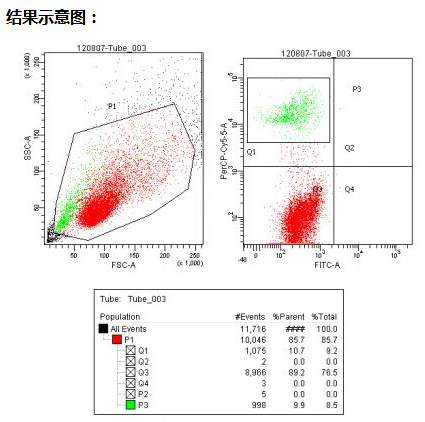
流式细胞检测服务
流式细胞检测服务流式细胞检测技术是利用流式细胞仪,以高能量激光照射高速流动状态下被荧光色素染色的单细胞或微粒,测量其产生的散射光和发射荧光的强度,从而对细胞的物理、生理、生化、免疫、遗传、分子生物学性状及功能状态等进行定性或定量检测的一种现代细胞分析技术。 实验步骤: 1. 用t25培养瓶培养各细胞株,代细胞生长达到80-90%融合后,弃掉旧培养液 2. 用2ml的pbs洗细胞两次后,加入胰酶消化细胞,显微镜下观察细胞变圆即可,加入4ml的完全培养液终止消化 3. 用移液器轻轻吹下细胞,将细胞混合液移入15ml离心管中,1000r/m离心10min,离心结束后,弃掉培养液上清,用 5mlpbs 悬浮细胞沉淀,1000r/m离心10min 4. 弃上清后用500ul pbs悬浮细胞沉淀。之后分别加入抗体,4℃条件下孵育30min用流式细胞仪进行流式分析 结果示意图: 服务周期:服务内容说明价格∕元实验周期流式检测不包括试剂盒费用50元/样/抗体3个工作日温馨提醒: 1. 流式分选建议用直标抗体 2. 提供105细胞
厂商
2018.05.24
公司名称: 上海沪震实业有限公司
公司地址: 上海市杨浦区密云路1018号复旦科技园808室 联系人: 刘经理 邮编: 200613
仪器信息网APP

展位手机站