视频号

抖音号

哔哩哔哩号

前沿资讯手机看

fighting!!!


分享到微信朋友圈
打开微信,点击底部的“发现”,
使用“扫一扫”即可将网页分享到朋友圈。
第二十届北京分析测试学术报告会暨展览会(BCEIA 2023) 将于2023年9月6-8日在北京·中国国际展览中心(顺义馆)召开。BCEIA作为展示国际新技术、新仪器、新设备的窗口,一直以来受到国内外众多专家、学者、科技人员的关注,同时,学术报告会作为BCEIA重要组成部分,始终面向世界科技前沿。BCEIA 2023将举办大会报告、分会报告、高峰论坛、同期会议、墙报展等多场精彩学术活动,邀请国内外行业顶尖学者及学术带头人,分享最具前瞻性的研究进展,针对学科关注度最高的技术及应用进行研讨和交流。
2023年9月7-8日,BCEIA2023学术报告会——光谱学分会将在学术会议区E-206会议室举行,聚焦“高灵敏光谱分析与成像”主题,围绕分子及纳米光谱、光谱分析与材料、高分辨光学成像、光谱仪与显微镜等主题方向,邀请到19位国内色谱领域资深科学家及青年才俊带来精彩报告。
特邀报告人

报告摘要
Surface-enhanced Raman spectroscopy (SERS) has unique advantages for in vivo analysis, but still possesses significant challenges. Aiming to the key issues for in vivo SERS analysis, including complex environment, low molecular content and intermolecular interdependence, a series of novel semiconductor Raman substrates were uniquely constructed for highly sensitive, selective and multi-channel SERS analysis of molecules associated with Alzheimer's disease.
First, by regulating the semiconductor energy level structure, we proposed a new SERS method that enhances the Raman signal by promoting charge transfer through level matching and heterojunction blocking of electron-hole recombination, resulting in a 4-order of magnitude enhancement of the SERS enhancement factor to 1010 and establishing a highly sensitive in vivo analysis method. Secondly, we proposed a new strategy for triple recognition of molecular specificity, level matching, and fingerprint peaks, establishing a highly selective Raman analysis method for in vivo analysis, a SERS optophysiological probe was created for real-time mapping and recording of chemical and electrical signals without cross-talk in the live brain. Moreover, it was the first time that a Raman fiber photometry was built up for real-time tracking and simultaneous quantitation of multiple molecules in mitochondrial across the brain of free-moving animals. Meanwhile, a highly selective non-metallic Raman probe was created through triple-recognition strategies of chemical reaction, charge transfer, and characteristic fingerprint peaks, for monitoring and quantifying of local mitochondrial O2•-, Ca2+ and pH in six brain regions upon hypoxia. It was discovered that hypoxia-induced O2•- burst was regulated by ASIC1a, leading to mitochondrial Ca2+ overload and acidification.
专家简介
田阳,华东师范大学特聘教授,现任华东师范大学化学与分子工程学院院长。2013年曾获国家杰出青年基金资助;获日本化学会“The distinguished lectureship award”,中国分析测试协会一等奖(第一完成人),中国化学会女分析化学家,上海市自然科学奖一等奖(第一完成人);受邀在神经学和神经科学等国际国内做大会、主题或邀请报告36次。目前担任Chemical Communications副主编和《高等化学学报》副主编。田阳教授长期从事活体电信号的化学表达分析领域研究,在发展生物化学分子(如酶、蛋白等)的精准分析测量策略、建立长时程稳定的高空间分辨成像方法、及开拓高速成像分析新仪器等方面开展了深入和系统的工作。
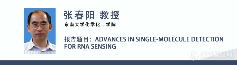
报告摘要
Single-molecule detection enables the measurement of molecules at the single-molecule level, and it can be used to study the conformational changes and interaction between the molecules, holding great potential in biochemical analysis and biomedical research. In comparison with the conventional ensemble measurements, single-molecule detection possesses the advantages of ultrahigh sensitivity, good selectivity, rapid analysis, and low sample consumption. Single-molecule detection can be used as an ideal analytical approach to quantify the low-abundant biomolecules with rapidity and simplicity. We demonstrate the applications of single-molecule detection-based biosensors for sensitive detection of various target biomolecules such as long noncoding RNAs (lncRNAs), microRNAs (miRNAs), and circular RNAs (circRNAs). The biosensors show extremely high sensitivity. Moreover, these biosensors enable simultaneous measurement of multiple endogenous RNAs at the single-cell level, and it may discriminate the expressions of various RNAs in lung tumor tissues and the healthy tissues, offering a promising platform for clinical diagnosis and biomedical research.
专家简介
Chun-yang Zhang obtained his PhD degree from Peking University, China, in 1999. During 1999–2008, he worked at Tsinghua University, China; Emory University, USA; The Johns Hopkins University, USA; and The City University of New York, USA. In 2009, he joined as a professor in the Shenzhen Institute of Advanced Technology, Chinese Academy of Sciences, China. During 2015-2023, he worked as the dean of college of Chemistry, Chemical Engineering and Material Science in Shandong Normal University, China. In 2023, he joins in Southeast University, China. He is the recipient of the China National Fund for Distinguished Young Scientists. His research focuses on analytical chemistry, bioelectrochemistry, bionanotechnology and single-molecule detection.

报告摘要
Marine plankton play important role in ocean biogeochemistry, and their observation is of fundamental significance for oceanographic research and coastal environment monitoring. However, current marine plankton observation still relies heavily on traditional manual net sampling and optical microscopy inspection, which has long been notoriously slow and labor intensive. Developing automated and online approaches for this task is expected to satisfy the urgent needs from marine scientists for research and government departments for operational oceanographic coastal seawater environment monitoring. The advent and application of in situ optical imaging have enabled more direct observations of marine plankton in different tempo-spatial scales, greatly promoted our understanding of marine plankton ecology. However, existing underwater plankton cameras compromise between their imaging resolution and field of view (FOV) for in situ observations. In order to enlarge the sampling volume in single frame acquisition, they usually adopt lower magnifications to enable larger FOV but sacrifice the resolution. This will inevitably lead to a decreased imaging resolution, leading to insufficiency to obtain enough image details for the relatively small plankton targets and hence inaccuracy for subsequent species identification and quantification.
In this talk, the speaker will report some recent developments by his team on in situ plankton imaging technologies. Particularly, the talk will emphasize a deep learning-based super-resolution in situ plankton imaging technology. This new technique is expected to enhance the existing plankton imageries and enable future underwater plankton imaging instruments for better in situ plankton observation and hence deeper our understanding of the marine plankton ecology.
专家简介
李剑平,男,博士,中国科学院深圳先进技术研究院正高级工程师,中国科学院大学博士生导师,深圳市海洋声光探测技术及装备工程研究中心主任。研究领域包括创新光学方法、先进光电仪器、机器视觉与机器学习在海洋观测中的应用。先后主持和参与了国家重点研发计划、国家自然科学基金、中国科学院、香港大学教育资助局(RGC)、广东省科技厅、深圳市科技创新委等研究项目。带领团队研制了水下浮游生物成像仪、走航式浮游植物成像流式细胞仪、海水叶绿素a、COD、BOD传感器等多种海洋观测探测仪器。在IEEE JOE,ICES JMS, FMARS,ECCV,ICCV, Optics Letters, Optics Express、Applied Optics等光学、海洋科学和机器视觉知名期刊和国际学术会议发表论文多篇,申请中国发明专利和实用新型专利43项,获得发明专利授权7项,实用新型专利授权6项;在ICCV, Ocean Optics, International Ocean Color Sciences, Focus on Microscopy等知名国际会议做主旨报告、口头报告多次。
李剑平博士是国际电子电气工程协会IEEE高级会员、美国光学学会Optica会员、国际光电工程师协会SPIE会员、国际海洋技术学会MTS会员、中国仪器仪表学会高级会员、中国海洋与湖沼学会海洋观测分会理事、中国海洋湖沼学会海洋腐蚀与污损专业委员会委员、深圳市人工智能学会会员、广东省自然资源厅赤潮专家库专家。长期担任Optics Letters, Optics Express, Biomedical Optics Express, Applied Spectroscopy, Applied Optics, Cytometry Part A等知名学术期刊论文审稿人,担任国际会议ICCV Computer Vision in the Ocean Workshop程序委员会委员和审稿人。

报告摘要
The advanced light source (ALS) analytical technologies have been expanded to dig into the underexplored behavior and fate of nanomedicines in vivo. It is increasingly important to further develop ALS-based analytical technologies with higher spatial and temporal resolution, multimodal data fusion, and intelligent prediction abilities to deeply unlock the potential of nanomedicines. In this presentation, we focus on several selected ALS analytical technologies, including imaging and spectroscopy, and provide an overview of the emerging opportunities for their applications in exploring the biological behavior and fate of nanomedicines. Improved ALS imaging and spectroscopy techniques will accelerate a profound understanding of the biological behavior of new nanomedicines.
专家简介
王亚玲,国家纳米科学中心研究员,广州市新发传染病疫苗研发技术创新促进会理事,主要研究方向为基于先进光源的纳米生物分析方法、新型纳米佐剂开发及产业化研究。近年来,在Nature protocols, Acc. Chem. Res., ACS Cent. Sci., Nano Today, Anal. Chem., ACS Nano,等期刊上发表70余篇论文,申请发明专利18项,获授权国家发明专利3项。作为首席科学家承担了科技部政府间科技合作重点专项,主持了国家重点研发计划大科学装置专项课题1项,国家自然科学基金青年、面上、重点项目子课题各1项,作为项目骨干参加中科院先导B项目、纳米生物效应及分析方法等相关十多个项目研究。

报告摘要
Molecular sensing and imaging have become powerful tools in both fundamental research and clinical diagnosis because they enable not only to quantify but also to track biological molecules of interest. During the past years, we are dedicated to developing new strategies that enable spatiotemporally selective molecular sensing with higher precision. For example, by designing light-activatable sensors and combining it with upconversion nanotechnology, spatiotemporally controlled imaging of metal ions in mitochondria was achieved. In addition, activatable sensors could be triggered by endogenous cues. By introducing peptide nucleic acid to bridge the gap between DNA and peptides, we demonstrated that DNA self-assembly could be specifically triggered by specific protease, thus generating fluorescence signal output for real-time monitoring of tumor response to drug treatment. Furthermore, we designed an enzyme-mediated signal amplification strategy that enables to selectively enhance the signal of inflammation-associated molecules in disease tissues, and thus allowing for molecular imaging with a high signal-to-background ratio.
专家简介
李乐乐,国家纳米科学中心研究员,博士生导师。于北京大学获博士学位,随后在美国伊利诺伊大学香槟分校、麻省理工学院和哈佛大学从事博士后研究。先后获得国家杰青、国家优青、海外高层次青年人才计划等项目支持。研究方向为活细胞和在体分子成像方法与疾病诊断技术,相关工作以通讯作者发表在Nat. Biomed. Eng.、Acc. Chem. Res.、J. Am. Chem. Soc.和Angew. Chem. Int. Ed.等期刊。
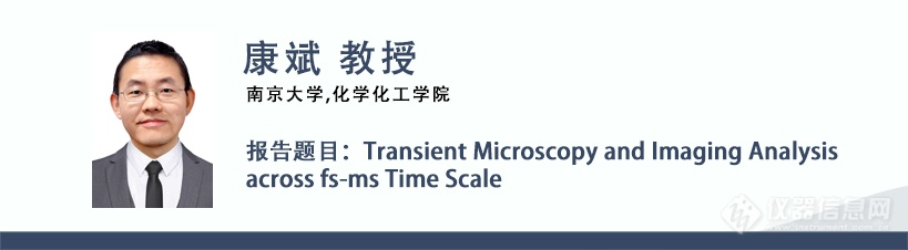
报告摘要
The transient microscope based on the Pump-Probe principle combines time-resolved detection and microscopic imaging, which enable to study the spatiotemporal evolution of microscopic particles and energy in the space and time domains. This report will share with you our research progress in recent years in the construction of transient microscopic imaging methods and instruments across time scales from fs to ms based on the Pump-Probe principle. Including: femtosecond time-resolved wide-field transient absorbance microscopy and measurement of various energy-carrying carriers in materials; nanosecond time-resolved transient photoelectrochemical microscopy and measurement of electric double layer formation kinetics at the nanoscale; microsecond time-resolved transient heat dissipation microscopy for the measurement of heat transfer and dissipation within single cells. The above several research cases show our attempts and thinking in expanding the connotation and extension of Pump-Probe technology. For the purpose of throwing bricks and starting jade, we discuss with colleagues the new scenarios of ultrafast spectroscopy and imaging technology applications.
专家简介
康斌,南京大学化学化工学院教授、博士生导师。研究兴趣为发展高时空分辨的成像测量方法、技术及仪器装置,诠析微纳化学系统及生物系统中的基本传质、传电及传能过程及其动力学。承担了国家自然科学基金委原创探索研究计划、国际合作中俄项目、面上项目等。受聘为俄罗斯科学院海外专家,共建莫斯科“生物医学光子学”国际实验室并任联合主任(co-director),担任日本北海道大学客座教授。

报告摘要
Optoelectronic devices made from functional nanomaterials are promising for biosensing and modulation at cellular, tissue-, and other levels. Key design principles for such devices include the development of efficient optoelectronic materials and their integration with biological samples via the biotic/abiotic interfaces. In this work, we first develop direct patterning methods for quantum dots and other functional nanomaterials. These methods rely on ligand crosslinking and other reactions, forming microscale quantum dot patterns without using traditional photoresists. Patterning nanomaterials with different optical and electrical properties leads to an array of multifunctional devices, whose sizes are about several microns and comparable with the sizes of cells. We then integrate these devices with cells or tissues via a layer of two dimensional metal organic frameworks. The optoelectronic devices form mechanically robust interfaces with cells and tissues. The favorable charge transport behavior at the interfaces make such bioelectronics devices effective in monitoring regional tissue oxygen saturation in awake animals, and optically modulating the neuronal activities of neurons and the sciatic nerves of rats.
专家简介
张昊自2019年起加入清华大学化学系,担任副教授、博士生导师。本科(2007年)和硕士(2010年)毕业于清华大学化学系,博士(2015年)毕业于美国芝加哥大学化学系,2016–2018年在美国西北大学材料系进行博士后研究。主要研究方向为功能材料的直接光刻图案化与3D打印以及柔性生物电子器件。
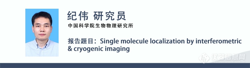
报告摘要
Remarkable progress in Single molecule localization microscopy (SMLM) has been made in the past decade. Here we developed interferometric and cryogenic imaging which exhibit excellent localization precision performances compared to conventional SMLMs. We introduced interferometric SMLMs named ROSE and ROSE-Z. A fluorescence molecule is located by the intensities of multiple excitation patterns of an interference fringe, providing improvement in the localization precision compared to the conventional centroid fitting method at the same photon budget. We demonstrate this technique by resolving a nanostructure down to 5 nm. We also built an ultra-stable super-resolution cryo-FM that exhibits excellent thermal and mechanically stability. We have demonstrated the super-resolution imaging capability of this system. The results suggest that our system is particularly suitable for SMLM and cryogenic super-resolution correlative light and electron microscopy. Based on the cryo-fluorescence imaging technique we developed, we build a cryogenic correlated light, ion and electron microscopy (cryo-CLIEM) that is capable of preparing cryo-lamellae under the guidance of three-dimensional confocal imaging. Moreover, we demonstrate a workflow to preselect and preserve nanoscale target regions inside the finished cryo-lamellae. By successfully preparing cryo-lamellae that contain a single centriole or contact sites between subcellular organelles, we show that this approach is generally applicable, and shall help in innovating more applications of cryo-ET.
专家简介
Dr. Wei Ji is a Principal Investigator at the Institute of Biophysics (IBP), Chinese Academy of Sciences (CAS). He obtained his Ph.D at IBP in 2010 and then focused on developing new superresolution microscopy and correlative light and electron microscopy techniques. His recent publications on interferometric single-molecule localization microscopy and integrated multimodality microscopy for target-guided cryo-lamellae preparation can be found in Nature Methods journal.
报告摘要
With the help of chemometrics (especially the optimization and combination of various algorithms), a multi-level infrared spectrum macro fingerprint analysis method for complex system was established. Based on the infrared spectra of hundreds of thousands of food, health care products and traditional Chinese medicine, the multi-molecular vibration theory is extended on the basis of single molecular vibration theory, which lays a theoretical foundation for "multi-level infrared spectrum macro fingerprint analysis". On the basis of genomics, proteomics, metabonomics and metalomics, the basic concept of "macrogenomics" was proposed. Combining the “multi-component ecological metabolism theory” , “multi-component competitive adsorption-diffusion theory” and “Multilevel Infrared Spectral Macro-Fingerprints Analysis” of mixture system, and following the three technical routes of " analysis without separating, analysis while separating and analysis while combining " , the growth and metabolism laws of animals and plants at the molecular spectrum level were revealed , the mechanism of human etiology, pathogenesis, health preservation and internal material transformation prevention and control were explained.
专家简介
1978年元月毕业于清华大学工程物理系后留校工作至今。1979-1980年从事基础物理和无机化学教学工作,1981-1983年从事原子光谱分析工作。1986年至今,从事红外光谱分析工作。主要研究领域:分子振动光谱学、化学计量学、中药和食品分析化学。主要学术贡献:在基因组学、蛋白组学、代谢组学等组学的基础上提出了“宏观组学”的基本概念。在国际上首次提出并建立了针对复杂混合物体系的“多级红外光谱宏观指纹分析法”。目前已发表SCI论文近200篇,获中国发明专利3项,已出版中英文学术专著4部,多次获得中国分析测试协会CAIA奖。多次在国内外举行的国际会议上作邀请报告,作为合作主席主持了“第4届二维相关光谱国际会议”。作为组委会主席筹办了“2018全国光谱大会”。 兼任北京理化分析测试技术学会副理事长,光谱分会理事长。《光谱学与光谱分析》副主编。美国药典委员会(USP)脱脂乳粉顾问组专家成员(2014)。
报告摘要
Long-term subcellular intravital 3D imaging in mammals is vital to study diverse intercellular behaviors and organelle functions during native physiological processes. However, optical heterogeneity, tissue opacity, and phototoxicity pose great challenges, leading to the tradeoff between the field of view, resolution, speed, and sample health. In this talk, I will discuss our recent work in multiscale intravital fluorescence microscopy based on computational imaging methods. Various large-scale fast subcellular processes are observed, including brain-wide neural recoding in mice at single resolution, 3D calcium propagations in cardiac cells, human cerebral organoids, and Drosophila larval neurons, membrane dynamics in zebrafish embryos, and large-scale cell migrations during immune response and tumor metastasis, enabling simultaneous in vivo studies of morphological and functional dynamics in 3D.
专家简介
Jiamin Wu is an assistant professor in the Department of Automation at Tsinghua University, and PI at the IDG/McGovern Institute for Brain Research, Tsinghua University. His current research interests focus on computational imaging and system biology, with a particular emphasis on developing mesoscale optical setups for observing large-scale biological dynamics in vivo. In the recent 5 years, He has published more than 40 journal papers in Nature, Cell, Nature Photonics, Nature Biotechnology, Nature Methods, and so on. He has served as the Associate Editor of PhotoniX and IEEE Transactions on Circuits and Systems for Video Technology.
报告摘要
Cell analysis is of great significance for understanding the essence and law of life, as cell is the basic structural and functional unit of organisms. To approach the true cell activities in vivo, living cell analysis in vitro requires simultaneous monitoring of cell responses through quantitative and controllable methods in simulated cell microenvironment. In order to monitor the biochemical characteristics of cells, liquid crystal biosensor was introduced into microfluidic chip. We synthesized liquid crystal elastomer microspheres functionalized with horse-radish peroxidase (LCEM-HRP), which can be immobilized directly on the surface of individual cells cultured in 2D microfluidic chip, to monitor the real-time releasing of H2O2 at the single-cell level. [1] The LCEM-HRP could report H2O2 through a concentric-to-radial transfiguration. The level of the transfiguration of LCEM-HRP revealed the different amounts of H2O2 released from cells. The cell lines and cell-cell heterogeneity were explored from different configurations of LCEM-HRP. This method realizes in situ real-time imaging of unstable molecules released from living single cells with high spatial-temporal resolution, high selectivity and high sensitivity. It provides a new idea for real-time imaging of signal molecules released from cells to the microenvironment.
专家简介
林玲,北京工商大学教授。2016年于日本东京大学工学部获得博士学位,并在博士期间研发微/纳流控系统用于活体单细胞微量样品的检测。回国后主要从事微流控芯片上细胞代谢产物的研究,利用微流控芯片构建细胞共培养模型,结合荧光成像及高灵敏度质谱检测技术,系统地研究了细胞在不同微环境条件下,药物诱导对特定细胞的作用机制, 为细胞微环境药物刺激响应等生命过程提供理论依据。近五来已发表SCI 学术论文50余篇,其中通讯或第一作者论文发表在Chem. Soc. Rev., Angew. Chem. Int. Ed., Chem. Sci., Anal. Chem.等国际学术期刊,并参著中英文专著3章。作为负责人获得国家自然科学基金青年、面上项目资助。

报告摘要
Classic nanomaterials characterization techniques can only give the morphology, structure, size, energy state, but the information of the microscopic activity on the micro level. Based on the single molecule fluorescence microscopy, this paper introduces some works on how to characterize the catalytic reactivity at single particle level. 1) developed the dynamic super resolution imaging microscopy and electrochemical-single molecule fluorescence microscopy to implement the dynamic tracking the active sites. 2) The study on dynamic of active sites and high-throughput single particle analysis of the electrochemical system were realized. The mechanism of quantum effect and synergistic effect is revealed at the single particle scale. These studies provide characteristic mechanism information for the analysis of active structure-activity relationship of nanoparticles.
专家简介
张玉微博士,教授,广州大学百人A计划教授;广州市高层次人才,中国科学院青年创新促进会会员。广东省杰出青年基金获得者,国家自然科学基金委优秀青年基金获得者,广州市青年科技工作者协会秘书长、副理事长。
从事能源、环境等领域纳米材料的荧光显微分析工作,近年来在PNAS、Nat. Commun.、JACS、Angew. Chem. Int. Ed.、Adv. Mater.、Anal. Chem.、ACS Nano等杂志发表SCI 论文50余篇,他引2000 余次。参与撰写英文专著两部《Rotating Electrode Methods and Oxygen Reduction Electrocatalysts》《Single Particle Nanocatalysis》。申请专利26 项,其中12 项已获授权。论文被包括Nature、Chem. Rev.、PNAS、 JACS 等刊物予以正面引用或评述, 相关工作获得包括Nature Energy、中国科学院官网、光明科技、搜狐科技等多家科技媒体的积极评价。科研工作被广州日报、学习强国等媒体平台报道。

报告摘要
Spatial omics provides spatial location and interaction network information between cells at the gene expression level. It is an important tool for in-depth study of tissue/cell functions, microenvironment interactions, developmental processes, disease pathology, etc., which are essential for clinical and pharmaceutical research and development. In recent years, spatial transcriptomics triggered a research boom not only in academia but also in industry. In principle, spatial transcriptomics can be categorized into two types: imaging based (e.g. FISH) and capturing based. The second type is characterized with unbiased capturing, which is more suitable for scientific discovery. However, it also suffers from limited spatial resolution, low capture efficiency, and the high technical barriers for DNA chip manufacturing.
We are developing massively parallel DNA sequencing techniques and sequencing-based high throughput detection platforms such as spatial omics tools. An ultra-high density DNA chip manufacturing technology was developed, which is suitable for spatial transcriptomics studies. Our strategy uses oligonucleotide arrays on DNA chips to capture tissue RNA and then correlate spatial barcodes with genetic sequences through sequencing. The key to this technique is the formation of a high-density, high-resolution DNA capture probe lattice on the capture chip. We used special technology to form these lattices on the glass surface so that the spacing between the capturing addresses can reach below 1 micron. The capture probes can be as high as 18 thousand per square micrometers, greatly promoting the capture efficiency. Based on this technology, we further demonstrate high-resolution spatial omics data, including the studies of mouse testes, mouse brains, mouse kidney, and other tissues.
专家简介
Drs. Marc Porter, Ning Fang, and Edward Yeung from 2006 to 2011. He served as a faculty member in North Carolina State University from 2011-2019. His research focuses on super resolution optical imaging and surface chemistry. In 2020, he co-founded Shenzhen Salus Biomed Co. Ltd., which is a biotech company aimed at developing massively parallel DNA sequencing techniques and sequencing-based high throughput detection techniques. Currently, he serves as the Chief Scientific Officer in Salus Biomed.

报告摘要
Terahertz spectral imaging is an important field of THz science and technology. In this presentation, I will introduce our recent work to demonstrate three different THz time-domain spectral imaging techniques by which biological samples were investigated on different spatial resolutions from millimeters, micrometers to nanometers. First, we employed a conventional far-field THz spectroscopy system with a millimeter resolution to image mouse skin tissue with melanoma. It was found that the melanoma could be unambiguously identified from the normal tissue in the frequency region of 0.6 - 2.0 THz. Second, we developed a home-built photoconductive antenna microprobe-based THz near-field system with a calibrated spatial resolution of five microns, by which mouse brain tissue slices and single cells were successfully studied and useful information were retrieved. Finally, we achieved to image individual protein molecules on the nanometer scale using a THz scattering-type scanning near-field optical microscope with the aid of self-designed high-performance THz probes and graphene-based substrate for biomolecules. Collectively, our results prove the capability of studying biological samples at different spatial resolution levels, manifesting that THz imaging techniques are holding a promising future in biomedical and biological fields.
专家简介
中国科学院重庆绿色智能技术研究院研究员、超分辨光学研究中心主任,重庆市高分辨三维动态成像工程技术研究中心主任,中国生物物理学会太赫兹生物物理分会副秘书长。主要从事太赫兹光谱成像仪器研制及应用研究,2021年获全国太赫兹生物物理优秀工作者荣誉称号,先后主持承担国家重点研发计划课题、国家自然科学基金、中国科学院科研仪器研制等多个重要研究任务,在领域重要期刊发表SCI论文80余篇,拥有受权发明专利10余项。

报告摘要
Membraneless organelles (MLOs) are multidimensional complex systems containing biomacromolecules including proteins, RNAs and DNAs. Formation of these systems are usually driven by phase separation within cells. Artificial intelligence provides novel insights in decoding MLOs and phase separation mechanisms. This report will mainly introduce the prediction methods for phase separation proteins and their applications in drug target discovery and interference, including PhaSePred, the phase separation protein prediction tool integrating multi-model features, and DropScan, the computational method for small molecule drugs targeting aberrant phase separation.
专家简介
北京大学基础医学院研究员,教育部青年长江学者。主要研究方向为生物分子相分离的智能解析与应用,围绕相分离发展了系列预测分析方法,从相分离角度探索潜在药物靶标。以通讯作者(含共同)在PNAS、Nature Chemical Biology、Genome Biology期刊发表SCI论文近30篇。作为课题负责人承担国家重点研发计划蛋白质专项,作为负责人承担国家自然科学基金4项。担任中国人工智能学会生物信息学与人工生命专委会委员、中国自动化学会智能健康与生物信息专委会委员、中国计算机学会生物信息学专委会委员、生物物理学会生物大分子相分离与相变分会委员等。

报告摘要
Here we present an overview of our recent works in live-cell superresolution (SR) microscopy. Over the past five years, we have developed several innovative techniques to improve the resolution and accuracy of live-cell imaging.
Our first breakthrough was the development of a structured illumination microscopy technique based on the continuity of biological structures embedded in Hessian matrices (Hessian-SIM). Hessian-SIM significantly reduces the photon dosage required for SR microscopy while suppressing reconstruction artifacts induced by random noise. Additionally, we demonstrated that the high sensitivity of this method allows for the use of sub-millisecond excitation pulses followed by dark recovery times, reducing photobleaching and enabling hour-long time-lapse SR imaging with common fluorescent probes in live cells (Nat. Biotechnol. 2018).
To enable holistic SR imaging, we developed a dual-mode microscopy technique that combines SIM with label-free three-dimensional optical diffraction tomography (ODT). By providing a holistic view of organelles and simultaneously highlighting molecules, this method is ideal for studying organelle interactomes. We demonstrated that the ODT module can resolve mitochondria, lipid droplets, the nuclear membrane, chromosomes, the tubular endoplasmic reticulum, and lysosomes (Light Sci Appl. 2020).
To further push the resolution limit of live-cell SR imaging, we developed a two-step iterative deconvolution algorithm based on continuity and sparsity of fluorescence signals (Sparse deconvolution), which extends resolutions beyond the physical limits of optical systems. Sparse-SIM achieving ~60 nm resolution at a 564 Hz frame rate, resolving dynamics of ring-shaped nuclear pores over an hour in live cells. The algorithm can also be used to improve resolutions of other fluorescence microscopes, such as confocal, STED, and lightsheet microscopes. Thus this mathematical path to improve microscopic resolution may have broad implications (Nat. Biotechnol. 2022).
Finally, for live-cell SR imaging to be quantitative, the completeness of delicate structures and the linearity of fluorescence signals are required in addition to resolution. To make live-cell SIM microscopy more quantitative, we proposed a physical model-based background removal method (BF-SIM). BF-SIM preserves intricate and weak structures down to sub-70 nm resolution while maintaining signal linearity, enabling us to discover novel, dynamic actin structures in live cells (Nat. Commun. 2023).
专家简介
Liangyi Chen is Boya Professor of Peking University. He obtained his undergraduate degrees Biomedical engineering in Xi’an JiaoTong University, then majored in Biomedical engineering in pursuing PhD degree in Huazhong University of Science and Technology. His lab focused on two interweaved aspects: the development of new imaging and quantitative image analysis algorithms, and the application of these technology to study how glucose-stimulated insulin secretion is regulated in the health and disease at multiple levels (single cells, islets and in vivo) in the health and disease animal models. The techniques developed included ultrasensitive Hessian structured illumination microscopy (Hessian SIM) for live cell super-resolution imaging, the Sparse deconvolution algorithm for extending spatial resolution of fluorescence microscopes limited by the optics, Super-resolution fluorescence-assisted diffraction computational tomography (SR-FACT) for revealing the three-dimensional landscape of the cellular organelle interactome, two-photon three-axis digital scanned lightsheet microscopy (2P3A-DSLM) for tissue and small organism imaging, and fast High-resolution Miniature Two-photon Microscopy (FHIRM-TPM) for Brain Imaging in Freely-behaving Mice. He is also recipient of the National Distinguish Scholar Fund project from National Natural Science Foundation of China.

报告摘要
Nanoimprint lithography (NIL) was invented 90s of last century, which is a high-resolution fabrication lithography technology. It has been widely used for the fabrication of many different kinds of micro/nano structures, for example the gratings and 2D gratings, which is one of the most critical optical components for the spectrum generation. The presentation will briefly introduce the development of nanoimprint lithography and how it will be used for the production of the slanted grating, the brazed grating and other special gratings et al.
专家简介
材料科学姑苏实验室纳米压印先进制研发中心负责人,国家级引进人才。国内最早从事纳米压印研究的科研工作者之一,长期从事以纳米压印技术为核心工艺的先进制程研究。早年留学瑞典期间,深入研究纳米压印基础理论和工程技术,毕业后以纳米压印专家加盟国际技术领先的Obducat公司,先后担任SMASH、ANIP等欧盟科研项目的技术负责人,以及LED衬底PSS(蓝宝石图案化衬底)、面板级铝线栅WGP、AR/VR光学元器件等产业化项目的技术负责人,实现纳米级器件低成本高通量工程制造。21年归国全职加入材料科学姑苏实验室,主要从事纳米印压制程能力开发,包括AR/VR光学元器件、超表面器件、复杂曲面精细结构制造等方面的工业化生产设计等。22年创立新维度微纳科技有限公司,致力于纳米压印技术的产业化推广。

报告摘要
We have proposed the radical-triggered luminescence to monitor the radical behaviours during polymer degradation without/with the addition of inorganic additives. Taking polyethylene (PE) as an example, the radical-triggered luminescence showed a single sigmoidal dynamic curve with the emissions from ROO•, manifesting the exponential proliferation for the degradation evolution. Alternatively, upon the addition of layered double hydroxides (LDHs) with positively charged Al centers, the degradation pathways could be modulated into a double sigmoidal dynamics, which assigned to a main product of RO• with the activation energy of 40.2 kJ/mol and a small amount of ROO• with 76.3 kJ/mol, respectively. Accordingly, the polymers with the additive-regulated pathways could exhibit prominently anti-degradation behaviours. The proposed mechanism for the regulated degradation pathways by LDHs was further proved with the addition of positively charged LDHs with varied metal compositions, and negatively charged SiO2 particals. Moreover, the established luminescence monitoring and LDHs regulation methods have strong universality, and achieve the anti-aging effect on polypropylene (PP), polystyrene (PS), polytetrafluoroethylene (PTFE) and other polyolefin systems. This work is beneficial for the deep understanding of the radical mechanisms during polymer degradation, and for the rational design of anti-degradation polymers.
专家简介
2021.07至今在郑州大学化学学院工作。现任Analytical Methods副主编,Industrial Chemistry & Materials副主编,《分析试验室》副主编,Trends in Analytical Chemistry,Chinese Chemical Letters,《光谱学与光谱分析》等期刊编委。主要研究方向包括危化品快速检测方法、材料结构光谱表征新方法、纳米材料化学发光分析方法及复合材料早期老化荧光检测及其寿命预测。主持国家基金委重大项目、重点项目、面上项目及国家“973”计划等项目10余项,近五年以通讯作者在Angewandte Chemie International Edition,Nature Communications,Science Advances,Analytical Chemistry等期刊发表论文100余篇,获批中国发明专利15项,专利技术转化2项。获批中国石油和化学工业联合会团体标准1项和企业标准3项。获得2015年高等学校自然科学奖二等奖、2020年中国分析测试协会科学技术奖一等奖等奖项。
报告摘要
Identification and detection of proteins by mass and binding are routinely used in biological research, but optical detection at the single-molecule level have been challenging due to the shot noise limit imposed by the quantum nature of light. Mass photometry was first invented with the interfereometric scattering microscopy, enabling optical mass detection of single macromolecules. However, due to the shot-noise limit, it typically requires high power illumination that could damage the biological specimen. We report that integral imaging with surface plasmon polaritons allows single-protein detection with a signal-to-noise ratio an-order-of-magnitude beyond the shot-noise limit. Therefore, our integral microscopy allows quantitative mass imaging and binding analysis of single unlabeled protein molecules with a three-orders-of-magnitude reduction in the light intensity. It also enables highly specific protein detection at the subpicomolar concentration level that would not otherwise be achievable.
专家简介
2011年博士毕业于浙江大学,并先后在香港科技大学和美国亚利桑那州立大学开展研究工作。2017年加入上海交通大学生物医学工程学院,主要研究方向为光学生物传感技术及仪器。在PNAS等顶级期刊发表多篇学术论文,承担基金委国家重大科研仪器研制项目、十四五国家重点研发计划专项课题、基金委面上项目等研究任务。
以上报告内容由BCEIA2023组委会提供

欢迎扫码报名参加BCEIA2023
[来源:仪器信息网] 未经授权不得转载

多元化产品线布局,坚持技术突破与创新——访元析仪器生命科学部门经理章宏飞
2023.07.20

全日程公布!高光谱技术在农业领域的最新应用进展网络研讨会8月8日召开
2023.08.02


守科研梦,筑未来路——HORIBA服务万里行·北大分析测试中心站圆满完结
2024.07.11
2024.07.11

28年致力于农畜产品品质无损检测技术的发展——访中国农业大学彭彦昆教授
2024.07.10
品牌合作伙伴


版权与免责声明:
① 凡本网注明"来源:仪器信息网"的所有作品,版权均属于仪器信息网,未经本网授权不得转载、摘编或利用其它方式使用。已获本网授权的作品,应在授权范围内使用,并注明"来源:仪器信息网"。违者本网将追究相关法律责任。
② 本网凡注明"来源:xxx(非本网)"的作品,均转载自其它媒体,转载目的在于传递更多信息,并不代表本网赞同其观点和对其真实性负责,且不承担此类作品侵权行为的直接责任及连带责任。如其他媒体、网站或个人从本网下载使用,必须保留本网注明的"稿件来源",并自负版权等法律责任。
③ 如涉及作品内容、版权等问题,请在作品发表之日起两周内与本网联系,否则视为默认仪器信息网有权转载。
![]() 谢谢您的赞赏,您的鼓励是我前进的动力~
谢谢您的赞赏,您的鼓励是我前进的动力~
打赏失败了~
评论成功+4积分
评论成功,积分获取达到限制
![]() 投票成功~
投票成功~
投票失败了~