搜全站
搜展位

广州竞赢化工科技有限公司
 关注
关注
已关注
金牌16年

![]() 已认证
已认证
粉丝量 0
400-860-5168转1729
仪器信息网认证电话,请放心拨打
解决方案

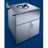
等离子增强原子层沉积系统沉积高均匀性和高保型性介电薄膜
应用领域
电子/电气检测样品
电子元器件产品检测项目
超薄膜沉积★超薄,纳米尺度介电薄膜与金属/金属性薄膜是MEMS/NEMS器件、其它IC部件,传感器,光学器件或催化剂关键部件
★IC业中的高精度30器件, 如高深宽比沟槽与穿透性硅通孔, ALO工艺是唯可以在这些器件上实现高保形,平整,无缺陷,无针孔的薄膜材料。
★可规模化生产的ALO工艺, 几种金属/金属性材料与介电材料: Pt, Ir, Ru, Cu, Ag, Au, TiN, AIN, TiAIN, ln203与Al203.
★沉积工艺可选:传统热ALO或者等离子增强ALD。
共1台
电镜负染色蛋白质案例
应用领域
生物产业检测样品
其他检测项目
电镜负染色载玻片的半边涂上真空油脂,并放在铜网旁边。注意:涂有真空油脂的载玻片在镀碳之后不会变色,通过对比两者的颜色可以估测碳膜的厚度。

暂无关联产品
Immunohistochemical staining of plastic
应用领域
检测样品
检测项目
Abstract
Aims-To investigate (1) whether adequate
immunohistochemical staining can
be achieved on sections cut from plastic
embedded bone marrow trephine biopsy
specimens after microwave heating in citrate
buffer; and (2) whether this immunohistochemical
staining is comparable with
that achieved on routine sections cut from
paraffin wax embedded trephine biopsy
specimens after decalcification procedures.
Methods-Sixty five consecutive bone
marrow trephine biopsy specimens of
more than 1 cm in length were divided
transversely into two equal parts. One part
was processed in paraffin wax foliowed by
decalcification. The other part was embedded
in the epoxyresin Polarbed 812 followed
by the cutting of 1 pm sections. Both
parts underwent immunohistochemical
staining by an identical panel of antibodies.
With Polarbed 812 plastic embedded
sections, microwave heating in
citrate buffer was undertaken before the
application of antisera.
Results-On sections cut from plastic embedded
material, immunohistochemical
staining was generally satisfactory, easy
to interpret and comparable with that
achieved with paraffin wax embedded
material. Exceptions were antibodies
to neutrophil elastase and CD61 where
immunostaining was consistently negative
on plastic embedded sections. Immunohistochemical
staining for CD20 was
consistently more reliable on plastic
embedded sections.
Conclusions-The results provide evidence
that, with few exceptions, satisfactory
immunohistochemical staining is possible
on plastic embedded bone marrow trephine
biopsy specimens after microwave
heating in citrate buffer. This, combined
with the advantage of superior cellular
morphology with semi-thin (1 pm) sections
of plastic embedded material, make
such embedding procedures the preferred
method for the processing ofbone marrow
trephine biopsy specimens.
共1台
Microwave-assisted immunostaining: a new approach yields
应用领域
检测样品
检测项目
Abstract
Advances in microwave technology permitted the development of new antigen labeling techniques. The recent microwave development
of a true variable wattage unit designed for laboratory use and an apparatus for dampening standing wave radiation patterns have allowed
investigators to better control the conditions within a microwave cavity. Thus, operating limits thought to be endemic to microwave-assisted
protocols could be effectively mitigated. Standard protocols for histochemistry call for prolonged incubations and numerous rinses that add
considerable time to the procedure. Here, we present microwave-assisted staining protocols for floating rat brain sections and cultured rat
hippocampal cells. Acetylcholinesterase (ACHE) histochemistry and immunocytochemistry were conducted inside a specially designed and
configured laboratory microwave oven. As a control additional tissue sections were stained on the bench and treated in the same manner as those
in the microwave. Labeling was minimal in the control tissue, but specific, high contrast staining was present in the microwave group. Tissues
were evenly stained with minimal background, and anatomical structures were easily detected. Also, the differences between lesioned and
intact sides of the brain were obvious and agreed with previous observations. Microwave-assisted methods resulted in significantly shorter
protocol times (∼10-fold) resulting in staining patterns of equal or superior quality to those obtained using conventional methods.
© 2004 Elsevier B.V. All rights reserved.
共1台
Microwave Tissue Processing Techniques:Their Evolution and Understanding
应用领域
检测样品
检测项目
ABSTRACT
The microwave has always promised to shorten tissue processing times. However, historically much of the use of the microwave has produced inadequate and varied results. This science has been further
hampered by assumptions and misunderstandings as to how the microwave shortens processing times. This article discusses the
important variables and advances in microwave processing and also illustrates why it was not technically possible for the variables to have been discovered earlier. Examples of protocols which can be used to standardize microwave processing techniques to produce consistent high quality results are given. The new methodology is
contrasted with the methodology of the past 30 years to illustrate the new advances in microwave tissue processing.
KEYWORDS:
transmission electron microscopy, light microscopy, microwave processing, fixation, paraffin processing, immunolabeling, invivo
labeling
共1台
