
 关注
关注
已关注

![]() 已认证
已认证
粉丝量 0
400-860-5168转2831
仪器信息网认证电话,请放心拨打

核心参数
工作原理: 推扫型
成像方式: 二元光学元件
使用状态: 地面/机载均适用
光谱范围: 46
光谱分辨率: 456
成像分辨率: 46
视场(TFOV): 456
空间分辨率(IFOV): 456
帧频: 45634
产品信息 
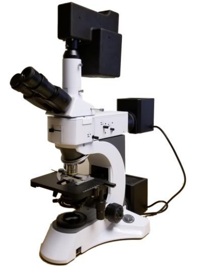
激光荧光显微高光谱成像系统
所属类别: ? 专用实验设备 ? 成像光谱仪/显微/荧光 高光谱成像仪
所属品牌:加拿大Photon etc公司
![]() 产品简介
产品简介
高速读取荧光高光谱
均化激光不会损伤细胞等样品
非逐点扫描
高速PL/EL Mapping
基于独特的体布拉格光栅滤波片技术(BTF)和光致发光成像技术,Photon etc公司最新推出的IMA激光荧光/光 致发光显微高光谱成像系统,采取革新的二维成像的方式,激光经过扩束后再经过匀化,将高斯分布的点激光扩展成平面均匀分布面激光,面激光均匀照射在样品 上,可以直接获得整个样品的荧光高光谱信息。从而获得分子结构方面的信息。有别于传统的激光荧光显微高光谱系统以逐点扫描的方式,而是一次性的获取整个样 品的光谱信息,故而只需要更短的成像时间以及具有更高的空间分辨率。
关键词:共焦荧光成像系统,共聚焦荧光成像系统,共焦荧光光谱成像系统,共焦荧光成像光谱仪系统,成像光谱仪,光致发光高光谱成像仪,激光荧光成像系统, 荧光显微成像系统
IMA荧光(EL/PL)显微高光谱成像仪
基于独特的体布拉格光栅滤波片技术(BTF)和光致发光成像技术,Photon etc公司最新推出的IMA激光荧光/光 致发光显微高光谱成像系统,采取革新的二维成像的方式,激光经过扩束后再经过匀化,将高斯分布的点激光扩展成平面均匀分布面激光,面激光均匀照射在样品 上,可以直接获得整个样品的荧光高光谱信息。从而获得分子结构方面的信息。有别于传统的激光荧光显微高光谱系统以逐点扫描的方式,而是一次性的获取整个样 品的光谱信息,故而只需要更短的成像时间以及具有更高的空间分辨率。

设备原理图:

系统参数:
VIS | VU | |
光谱范围 | 400-1000nm | 900-1700nm |
光谱分辨率 | <2.5nm(最小可到0.2nm) | <4nm(最小可到0.4nm) |
图像分辨率 | 亚微米 | 亚微米 |
成像速度 | 20x20μm in 1s @100X | |
激发光源 | 488nm,515nm(可选其他波长) | 808,980nm(可选其他波长) |
CCD | 科学级CCD,背照式CCD,EMCCD等 | InGaAs相机 |
显微镜 | 倒置或正置 | 倒置或正置 |
物镜 | 20X,60X,100X | 20X,60X,100X |
IMA的典型应用:
NANOPARTICLES IN CANCER CELLS
 Dark field illumination is commonly used for the analysis of biological samples containing nanomaterials that significantly scatter light. When combined to hyperspectral imaging, it becomes an exceptional tool to also detect the composition and the location of nanomaterials embedded in cells. IMATM, Photon etc.’s hyperspectral imager, can be equipped with a highly efficient dark field condenser and generate high contrast images of biological samples.
The high throughput of Photon etc.’s hyperspectral filter allows the rapid acquisition of spectrally resolved high resolution images. Since the camera captures the whole area in the field of view, it is possible to collect spectral and spatial information in real time, with the possibility of recording spectrally resolved videos to follow the dynamics of cells and luminescent nanoscale components. PHySpecTM, Photon etc software, enables principal component analysis (PCA) in order to identify the smallest variations of single and aggregated nanoparticles.
With the purpose of showing the capabilities of IMATM to analyse nanomaterials in biological systems, a sample of MDA-MB-23 human breast cancer cells has been tagged with 60 nm gold nanoparticles (GNPs) and exposed to a dark field illumination on the entire field of view (Figure 1). With a 60×objective, an area of 150×112 μm was imaged, with a step of 2 nm and an exposition time of 2 s per wavelength. The complete analysis took only a few minutes, for more than one million spectra, each of them covering the whole visible spectrum.
Cells typically have a flat scattering spectrum, whereas GNPs show a sharp peak around 550 nm. Figure 2 illustrates the 550 nm image extracted from the dark field hyperspectral cube of the breast cancer. The GNPs are marked with a green colouring after PCA software processing. The magnification of a breast cancer cell (Figure 3a) and the spectra of the regions containing GNPs (some examples in Figure 3b) confirmed the presence of single 60 nm NPs (peak at 550 nm) and their aggregates (peaks red-shifted). The hyperspectral camera did not detect any GNPs in the areas between the cells.
CHARACTERISATION OF SOLAR CELLS USING HYPERSPECTRAL IMAGER

A new characterization method based on hyperspectral imaging recording spectrally resolved images allows the cartography of electroluminescence (EL) and photoluminescence (PL). From the data acquired, spatial variations of cell properties such as open circuit voltage and transport mechanisms were identified and characterized. Furthermore, the system was compared to a classical confocal microscope, showing significant gains in acquisition time.
Spectrally resolved images provide considerable advantages such as, absolute calibration of intensity, micrometer scale resolution, and excitation and detection on a surface (no information loss from lateral diffusion and roughness). In luminescence imaging, absolute calibration is a main concern and is here done in two steps: first, an absolute calibration at a determined point (spatially and spectrally) with a laser, and then a relative calibration on the whole space and the whole spectrum, with a calibrated lamp coupled to an integrating sphere.The images rendered by IMATM are spectrally resolved luminescence images from multicrystalline CIS solar cell, offering means of studying its spatial inhomogeneities. On high efficiency GaAs solar cells, we got absolute measurements of EL and successfully investigated reciprocity relations. Our next step is to record quantitative maps of CIGS physical properties from PL and EL images, such as VOC , transport parameters and more.
A confocal microscope coupled to a spectrometer provides similar data. The 532nm laser is focused onto the cell front contact, and the cartography of PL spectra is obtained by scanning the sample. The acquisition time with the imager is much faster. 150*150μm2 at 107 W/m2 would take hundreds of hours in confocal, but only 8min with IMA. Moreover, surface excitation and detection allow to get rid of diffusion and roughness troubles for quantitative analysis.
分享到 : 人人网 腾讯微博 新浪微博 搜狐微博 网易微博
相关产品  显微拉曼成像光谱仪
  光致发光成像光谱仪
  BPF低波数带通滤光片
  BNF低波数陷波滤波片
企业名称
上海昊量光电设备有限公司
企业信息已认证
企业类型
有限责任公司(自然人投资或控股)
信用代码
913101176727298067
成立日期
2008-04-03
注册资本
人民币100.0000万元整
经营范围
许可项目:货物进出口:技术进出口.(依法须经批准的项目,经相关部门批准后方可开展经营活动,具体经营项目以相关部门批准文件或许可证件为准)一般项目:光电设备及元器件、光纤光缆、光电传感器、仪器设备、电子产品、通讯器材研发、生产、销售;工业自动化设备及配件、机械设育、汽车零配件、化工产品(除危险化学品、监控化学品、烟花爆竹、民用爆炸物品、易制毒化学品)、建筑装饰材料、服装、纺织品及眼材料、计算机软硬件及辅助设备销售;软件开发;技术服务、技术开发、技术咨询、技术交流、技术转让、技术推广.(除依法须经批准的项目外,凭营业执照依法自主开展经营活动)
上海昊量光电设备有限公司
公司地址
上海市徐汇区虹梅路2007号远中产业园三期6号楼3楼
客服电话
公司名称: 上海昊量光电设备有限公司
公司地址: 上海市徐汇区虹梅路2007号远中产业园三期6号楼3楼 联系人: 昊量 邮编: 200235 联系电话: 400-860-5168转2831
仪器信息网APP

展位手机站