当前位置: 仪器信息网 > 行业主题 > >

牡荆苷

仪器信息网牡荆苷专题为您整合牡荆苷相关的最新文章,在牡荆苷专题,您不仅可以免费浏览牡荆苷的资讯, 同时您还可以浏览牡荆苷的相关资料、解决方案,参与社区牡荆苷话题讨论。

牡荆苷相关的耗材

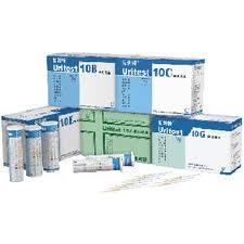
  • 京都SP-4430干式生化仪Mulit-PANEL-1六项常规复合试纸1
    适用于京都SP-4430干式生化分析仪规格:50枚/盒
  • SUPELCOGEL Pb(铅型)树脂型糖柱 液相色谱柱 (单糖、木糖/半乳糖、甘露糖分离)
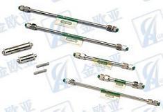
    液相色谱柱 SUPELCOGEL Pb(铅型)树脂型糖柱(单糖、木糖/半乳糖、甘露糖分离)产品描述SUPELCOGEL Pb 色谱柱中这种铅为反离子的树脂为单糖提供了最佳的选择性和分离效果。SUPELCOGEL Pb 色谱柱为木糖、半乳糖和甘露糖的分离获得了良好的分离效果,而它们在钙离子树脂的色谱柱中并不能完全分离。应用特点用于单糖、木糖/半乳糖、甘露糖分离,符合USP L34方法型号规格30cm*7.8mm,9&mu m
  • SUPELCOGEL Pb(铅型)树脂型糖柱 液相色谱柱(单糖、木糖/半乳糖、甘露糖分离)
    液相色谱柱 SUPELCOGEL Pb(铅型)树脂型糖柱(单糖、木糖/半乳糖、甘露糖分离)货号59335-U产品描述分析/色谱法、高效液相色谱、高效液相色谱法,高效液相色谱柱列的碳水化合物应用特点用于单糖、木糖/半乳糖、甘露糖分离,符合USP L34方法型号规格10cm*7.8mm,9&mu m
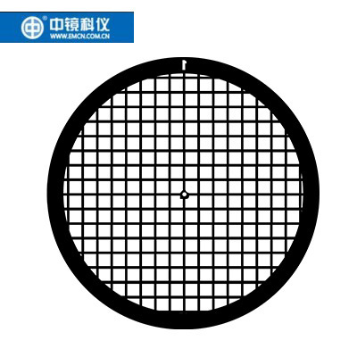
  • 中镜科仪 钼单孔环、钼光圈、钼网 透射电子显微镜载网
    单孔环孔径75至2000微米,材料有钛、铝、铂、钼、金、镍、铜,有国产和进口两种,主要用于研磨块状样品作载体。铜单孔环统一厚度是30um。包装100枚/瓶,中镜科仪生产,铜载网是最广泛使用的载网,无磁性。 商品编号商品名称厚度规格产地AZA600M钼单孔环0.05100枚/瓶中国AZA600M-3钼单孔环0.03100枚/瓶中国AZA800M钼单孔环0.05100枚/瓶中国AZA800M-3钼单孔环0.03100枚/瓶中国AZA1000M钼单孔环-1.0mm0.051.0mm,100枚/瓶中国AZA1000M-3钼单孔环-1.0mm0.031.0mm,100枚/瓶中国AZA1200M钼单孔环-1.2mm0.051.2mm,100枚/瓶中国AZA1200M-3钼单孔环-1.2mm0.031.2mm,100枚/瓶中国AZA1500M钼单孔环-1.5mm0.051.5mm,100枚/瓶中国AZA1500M-3钼单孔环-1.5mm0.031.5mm,100枚/瓶中国AZA1800M钼单孔环-1.8mm0.051.8mm,100枚/瓶中国AZA1800M-3钼单孔环-1.8mm0.031.8mm,100枚/瓶中国AZA2000M钼单孔环-2.0mm0.052.0mm,100枚/瓶中国AZA2000M-3钼单孔环-2.0mm0.032.0mm,100枚/瓶中国
  • D-甘露醇分析柱
    近期政府公布了新的可使用的食品添加剂的列表,同时也对这些生产和使用的企业公布了新的食品添加剂的国家检测标准。其中有一种很常用的食品添加剂:D-甘露糖醇,其国家新检测标准中规定必须使用伯乐公司的100× 7.8mm的快速糖分析柱(货号:1250105)D-甘露糖醇是低热量甜味剂,胶姆糖和糖果的防粘剂,营养增补剂及组织改良剂,保湿剂,使用范围很广,包括所有的糖果、酥糖、大部分的饼干,以及大部分的甜味的食品。 货号规格mm应用粒径离子形式交联率pH范围125-0105快速糖分析柱 100× 7.8葡萄糖、半乳糖、蔗糖、果糖9&mu m铅8%5-9国家指定标准
  • 人参皂苷正相色谱柱 TSKGEL Amide-80HR
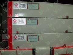
    北京绿百草科技供应TOSOH正相/亲水色谱柱TSKGEL Amide-80HR,货号21982,4.6*250mm。TSKGEL Amide-80HR,粒径5&mu m,孔径100A。Amide-80HR以硅胶为基体,键合了氨基甲酰基的色谱柱,是对TSKgel Amide-80 5µ m进行了改良,提高了分离能力和耐久性,是一款高性能正相/亲水作用色谱柱。TSKGEL Amide-80主要分析糖类、多肽、核酸、亲水性药物等,日本药典中推荐使用TSKGEL Amide-80HR分离人参皂苷。北京绿百草科技可以提供Amide-80详细信息,还有相应的保护柱提供。
  • DR/DD型植物茎干半径/直径变化传感器
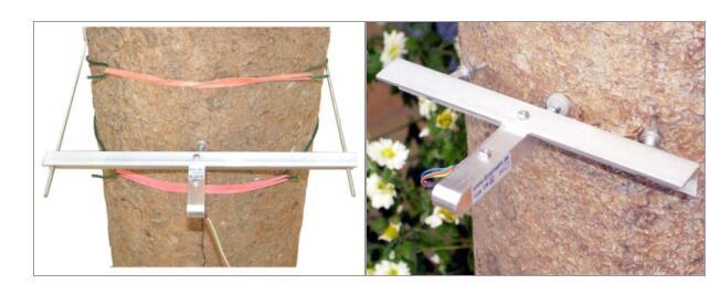
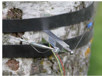
    在植物和环境的关系的研究中,一般环境指标,如气象因子、土壤等,可以每小时、每天高密度连续观测,但植物指标往往一年仅测定一个或数个,如生长量,产量,年轮宽度等等,其结果是一个因变量对数十个或数百个自变量,无法准确确定究竟哪个环境因素影响植物的生长。如果用茎干变化测量系统可以连续取得植物相关的数据,则可以大大提高植物和环境关系研究的可行性和准确性。优点:此仪器可定位精确观测植物茎干的变化, 数据可以直读, 也可用数采自动记录;专用配套小数采自带的电源可连续测量2年;优点:精度高, 廉价, 安装方便, 性能稳定, 测量时传感器不需要电源,几乎无需维护措施,特殊尺寸可以定制。技术参数:型号DD型DR型测量范围测直径变化,适于直径变化在0~20 cm 的植物,大于20 cm需特制;不伤害植物。测半径变化,适于直径8 cm 以上的植物,茎杆上要钻两个4 mm 的小孔。扩张范围11 mm,测量对象变化超过11mm后需要重新调节标准配置传感器,固定框架,2 m电缆。传感器,固定框架,2 m电缆。安装工具万用表,两个小扳手,电缆固定带。万用表,两个小扳手,电缆固定带,手摇钻或电钻(直径4mm), 树体伤保护胶。尺寸及重量27×24×1.5 cm,65 g14×15×1.5 cm,60 g读取数据需要读数表或数据采集器测量精度5mm (植物半径日变化0~300mm)温度系数0.1 mm/℃ (温度变化1℃, 变化小于0.1mm)适用环境温度-30~40°C, 湿度0~100%输出方式模拟输出 0~50 kΩ,不耗电。外壳材料表面强化铝,不锈钢电缆长度2 m,电缆可以延长到200 m 产地:德国
  • 类器官Cervical cancer Organoid Kit(宫颈癌)
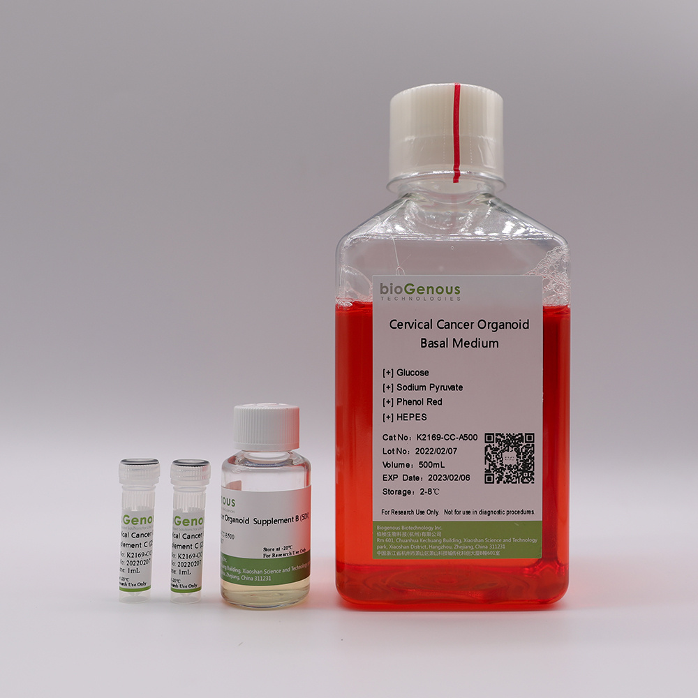
    类器官(Organoids)是指将成体干细胞或多能干细胞在体外三维培养形成的具有一定空间结构的组织类似物。类器官在组织结构、细胞类型、自我更新能力和功能等方面与来源组织高度一致,从而在发育生物学、疾病造模、精准医学、药物研发、基因和细胞疗法、感染和免疫以及再生医学等生物医学的多个领域展现出独特的优势。产品介绍Product Description:bioGenousTMCervical Cancer Organoid Kit is a chemically defined cell culture medium for the establishment and maintenance of human cervical cancer organoids. Patient-derived cancer organoids recapitulate the genomic and pathological features of original tumors and therefore hold great promise for medical research and precision medicine.技术参数Product Information:ComponentComponent Cat#VolumeStorage & StabilitybioGenousTMCervical Cancer Organoid Basal MediumK2169-CC-A100/A500100mL/500mL4℃,12 monthsbioGenousTMCervical Cancer Organoid Supplement B (50x)K2169-CC-B100/B5002mL/10mL-20℃,avoid repeated freeze-thaw cycles, 12 monthsbioGenousTMCervical Cancer Organoid Supplement C (250x)K2169-CC-C100/C5000.4mL/2mL-20℃, avoid repeated freeze-thaw cycles, 12 monthsPreparation of Cervical Cancer Organoid Complete MediumUse a sterile technique to prepare the cervical cancer organoid complete medium. The following example is for preparing 10mL complete medium. If preparing other volumes, adjust accordingly.1.Thaw Cervical Cancer Organoid Supplement B(50x) and Cervical Cancer Organoid Supplement C(250x) on ice. Mix thoroughly.NOTE:Once thawed, use immediately or aliquot and store at -20°C for not more than 10 months. After thawing the aliquots, use immediately. Do not re-freeze.2.Add 200μL Cervical Cancer Organoid Supplement B(50x) and 40μL Cervical Cancer Organoid Supplement C(250x) to 9.76mL Cervical Cancer Organoid Basal Medium. Mix thoroughly.NOTE:If not used immediately, store the complete medium at 2-8°C for not more than 2 weeks. The Cervical Cancer Organoid Supplement B contains fungicide and antibiotics (50x).Protocol for Establishment ofPatient-Derived Cervical Cancer OrganoidsCAUTIONStudies involving primary human tissue material must follow all relevant institutional and government regulations. Informed consent must be obtained from all subjects before the collection of the primary human tissue material.Establishment of Organoids from Primary Tissue1.Collect primary human cervical cancer tissue pieces in ice-cold Primary Tissue Storage Solution (K601005) with conical tubes. Keep tissue samples at 4°C until the start of the isolation.2.Assess whether the obtained tissue pieces consist purely of epithelium. If fat or muscle tissues are present, remove these non-epithelial components as much as possible using surgical scissors or scalpels and forceps under a dissection microscope. If no fat or muscle tissues are present, continue to the next step immediately.3.Rinse the tissuewith Cancer Organoid Basal Medium (B213152) or DPBS twice.4.Mince the tissue into small fragments of 1-3 mm3in a cell culture dish using surgical scissors or scalpels.5.Digest the tissue fragments with 10mL of Tumor Tissue Digestion Solution (K601003) in a 15mL conical tube at 37°C, with variable incubation times ranging from 30 min to 1 h. Carefully monitor the digestion process, mixing the content of the tube every 5-10 min by shaking vigorously or pipetting the mixture up and down. The digestion process could be finished when most of tissue fragments are able to pass through the 1mL pipette tips.6.Add FBS to the tissuedigestionmixture to a final concentration of 2%, and filter using a 100 μm cell strainer.7.Collect and centrifuge the filtered cells at 250g for 3 min at 4 °C. In case of a visible red pellet, aspirate the supernatant, and resuspend the pellet using 2mL of Red Blood Cell Lysis Solution (E238010) to lyse the erythrocytes at room temperature for 1 min and centrifuge at 250g for 3 min at 4°C.8.Aspirate the supernatant and resuspend the pellet in Cancer Organoid Basal Medium, centrifuge at 250g for 3 min at 4°C,repeat this step once more time.9.Aspirate the supernatant and resuspend the pellet in Matrigel. The Matrigel should be kept on ice to prevent it from solidifying. Perform the process as quickly as possible. The amount of Matrigel used depends on the size of the pellet. Approximately 10,000 cells should be plated in 25 μL of Matrigel.CRITICAL:Do not overly dilute the Matrigel (Matrigel should be 70% (Matrigel vol/Total vol)), as this may inhibit the proper formation of the solid droplets.10.Plate the Matrigel containing organoids at the bottom of 24-well cell culture plates in droplets of ~30μL each around the center of the well..CRITICAL:Once the organoids are resuspended in Matrigel, proceed with plating as quickly as possible, as the Matrigel may solidify in the tube or pipette tip. Do not let the Matrigel touch the wall of wells.11.Place the culture plate into a humidified incubator at 37 °C and 5% (vol/vol) CO2for15-25 minto let the Matrigel solidify.12.Prepare the required amount of cervical cancer organoid complete medium.13.Once the Matrigel droplets have solidified (15-25 min), open the plate and carefully add 500 μL of organoid complete medium to each well.CRITICAL:Do not add the medium directly on top of the Matrigel droplets, as this might disrupt the droplets.14.Place the culture plate in a humidified incubator at 37 °C and 5% (vol/vol) CO2.15.Change the medium every 3-4 d by carefully aspirating the medium from the wells and replacing it with a fresh, pre-warmed organoid complete medium.16.Closely monitor the organoid formation. Ideally, patient-derived cervical cancer organoids should be passaged for the first time between 7 and 10 d after the initial plating.Splitting and Passaging of Organoids17.Pipette up and down to disrupt the Matrigel and transfer the organoid suspension to a 1.5 mL conical tube.18.Pipette the organoid suspension up and down to mix thoroughly by pipetting against the bottom of the tube to create pressure, which will aid the removal of Matrigel.19.Centrifuge organoids at 250g for 3 min at room temperature.20.Aspirate the supernatant and split organoids using either Organoid Dissociation Solution (E238001) or by mechanical disruption. For Organoid Dissociation Solution-based cell dissociation, resuspend the pellet in 0.2 mL of Organoid Dissociation Solution, pipette up and down and incubate at 37 °C until organoids fall apart. Pipette up and down with a filter tip for ≥8 times every 2 min to aid in the disruption of the organoids. Closely monitor the digestion to keep the incubation time inthe Organoid Dissociation Solution to a minimum. In case of mechanical disruption, resuspend the pellet in 1.5 mL ofCancer Organoid Basal Medium. Carefully pipette the organoid suspension up and down 30 times by pipetting against the bottom of the tube to create pressure, which will aid organoid disruption.CRITICAL:Do not dissociate in Organoid Dissociation Solution for 7 min, as this may result in poor organoid outgrowth or even loss of the culture. As a rule of thumb, digestion is complete if a mixture of small lumps of cells (consisting of 10–50 cells) can be observed.21.After shearing is complete, wash once by topping up with 1 mL ofCancer Organoid Basal Medium, and centrifuge at 250g for 3 min at room temperature.22.Aspirate the supernatant and resuspend the organoid pellet in 70% (vol/vol) Matrigel, and plate organoids in droplets at the bottom of a culture plate as described in Steps 10. After plating is complete, transfer the plate into a humidified incubator at 37 °C and 5% (vol/vol) CO2for 15–25 min.23.Pre-warm cervical cancer organoid complete medium at 37 °C.24.After the Matrigel droplets have solidified (15–25 min), carefully pipette pre-warmed medium into the wells.25.Place culture plates in a humidified incubator at 37 °C and 5% (vol/vol) CO2until the organoids are needed for further experiments.
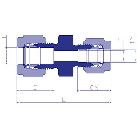
  • 变径接头螺母
    变径接头螺母本公司专业提供螺母,密封圈,螺帽盖,死堵,死堵头,适配接头,软管接头,两通、三通、直角接头和变径接头规格(不锈钢或黄铜):1/16英寸、1/8英寸、1/4英寸、M8×1等
  • Benson BP-100 Ca++ 300*7.8mm 甘露醇,山梨醇和木糖醇分析色谱柱-北京绿百草科技
    Benson BP-100 Ca++ 300*7.8mm 甘露醇,山梨醇和木糖醇分析色谱柱-北京绿百草科技 伯乐Aminex糖分析色谱柱125-0095 HPX-87P;125-0095 HPX-87C;125-0140 HPX-87H。 北京绿百草提供各种糖分析柱: Benson BP-100 Ca++ ,USP L19,单糖, 糖精, 果汁, 软饮料, 奶制品, 蔬菜, 医疗科技粒径;由磺化交联苯乙烯二乙烯基苯共聚物组成的强阳离子交换树脂,Ca型,粒径9um。其他色谱柱型号为: Sugar-D;BP-100,H+,USP L17;BP-100,Pb++,USP L34;BP-100,Ca++,USP L19;BP-200,Ca++,USP L19;BP-100,Ag+;USP L17,HC-75 H型;Hamilton USP L19,HC-75 Ca型;USP L34,HC-75 Pb型;HC-40; 了解更多信息请登陆北京绿百草:www.greenherbs.com.cn或010-51659766。
  • 北京/上海/广州液相色谱仪配件-柱塞杆
    北京/上海/广州液相色谱仪配件-柱塞杆南京科捷分析仪器有限公司是专业研究、开发、制造和销售色谱仪(气相色谱仪、液相色谱仪)、比表面积测定仪、高纯气体分析设备、色谱配件、色谱试剂以及其他分析仪器的高科技企业。以下是各柱塞杆详细介绍:安捷伦系列柱塞杆:产品名称规格型号相关配置产地柱塞杆Agilent1050/11005063-6589安捷伦柱塞杆Agilent10505062-244安捷伦柱塞杆Agilent1050/11005062-8516安捷伦岛津系列的柱塞杆:(1)LC-10ADvp柱塞杆:228-35601-91(相关配置)(2)LC-10ATvp柱塞杆:228-35009-92TSP的柱塞杆:SP8800/8810柱塞杆:A3102-010北京/上海/广州液相色谱仪配件-柱塞杆南京科捷是专业研究、开发、制造和销售色谱仪(气相色谱仪、液相色谱仪)、高纯气体分析设备、色谱配件(气相进样针,微量注射器,毛细管柱,填充色谱柱,色谱柱,进样垫,气体三通阀,四通阀,六通阀,八通阀,十通阀,十二通阀,检测器灯,检测器灯,自动进样器,泵零件,检测灯,自动进样器,泵零件,检测灯,泵零件,检测灯, 自动进样器,泵零件,保护柱套,管路配件,国产液相色谱柱,进口液相色谱柱,SPE固相萃取柱,过滤膜,液相进样针,阀配件,柱塞杆,密封圈,氘灯,高效色谱级试剂)、色谱试剂、顶空,顶空进样器以及其他分析仪器的高科技企业。
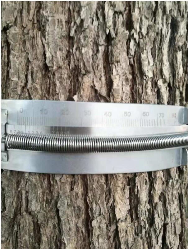
  • QT304树木生长尺
    用途:树木生长尺能方便、无损安装在被测量树木上,长期野外监测树木生长过程中的树径变化。材质:所有零件采用SUS304不锈钢,保持长期野外使用不锈蚀、不磨损。材料有较小的线胀系数(17.2*10-6/℃)。 技术参数:★适用树干周长范围:5-20cm,可根据用户要求定制★刻线采用激光蚀刻,刻度清晰,读取方便,在单面的刻度产生的摩擦力非常小,刻度不会消失。★ 采用π为单位,可直接读取树木直径以及直径变化量;可测的直径变化量:0-5π厘米, 以0.05π厘米为绝对刻度, 测量带与游标无摩擦,读数精度为0.01π厘米。★主尺长度≤20cm,重量≤12.3g。辅助尺长度:150mm-3200mm。★采用美国定制弹簧。弹簧材质:SUS304 - WPB高张力弹簧,初张力:≥150g,弹性系数:≤8g/mm。可以根据客户的要求设计定制不同力学性能的弹簧。★干摩擦系数:≤0.45;对树木无伤害,可以做到无损长期监测。产地:中国
  • 推杆
    推杆 -- 用于CTC自动进样针Replacement Plungers (SGE) for CTC Autosampler Syringesd订货信息:catalog #volumeunits2390725 &mu Lea.2390025 &mu Lea.
  • 京都SP-4430干式生化仪MG镁试纸
    适用于京都SP-4430干式生化分析仪规格:50枚/盒;
  • 进口铜网镍网钼网(电镜耗材)
    进口铜网镍网钼网, 规格有,50目,100目,150目, 200目,300目,400目 mesh (100个/盒)直径:3.05mm
  • Shodex OHpak SB-804 HQ (8.0×300mm)重组乙型肝炎疫苗和重组乙型肝炎疫苗(汉逊酵母)专用
    Shodex OHpak SB-804 HQ聚合物基质填料水溶性SEC(GFC)色谱柱在2010年药典第三部的应用1.药品名称:重组乙型肝炎疫苗(CHO 细胞)Recombinant Hepatitis B Vaccine(CHO Cell)药品类别:预防类测试项目:CHO细胞表达的乙型肝炎病毒表面抗原 (HBsAg)色谱柱要求:亲水树脂体积排阻色谱柱,排阻分子低于1000KD药典推荐的流动相:0.05mol/L 磷酸盐缓冲液,PH6.8分析要求:HBsAg 不低于 95.0%2.药品名称:重组乙型肝炎疫苗(汉逊酵母)Recombinant Hepatitis B VaccineVaccine(Hansenula polymer pha)药品类别:预防类测试项目:CHO细胞表达的乙型肝炎病毒表面抗原 (HBsAg)色谱柱要求:亲水甲基丙烯酸树脂体积排阻色谱柱,排阻极限10000KD药典推荐的流动相:含0.05%叠氮化钠的1mM/LPBS(PH7.0) 上样量10uL分析要求:HBsAg 不低于99.0%OHpak SB-804 HQ (GFC分离)分析肝素。Sample : HeparinColumn : Shodex OHpak SB-804 HQ (8.0mmI.D. x 300mm)Eluent : 0.1M NaClFlow rate : 1.0mL/minDetector : Shodex RIColumn temp. : 40℃订货号产品名称塔板数(TPN/column)排阻限(普鲁兰)粒径(μm)规格(mm)内径 x 长F6429100F6429101F6429102F6429103F6429104F6429105F6429106F6709430OHpak SB-802 HQOHpak SB-802.5 HQOHpak SB-803 HQOHpak SB-804 HQOHpak SB-805 HQOHpak SB-806 HQOHpak SB-806M HQOHpak SB-G 12,000 16,000 16,000 16,000 12,000 12,000 12,000保护柱4,000 10,000 100,000 1,000,000 *(4,000,000) *(20,000,000)*(20,000,000)-86610131313108.0 x 3008.0 x 3008.0 x 3008.0 x 3008.0 x 3008.0 x 3008.0 x 3006.0 x 50
  • Benson BP-100 Ca++ 300*7.8mm 甘露醇,山梨醇和木糖醇分析色谱柱
    Benson BP-100 Ca++ 300*7.8mm 甘露醇,山梨醇和木糖醇分析色谱柱 伯乐Aminex糖分析色谱柱125-0095 HPX-87P;125-0095 HPX-87C;125-0140 HPX-87H。 北京绿百草提供各种糖分析柱: Benson BP-100 Ca++ ,USP L19,单糖, 糖精, 果汁, 软饮料, 奶制品, 蔬菜, 医疗科技粒径;由磺化交联苯乙烯二乙烯基苯共聚物组成的强阳离子交换树脂,Ca型,粒径9um。其他色谱柱型号为: Sugar-D;BP-100,H+,USP L17;BP-100,Pb++,USP L34;BP-100,Ca++,USP L19;BP-200,Ca++,USP L19;BP-100,Ag+;USP L17,HC-75 H型;Hamilton USP L19,HC-75 Ca型;USP L34,HC-75 Pb型;HC-40; 了解更多信息请登陆北京绿百草:www.greenherbs.com.cn或010-51659766。
  • 中镜科仪 50至400目进口镍网镀碳支持膜
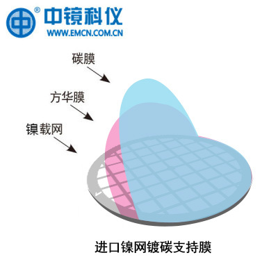
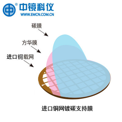
    普通碳支持膜是一种较常见并被广泛使用的一种支持膜,为两层支持膜结构,可以采用不同规格的载网做载体。从空间结构来讲,从下到上依次为载网,方华膜和碳膜,它是在一层有机方华膜上再覆盖一层碳膜。由于碳层具有较强的导电以及导热性,弥补了无碳方华膜的荷电效应以及热效应,增强了膜整体的稳定性,可满足大多数纳米材料(尤其是可分散的粉体材料)的一般形貌测试需要。但是由于这种支持膜本身的结构导致了整体支持膜的厚度较大,一般不太容易满足高质量的高分辨测试的要求。英国进口镍载网,中镜科仪生产,支持膜厚度10-20nm。中包装50枚/盒产品编号Prod.No.载网材质Material载网目数Mesh载网产地Made in支持膜产地Made in支持膜厚度Thickness包装UnitBZ100205Nb镍50目英国中镜科仪10-20nm50枚/盒BZ10021Nb镍100目英国中镜科仪10-20nm50枚/盒BZ100215Nb镍150目英国中镜科仪10-20nm50枚/盒BZ10022Nb镍200目英国中镜科仪10-20nm50枚/盒BZ10023Nb镍300目英国中镜科仪10-20nm50枚/盒BZ10024Nb镍400目英国中镜科仪10-20nm50枚/盒英国进口镍载网,中镜科仪生产,支持膜厚度10-20nm。大包装100枚/盒产品编号Prod.No.载网材质Material载网目数Mesh载网产地Made in支持膜产地Made in支持膜厚度Thickness包装UnitBZ100205Na镍50目英国中镜科仪10-20nm100枚/盒BZ10021Na镍100目英国中镜科仪10-20nm100枚/盒BZ100215Na镍150目英国中镜科仪10-20nm100枚/盒BZ10022Na镍200目英国中镜科仪10-20nm100枚/盒BZ10023Na镍300目英国中镜科仪10-20nm100枚/盒BZ10024Na镍400目英国中镜科仪10-20nm100枚/盒【存储】:室温避光干燥保存(建议放干燥器或者干燥箱内)、防污染、防震荡,未拆封保质期1年,拆封之后最好6个月内用完。【使用注意】:取用时采用高精尖镊子小心操作,防止产生弯折等破坏。存在有机膜,不能与有机溶剂接触。面向样品盒有字母的一面是铺膜的一面。
  • 类器官Head and Neck Squamous Cell Carcinoma Organoid Kit(头颈鳞癌)
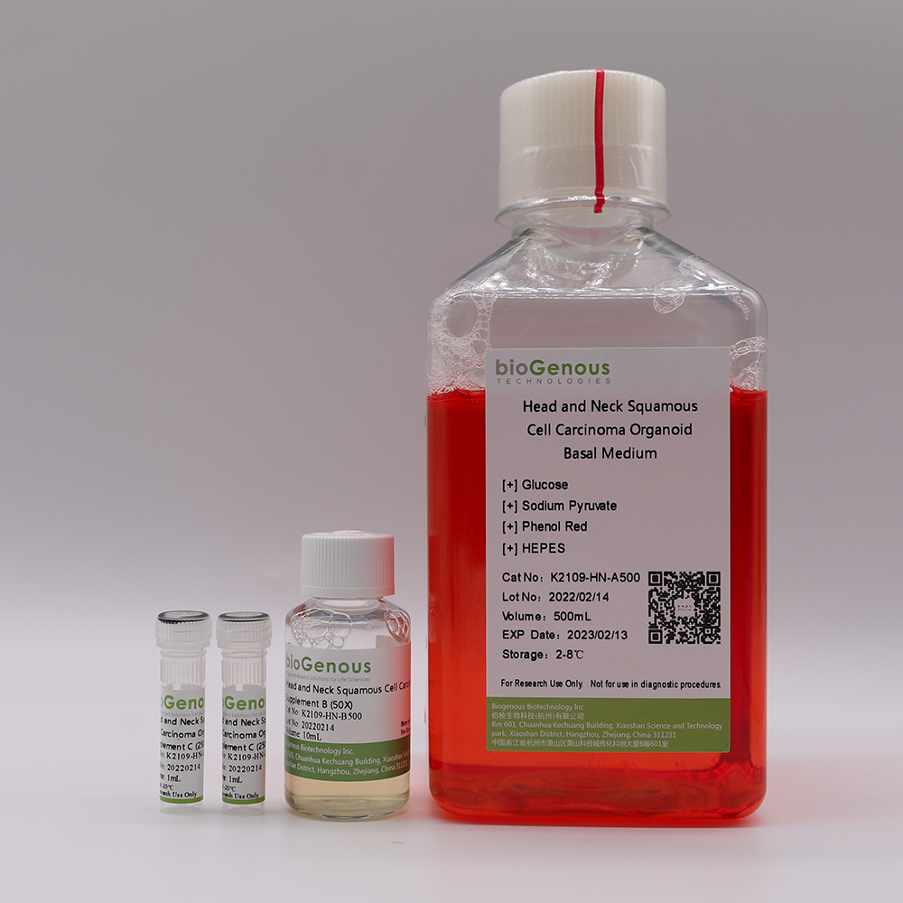
    类器官(Organoids)是指将成体干细胞或多能干细胞在体外三维培养形成的具有一定空间结构的组织类似物。类器官在组织结构、细胞类型、自我更新能力和功能等方面与来源组织高度一致,从而在发育生物学、疾病造模、精准医学、药物研发、基因和细胞疗法、感染和免疫以及再生医学等生物医学的多个领域展现出独特的优势。产品介绍Product Description:bioGenousTMHead and Neck Squamous Cell Carcinoma Organoid Kit is a chemically defined cell culture medium for the establishment and maintenance of human head and neck squamous cell carcinoma organoids. Patient-derived cancer organoids recapitulate the genomic and pathological features of original tumors and therefore hold great promise for medical research and precision medicine.技术参数Product Information:ComponentComponent Cat#VolumeStorage & StabilitybioGenousTMHead and Neck Squamous Cell Carcinoma Organoid Basal MediumK2109-HN-A100/A500100mL/500mL4℃,12 monthsbioGenousTMHead and Neck Squamous Cell Carcinoma Organoid Supplement B (50x)K2109-HN-B100/B5002mL/10mL-20℃,avoid repeated freeze-thaw cycles, 12 monthsbioGenousTMHead and Neck Squamous Cell Carcinoma Organoid Supplement C (250x)K2109-HN-C100/C5000.4mL/2mL-20℃, avoid repeated freeze-thaw cycles, 12 monthsPreparation of Head and Neck Squamous Cell Carcinoma Organoid Complete MediumUse a sterile technique to prepare the head and neck squamous cell carcinoma organoid complete medium. The following example is for preparing 10mL complete medium. If preparing other volumes, adjust accordingly.1.Thaw Head and Neck Squamous Cell Carcinoma Organoid Supplement B(50x) and Head and Neck Squamous Cell Carcinoma Organoid Supplement C(250x) on ice. Mix thoroughly.NOTE:Once thawed, use immediately or aliquot and store at -20°C for not more than 10 months. After thawing the aliquots, use immediately. Do not re-freeze.2.Add 200μL Head and Neck Squamous Cell Carcinoma Organoid Supplement B(50x) and 40μL Head and Neck Squamous Cell Carcinoma Organoid Supplement C(250x) to 9.76mL Head and Neck Squamous Cell Carcinoma Organoid Basal Medium. Mix thoroughly.NOTE:If not used immediately, store the complete medium at 2-8°C for not more than 2 weeks. The Head and Neck Squamous Cell Carcinoma Organoid Supplement B contains fungicide and antibiotics (50x).Protocol for Establishment ofPatient-Derived Head and Neck Squamous Cell Carcinoma OrganoidsCAUTIONStudies involving primary human tissue material must follow all relevant institutional and government regulations. Informed consent must be obtained from all subjects before the collection of the primary human tissue material.Establishment of Organoids from Primary Tissue1.Collect primary human head and neck squamous cell carcinoma tissue pieces in ice-cold Primary Tissue Storage Solution(K601005)with conical tubes. Keep tissue samples at 4°C until the start of the isolation.2.Assess whether the obtained tissue pieces consist purely of epithelium. If fat or muscle tissues are present, remove these non-epithelial components as much as possible using surgical scissors or scalpels and forceps under a dissection microscope. If no fat or muscle tissues are present, continue to the next step immediately.3.Rinse the tissuewith Cancer Organoid Basal Medium (B213152) or DPBS twice.4.Mince the tissue into small fragments of 1-3 mm3in a cell culture dish using surgical scissors or scalpels.5.Digest the tissue fragments with 10mL of Tumor Tissue Digestion Solution(K601003)in a 15mL conical tube at 37°C, with variable incubation times ranging from 30 min to 1.5 h. Carefully monitor the digestion process, mixing the content of the tube every 5-10 min by shaking vigorously or pipetting the mixture up and down. The digestion process could be finished when most of tissue fragments are able to pass through the 1mL pipette tips.6.Add FBS to the tissuedigestion mixture to a final concentration of 2%, and filter using a 100 μm cell strainer.7.Collect and centrifuge the filtered cells at 250g for 3 min at 4 °C. In case of a visible red pellet, aspirate the supernatant, and resuspend the pellet using 2mL of Red Blood Cell Lysis Solution (E238010)to lyse the erythrocytes at room temperature for 1 min and centrifuge at 250g for 3 min at 4°C.8.Aspirate the supernatant and resuspend the pellet in Cancer Organoid Basal Medium, centrifuge at 250g for 3 min at 4°C,repeat this step once more time.9.Aspirate the supernatant and resuspend the pellet in Matrigel. The Matrigel should be kept on ice to prevent it from solidifying. Perform the process as quickly as possible. The amount of Matrigel used depends on the size of the pellet. Approximately 10,000 cells should be plated in 25 μL of Matrigel.CRITICAL:Do not overly dilute the Matrigel (Matrigel should be 70% (Matrigel vol/Total vol)), as this may inhibit the proper formation of the solid droplets.10.Plate the Matrigel containing organoids at the bottom of 24-well cell culture plates in droplets of ~30μL each around the center of the well..CRITICAL:Once the organoids are resuspended in Matrigel, proceed with plating as quickly as possible, as the Matrigel may solidify in the tube or pipette tip. Do not let the Matrigel touch the wall of wells.11.Place the culture plate into a humidified incubator at 37 °C and 5% (vol/vol) CO2for15-25 min to let the Matrigel solidify.12.Prepare the required amount of head and neck squamous cell carcinoma organoid complete medium.13.Once the Matrigel droplets have solidified (15-25 min), open the plate and carefully add 500 μL of organoid complete medium to each well.CRITICAL:Do not add the medium directly on top of the Matrigel droplets, as this might disrupt the droplets.14.Place the culture plate in a humidified incubator at 37 °C and 5% (vol/vol) CO2.15.Change the medium every 3-4 d by carefully aspirating the medium from the wells and replacing it with a fresh, pre-warmed organoid complete medium.16.Closely monitor the organoid formation. Ideally, patient-derived head and neck squamous cell carcinoma organoids should be passaged for the first time between 7 and 10 d after the initial plating.Splitting and Passaging of Organoids17.Pipette up and down to disrupt the Matrigel and transfer the organoid suspension to a 1.5 mL conical tube.18.Pipette the organoid suspension up and down to mix thoroughly by pipetting against the bottom of the tube to create pressure, which will aid the removal of Matrigel.19.Centrifuge organoids at 250g for 3 min at room temperature.20.Aspirate the supernatant and split organoids using either Organoid Dissociation Solution (E238001) or by mechanical disruption. For Organoid Dissociation Solution-based cell dissociation, resuspend the pellet in 0.2 mL of Organoid Dissociation Solution, pipette up and down and incubate at 37 °C until organoids fall apart. Pipette up and down with a filter tip for ≥8 times every 2 min to aid in the disruption of the organoids. Closely monitor the digestion to keep the incubation time inthe Organoid Dissociation Solution to a minimum. In case of mechanical disruption, resuspend the pellet in 1.5 mL ofCancer Organoid Basal Medium. Carefully pipette the organoid suspension up and down 30 times by pipetting against the bottom of the tube to create pressure, which will aid organoid disruption.CRITICAL:Do not dissociate in Organoid Dissociation Solution for 7 min, as this may result in poororganoid outgrowth or even loss of the culture. As a rule of thumb, digestion is complete if a mixture of small lumps of cells (consisting of 10–50 cells) can be observed.21.After shearing is complete, wash once by topping up with 1 mL ofCancer Organoid Basal Medium, and centrifuge at 250g for 3 min at room temperature.22.Aspirate the supernatant and resuspend the organoid pellet in 70% (vol/vol) Matrigel, and plate organoids in droplets at the bottom of a culture plate as described in Steps 10. After plating is complete, transfer the plate into a humidified incubator at 37 °C and 5% (vol/vol) CO2for 15–25 min.23.Pre-warm head and neck squamous cell carcinoma organoid complete medium at 37 °C.24.After the Matrigel droplets have solidified (15–25 min), carefully pipette pre-warmed medium into the wells.Place culture plates in a humidified incubator at 37 °C and 5% (vol/vol) CO2until the organoids are needed for further experiments.
  • 用于硝化甘油的GDX-103采样管
    使用乙醇浸泡的溶剂解析GDX-103管在高温干燥去水后可用于硝化甘油吸附。特点?? 高解析效率:95% with 三甲胺?? 低阻力:4kPa@200ml/min,能使绝大部分采样器在该流量段下实现恒流Use for ? 工作场所空气中有毒物质测定 炸药类化合物 GBZ/T 160.80 3-2004 硝化甘油填料与克重:100mg/50mg 目数:40-60 外径×长度:6×120 最小包装:100支/盒
  • DC2茎干周长生长测量仪
    DC2是C1改进版本。钢丝绳的拉力改为径向设置。这样钢丝绳对树的压力和树径无关,不同树径的测量结果可直接比较。传感器通过一个由热膨胀系数最低的合金制成的钢丝绳固定在树上。用小塑料环减少钢丝绳对树的压力,并减少摩擦阻力,大大提高灵敏度。优点适合于所有直径大于 5厘米的树钢丝绳对树的压力和树径无关,不同树径的测量结果可直接比较钢丝绳的拉力根据树径自动调整,反应敏感 对植物无损伤 可抗拒风,雪,下跌小树枝和小果实的影响,保证稳定测量易于安装技术参数名称DC2周长生长测量仪2 型适用于树杆直径 5厘米传感器测量范围15 毫米复调测量范围无限准确度± 2微米(12位数采)分辨率微米线性系数2%传感器的温度系数微米/度钢丝绳热膨胀系数工作条件温度范围 -30~40 °C, 湿度范围 0~100%产地:德国
  • TSKgel SuperMultiporePW-N 色谱柱
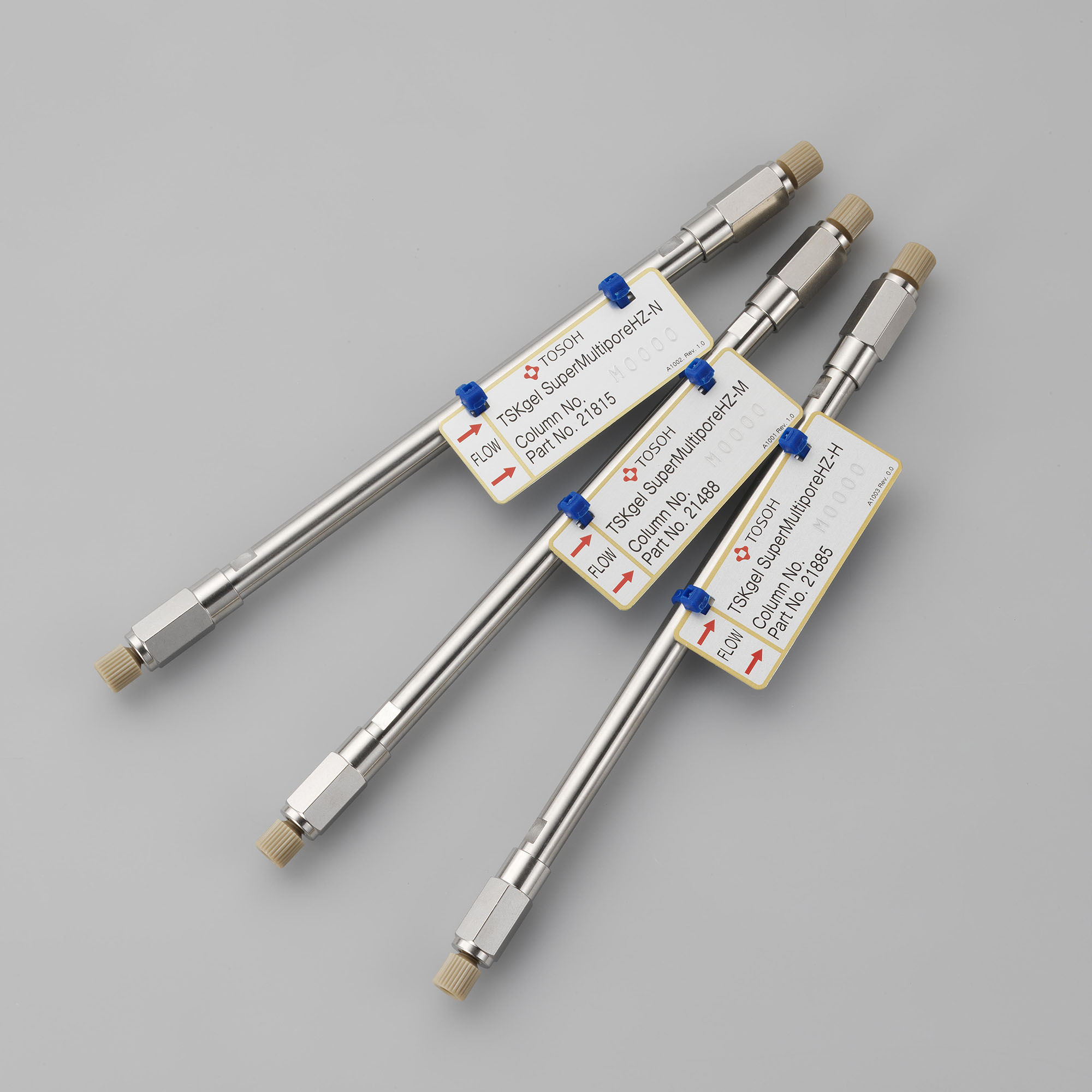
    TSKgel SuperMultiporePW 色谱柱是分离大多数水溶性合成聚合物及多糖类物质的首选色谱柱。该系列色谱柱装填有小粒径、细孔多分散型填料的半微型色谱柱(6.0 mm ID X 15 cm),可实现超高速、高效分离,节省溶剂。此类色谱柱的校正曲线线性优异、没有拐点,避免了色谱图中出现不正常的凹凸现象。根据样品分子量和分子量分布情况的不同,该系列色谱柱共有3中不同规格可供选择。TSKgel SuperMultiporePW色谱柱适用于分析中性低聚物,在一半的分析时间内即可达到与常规的30cm色谱柱相同的分离性能。药品和保健食品中使用的肝素和硫酸软骨素为酸性多糖,可使用TSKgel SuperMultipore PW色谱柱,以硝酸钠等洗脱液进行快速分离。TSKgel SuperMultiporePW-N填料粒径4μm,分子排阻界限为1.2×10⁵(PEO、PEG)。
  • TSKgel SuperMultiporePW-H 色谱柱
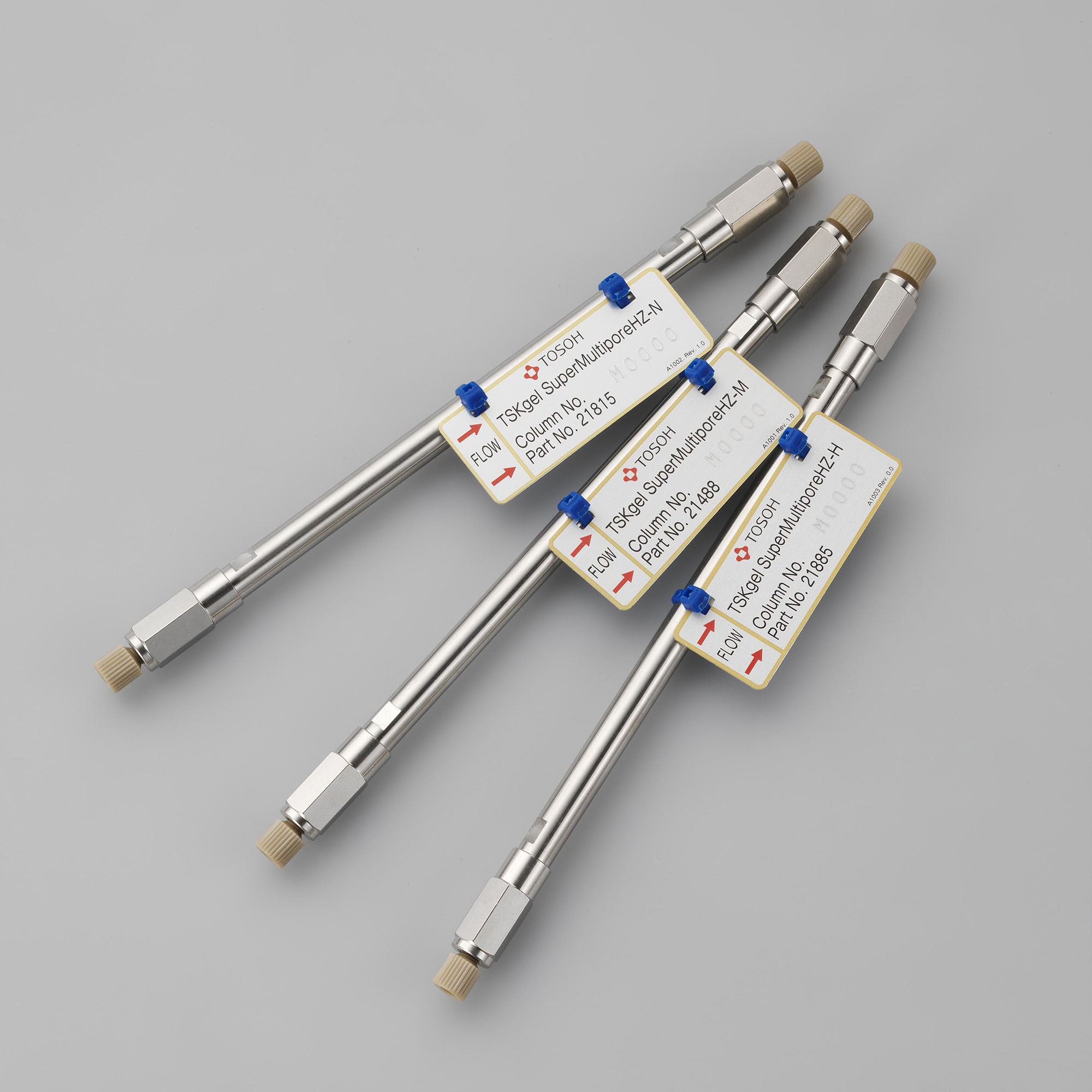
    TSKgel SuperMultiporePW 色谱柱是分离大多数水溶性合成聚合物及多糖类物质的首选色谱柱。该系列色谱柱装填有小粒径、细孔多分散型填料的半微型色谱柱(6.0 mm ID X 15 cm),可实现超高速、高效分离,节省溶剂。此类色谱柱的校正曲线线性优异、没有拐点,避免了色谱图中出现不正常的凹凸现象。根据样品分子量和分子量分布情况的不同,该系列色谱柱共有3中不同规格可供选择。TSKgel SuperMultiporePW色谱柱适用于分析中性低聚物,在一半的分析时间内即可达到与常规的30cm色谱柱相同的分离性能。药品和保健食品中使用的肝素和硫酸软骨素为酸性多糖,可使用TSKgel SuperMultipore PW色谱柱,以硝酸钠等洗脱液进行快速分离。TSKgel SuperMultiporePW-H填料粒径5μm,分子排阻界限为1×10⁷(PEO、PEG)。
  • TSKgel SuperMultiporePW-M 色谱柱
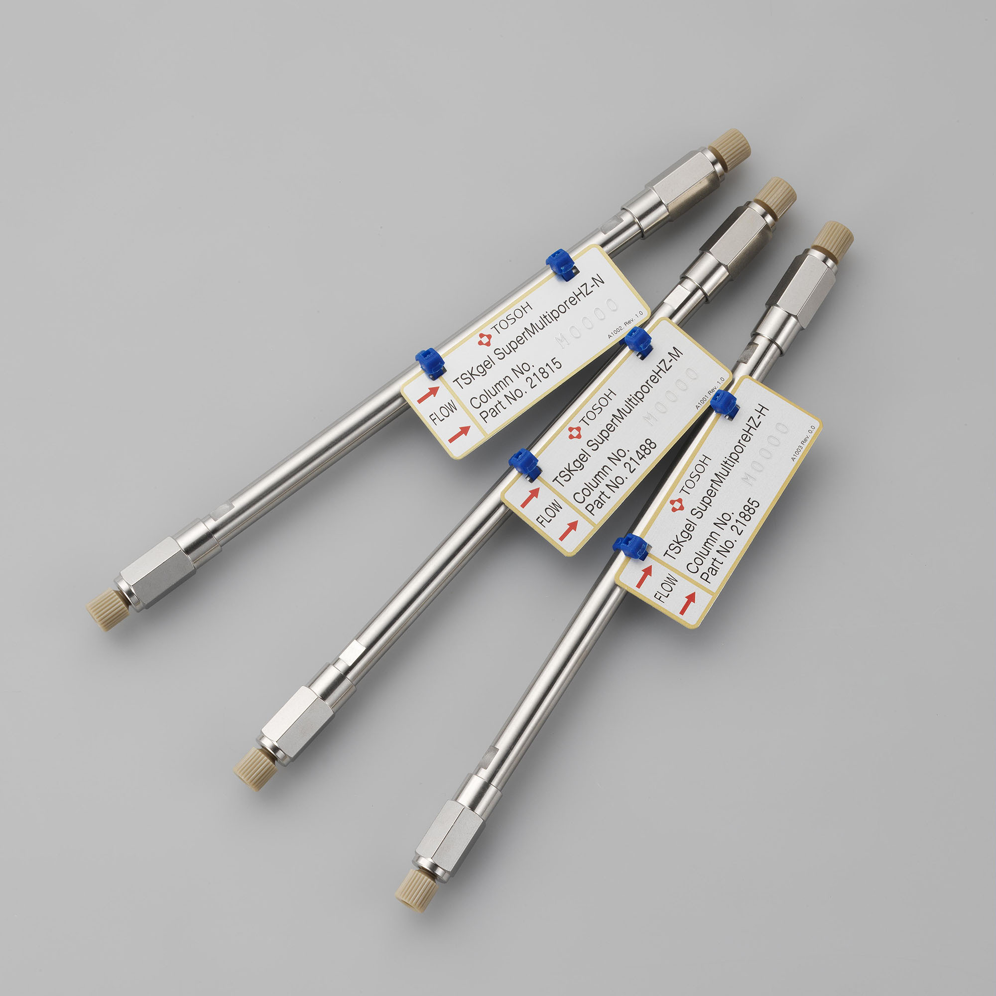
    TSKgel SuperMultiporePW 色谱柱是分离大多数水溶性合成聚合物及多糖类物质的首选色谱柱。该系列色谱柱装填有小粒径、细孔多分散型填料的半微型色谱柱(6.0 mm ID X 15 cm),可实现超高速、高效分离,节省溶剂。此类色谱柱的校正曲线线性优异、没有拐点,避免了色谱图中出现不正常的凹凸现象。根据样品分子量和分子量分布情况的不同,该系列色谱柱共有3中不同规格可供选择。TSKgel SuperMultiporePW色谱柱适用于分析中性低聚物,在一半的分析时间内即可达到与常规的30cm色谱柱相同的分离性能。药品和保健食品中使用的肝素和硫酸软骨素为酸性多糖,可使用TSKgel SuperMultipore PW色谱柱,以硝酸钠等洗脱液进行快速分离。TSKgel SuperMultiporePW-M填料粒径5μm,分子排阻界限为1×10⁶(PEO、PEG)。
  • GilderGrids 100-200目方孔钼网 英国进口 中镜科仪
    25枚/瓶,英国进口,钼载网可用于高温惰性环境中的研究,负载的样品不会发生变形。产品包装实拍图 编号材料Material载网目数Mesh Size中心距μmPitch肋宽μmBar Width孔径μmHole Size包装Unit产地Made inAG100M钼1002505020025枚/瓶英国 编号材料Material载网目数Mesh Size中心距μmPitch肋宽μmBar Width孔径μmHole Size包装Unit产地Made inAG200M钼200125359025枚/瓶英国
  • 石墨赶酸仪,实验室石墨消解仪
    OA-360智能石墨赶酸仪被广泛应用于实验室样品消解的赶酸处理,采用包裹式加热,具有可设置多段温度赶酸的智能程序功能,支持孔数、孔径及孔深定制,实现高效赶酸处理。可作为各种微波消解仪配套赶酸设备,解决微波消解赶酸难的问题。产品特点1、多孔设计,赶酸更快批量处理能力强,大大缩短赶酸时间;标配36位,可根据消解罐大小定制。2、高纯石墨块包裹式加热高纯石墨体具有良好的导热性,加热均匀,平行性好。立体包裹式加热,热量损失更少,效率更高。3、远程控制,程序升温采用PID程序温控系统,控温精度±0.2℃;实现多段升温、保持时间、升温时间,自动停止加热。4、工步指示灯程序工步显示灯,指示当前实验样品消解步骤,状态一目了然。5、高品质工艺,耐腐蚀石墨体及整机壳体喷涂特氟龙涂层,易清洁、耐腐蚀;整机外围无金属部件,可在强酸强碱等恶劣环境中放心使用。6、真实的样品温度石墨赶酸仪可选配外置温度探针,保证了样品的真实温度,使样品赶酸更加到位。应用领域环境监测:污水、饮用水、淤泥、矿泥、排污、土壤等农业食品检验:大米、奶粉、鱼类、蔬菜、烟草、植物、化肥等产品质量控制:化妆品、副食品、工业制品等科学研究:实验分析、项目开发等疾病预防控制:生物样品、人体毛发等
  • 中镜科仪 50至400目进口铜网普通碳膜支持膜
    普通碳支持膜是一种zui常见并被广泛使用的一种支持膜,为两层支持膜结构,可以采用不同规格的载网做载体。从空间结构来讲,从下到上依次为载网,方华膜和碳膜,它是在一层有机方华膜上再覆盖一层碳膜。由于碳层具有较强的导电以及导热性,弥补了无碳方华膜的荷电效应以及热效应,增强了膜整体的稳定性,可满足大多数纳米材料(尤其是可分散的粉体材料)的一般形貌测试需要。但是由于这种支持膜本身的结构导致了整体支持膜的厚度较大,一般不太容易满足高质量的高分辨测试的要求。英国进口铜载网,中镜科仪生产,支持膜厚度10-20nm。中包装50枚/盒。产品编号Prod.No.载网材质Material载网目数Mesh载网产地Made in支持膜产地Made in支持膜厚度Thickness包装UnitBZ100205b铜50目英国中镜科仪10-20nm50枚/盒BZ1002075b铜75目英国中镜科仪10-20nm50枚/盒BZ10021b铜100目英国中镜科仪10-20nm50枚/盒BZ100215b铜150目英国中镜科仪10-20nm50枚/盒BZ10022b铜200目英国中镜科仪10-20nm50枚/盒BZ10023b铜300目英国中镜科仪10-20nm50枚/盒BZ10024b铜400目英国中镜科仪10-20nm50枚/盒英国进口铜载网,中镜科仪生产,支持膜厚度10-20nm。大包装100枚/盒。产品编号Prod.No.载网材质Material载网目数Mesh载网产地Made in支持膜产地Made in支持膜厚度Thickness包装UnitBZ100205a铜50目英国中镜科仪10-20nm100枚/盒BZ1002075a铜75目英国中镜科仪10-20nm100枚/盒BZ10021a铜100目英国中镜科仪10-20nm100枚/盒BZ100215a铜150目英国中镜科仪10-20nm100枚/盒BZ10022a铜200目英国中镜科仪10-20nm100枚/盒BZ10023a铜300目英国中镜科仪10-20nm100枚/盒BZ10024a铜400目英国中镜科仪10-20nm100枚/盒【存储】:室温避光干燥保存(建议放干燥器或者干燥箱内)、防污染、防震荡, 未拆封保质期1年,拆封之后最好6个月内用完。【使用注意】:取用时采用高精尖镊子小心操作,防止产生弯折等破坏。存在有机膜,不能与有机溶剂接触。面向样品盒有字母的一面是铺膜的一面。
  • 测定右旋糖酐20色谱柱TSK G3000PWXL
    北京绿百草科技专业提供测定右旋糖酐20分子量及分布的尺寸排阻色谱柱TSKGEL G3000PWXL。TSKGEL G3000PWXL色谱柱填料是亲水、刚性、球型多孔的甲基丙烯酸甲酯高聚物,化学和机械性能好,粒径7&mu m,孔径200A,PH值范围2-12,可以使用含有50%的混合有机溶剂为流动相。2010版中国药典推荐使用TSKGEL GPWXL测定右旋糖酐20分子量以及分子量的分布。
  • 类器官Hepatocellular Carcinoma Organoid Kit(肝实质细胞癌)
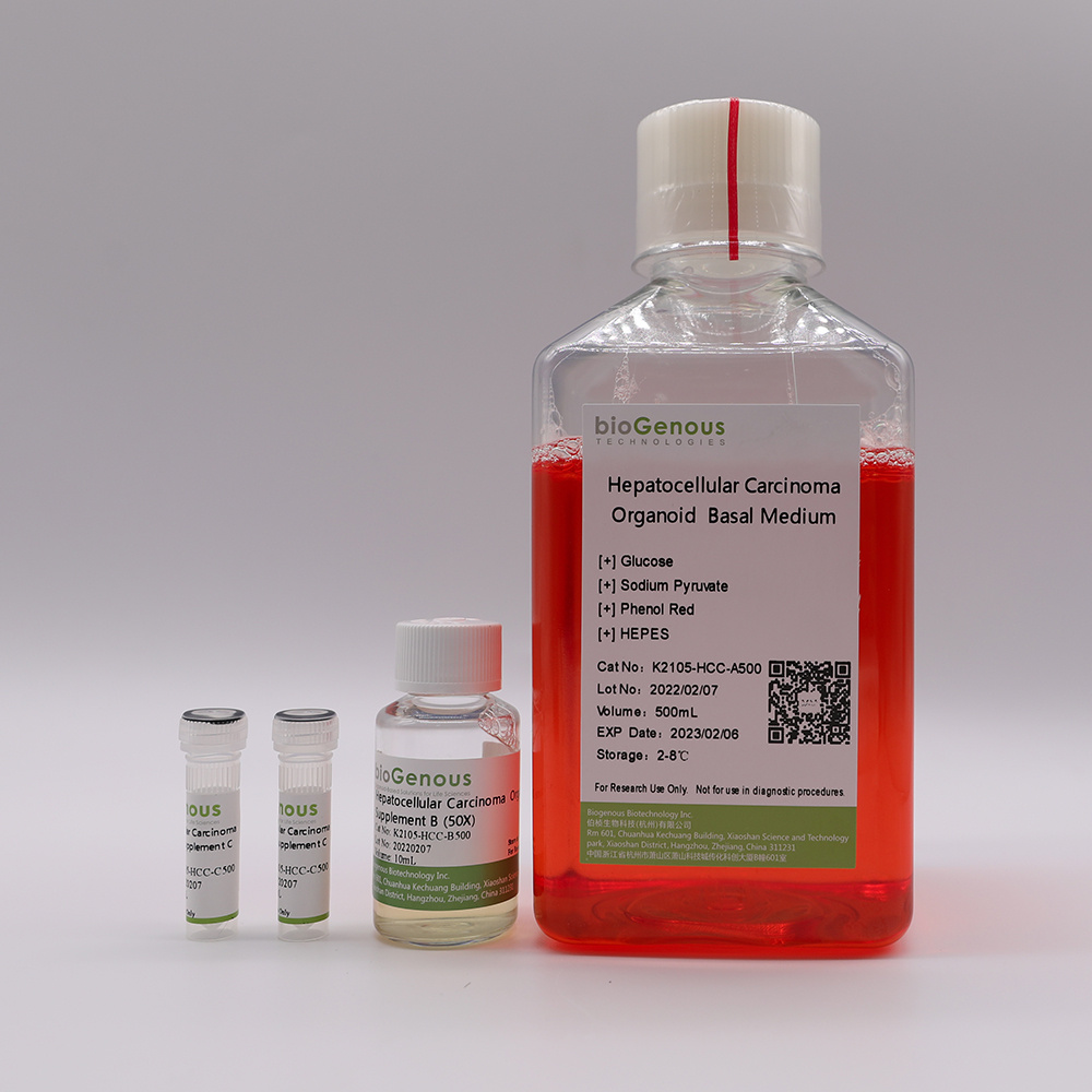
    类器官(Organoids)是指将成体干细胞或多能干细胞在体外三维培养形成的具有一定空间结构的组织类似物。类器官在组织结构、细胞类型、自我更新能力和功能等方面与来源组织高度一致,从而在发育生物学、疾病造模、精准医学、药物研发、基因和细胞疗法、感染和免疫以及再生医学等生物医学的多个领域展现出独特的优势。Product Description:bioGenousTMHepatocellular Carcinoma Organoid Kit is a chemically defined cell culture medium for the establishment and maintenance of human hepatocellular carcinoma organoids. Patient-derived cancer organoids recapitulate the genomic and pathological features of original tumors and therefore hold great promise for medical research and precision medicine.技术参数Product Information:ComponentComponent Cat#VolumeStorage & StabilitybioGenousTMHepatocellular Carcinoma Organoid Basal MediumK2105-HCC-A100/A500100mL/500mL4℃,12 monthsbioGenousTMHepatocellular Carcinoma Organoid Supplement B (50x)K2105-HCC-B100/B5002mL/10mL-20℃,avoid repeated freeze-thaw cycles, 12 monthsbioGenousTMHepatocellular Carcinoma Organoid Supplement C (250x)K2105-HCC-C100/C5000.4mL/2mL-20℃, avoid repeated freeze-thaw cycles, 12 monthsPreparation of Hepatocellular Carcinoma Organoid Complete MediumUse a sterile technique to prepare the hepatocellular carcinoma organoid complete medium. The following example is for preparing 10mL complete medium. If preparing other volumes, adjust accordingly.1.Thaw Hepatocellular Carcinoma Organoid Supplement B(50x) and Hepatocellular Carcinoma Organoid Supplement C(250x) on ice. Mix thoroughly.NOTE:Once thawed, use immediately or aliquot and store at -20°C for not more than 10 months. After thawing the aliquots, use immediately. Do not re-freeze.2.Add 200μL Hepatocellular Carcinoma Organoid Supplement B(50x) and 40μL Hepatocellular Carcinoma Organoid Supplement C(250x) to 9.76mL Hepatocellular Carcinoma Organoid Basal Medium. Mix thoroughly.NOTE:If not used immediately, store the complete medium at 2-8°C for not more than 2 weeks. The Hepatocellular Carcinoma Organoid Supplement B contains fungicide and antibiotics (50x).Protocol for Establishment ofPatient-Derived Hepatocellular Carcinoma OrganoidsCAUTIONStudies involving primary human tissue material must follow all relevant institutional and government regulations. Informed consent must be obtained from all subjects before the collection of the primary human tissue material.Establishment of Organoids from Primary Tissue1.Collect primary human hepatocellular carcinoma tissue pieces in ice-cold Primary Tissue Storage Solution (K601005) with conical tubes. Keep tissue samples at 4°C until the start of the isolation.2.Assess whether the obtained tissue pieces consist purely of epithelium. If fat or muscle tissues are present, remove these non-epithelial components as much as possible using surgical scissors or scalpels and forceps under a dissection microscope. If no fat or muscle tissues are present, continue to the next step immediately.3.Rinse the tissuewith Cancer Organoid Basal Medium (B213152) or DPBS twice.4.Mince the tissue into small fragments of 1-3 mm3in a cell culture dish using surgical scissors or scalpels.5.Digest the tissue fragments with 10mL of Tumor Tissue Digestion Solution (K601003) in a 15mL conical tube at 37°C, with variable incubation times ranging from 15 min to 45 min. Carefully monitor the digestion process, mixing the content of the tube every 5-10 min by shaking vigorously or pipetting the mixture up and down. The digestion process could be finished when most of tissue fragments are able to pass through the 1mL pipette tips.6.Add FBS to the tissuedigestionmixture to a final concentration of 2%, and filter using a 100 μm cell strainer.7.Collect and centrifuge the filtered cells at 250g for 3 min at 4 °C. In case of a visible red pellet, aspirate the supernatant, and resuspend the pellet using 2mL of Red Blood Cell Lysis Solution (E238010) to lyse the erythrocytes at room temperature for 1 min and centrifuge at 250g for 3 min at 4°C.8.Aspirate the supernatant and resuspend the pellet in Cancer Organoid Basal Medium, centrifuge at 250g for 3 min at 4°C,repeat this step once more time.9.Aspirate the supernatant and resuspend the pellet in Matrigel. The Matrigel should be kept on ice to prevent it from solidifying. Perform the process as quickly as possible. The amount of Matrigel used depends on the size of the pellet. Approximately 10,000 cells should be plated in 25 μL of Matrigel.CRITICAL:Do not overly dilute the Matrigel (Matrigel should be 70% (Matrigel vol/Total vol)), as this may inhibit the proper formation of the solid droplets.10.Plate the Matrigel containing organoids at the bottom of 24-well cell culture plates in droplets of ~30μL each around the center of the well..CRITICAL:Once the organoids are resuspended in Matrigel, proceed with plating as quickly as possible, as the Matrigel may solidify in the tube or pipette tip. Do not let the Matrigel touch the wall of wells.11.Place the culture plate into a humidified incubator at 37 °C and 5% (vol/vol) CO2for15-25 minto let the Matrigel solidify.12.Prepare the required amount of hepatocellular carcinoma organoid complete medium.13.Once the Matrigel droplets have solidified (15-25 min), open the plate and carefully add 500 μL of organoid complete medium to each well.CRITICAL:Do not add the medium directly on top of the Matrigel droplets, as this might disrupt the droplets.14.Place the culture plate in a humidified incubator at 37 °C and 5% (vol/vol) CO2.15.Change the medium every 3-4 d by carefully aspirating the medium from the wells and replacing it with a fresh, pre-warmed organoid complete medium.16.Closely monitor the organoid formation. Ideally, patient-derived hepatocellular carcinoma organoids should be passaged for the first time between 7 and 10 d after the initial plating.Splitting and Passaging of Organoids17.Pipette up and down to disrupt the Matrigel and transfer the organoid suspension to a 1.5 mL conical tube.18.Pipette the organoid suspension up and down to mix thoroughly by pipetting against the bottom of the tube to create pressure, which will aid the removal of Matrigel.19.Centrifuge organoids at 250g for 3 min at room temperature.20.Aspirate the supernatant and split organoids using either Organoid Dissociation Solution (E238001) or by mechanical disruption. For Organoid Dissociation Solution-based cell dissociation, resuspend the pellet in 0.2 mL of Organoid Dissociation Solution, pipette up and down and incubate at 37 °C until organoids fall apart. Pipette up and down with a filter tip for ≥5 times every 1 min to aid in the disruption of the organoids. Closely monitor the digestion to keep the incubation time inthe Organoid Dissociation Solution to a minimum. In case of mechanical disruption, resuspend the pellet in 1.5 mL ofCancer Organoid Basal Medium. Carefully pipette the organoid suspension up and down 30 times by pipetting against the bottom of the tube to create pressure, which will aid organoid disruption.CRITICAL:Do not dissociate in Organoid Dissociation Solution for 3 min, as this may result in poor organoid outgrowth or even loss of the culture. As a rule of thumb, digestion is complete if a mixture of small lumps of cells (consisting of 10–50 cells) can be observed.21.After shearing is complete, wash once by topping up with 1 mL ofCancer Organoid Basal Medium, and centrifuge at 250g for 3 min at room temperature.22.Aspirate the supernatant and resuspend the organoid pellet in 70% (vol/vol) Matrigel, and plate organoids in droplets at the bottom of a culture plate as described in Steps 10. After plating is complete, transfer the plate into a humidified incubator at 37 °C and 5% (vol/vol) CO2for 15–25 min.23.Pre-warm hepatocellular carcinoma organoid complete medium at 37 °C.24.After the Matrigel droplets have solidified (15–25 min), carefully pipette pre-warmed medium into the wells.25.Place culture plates in a humidified incubator at 37 °C and 5% (vol/vol) CO2until the organoids are needed for further experiments.
  • 类器官Cholangiocarcinoma Organoid Kit(肝内胆管癌)
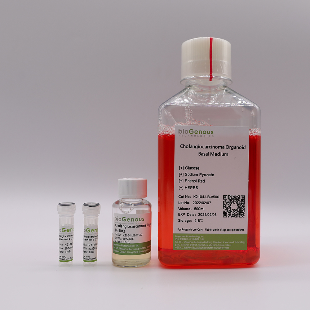
    类器官(Organoids)是指将成体干细胞或多能干细胞在体外三维培养形成的具有一定空间结构的组织类似物。类器官在组织结构、细胞类型、自我更新能力和功能等方面与来源组织高度一致,从而在发育生物学、疾病造模、精准医学、药物研发、基因和细胞疗法、感染和免疫以及再生医学等生物医学的多个领域展现出独特的优势。产品介绍Product Description:bioGenousTMCholangiocarcinoma Organoid Kit is a chemically defined cell culture medium for the establishment and maintenance of human cholangiocarcinoma organoids. Patient-derived cancer organoids recapitulate the genomic and pathological features of original tumors and therefore hold great promise for medical research and precision medicine.技术参数Product Information:ComponentComponent Cat#VolumeStorage & StabilitybioGenousTMCholangiocarcinoma Organoid Basal MediumK2104-LB-A100/A500100mL/500mL4℃,12 monthsbioGenousTMCholangiocarcinoma Organoid Supplement B (50x)K2104-LB-B100/B5002mL/10mL-20℃,avoid repeated freeze-thaw cycles, 12 monthsbioGenousTMCholangiocarcinoma Organoid Supplement C (250x)K2104-LB-C100/C5000.4mL/2mL-20℃, avoid repeated freeze-thaw cycles, 12 monthsPreparation of Cholangiocarcinoma Organoid Complete MediumUse a sterile technique to prepare the cholangiocarcinoma organoid complete medium. The following example is for preparing 10mL complete medium. If preparing other volumes, adjust accordingly.1. Thaw Cholangiocarcinoma Organoid Supplement B(50x) and Cholangiocarcinoma Organoid Supplement C(250x) on ice. Mix thoroughly.NOTE:Once thawed, use immediately or aliquot and store at -20°C for not more than 10 months. After thawing the aliquots, use immediately. Do not re-freeze.2.Add 200μL Cholangiocarcinoma Organoid Supplement B(50x) and 40μL Cholangiocarcinoma Organoid Supplement C(250x) to 9.76mL Cholangiocarcinoma Organoid Basal Medium. Mix thoroughly.NOTE:If not used immediately, store the complete medium at 2-8°C for not more than 2 weeks. The Cholangiocarcinoma Organoid Supplement B contains fungicide and antibiotics (50x).Protocol for Establishment ofPatient-Derived Cholangiocarcinoma OrganoidsCAUTIONStudies involving primary human tissue material must follow all relevant institutional and government regulations. Informed consent must be obtained from all subjects before the collection of the primary human tissue material.Establishment of Organoids from Primary Tissue1.Collect primary human cholangiocarcinoma tissue pieces in ice-cold Primary Tissue Storage Solution (K601005) with conical tubes. Keep tissue samples at 4°C until the start of the isolation.2.Assess whether the obtained tissue pieces consist purely of epithelium. If fat or muscle tissues are present, remove these non-epithelial components as much as possible using surgical scissors or scalpels and forceps under a dissection microscope. If no fat or muscle tissues are present, continue to the next step immediately.3.Rinse the tissuewith Cancer Organoid Basal Medium (B213152) or DPBS twice.4.Mince the tissue into small fragments of 1-3 mm3in a cell culture dish using surgical scissors or scalpels.5.Digest the tissue fragments with 10mL of Tumor Tissue Digestion Solution (K601003) in a 15mL conical tube at 37°C, with variable incubation times ranging from 30 min to 1.5 h. Carefully monitor the digestion process, mixing the content of the tube every 5-10 min by shaking vigorously or pipetting the mixture up and down. The digestion process could be finished when most of tissue fragments are able to pass through the 1mL pipette tips.6.Add FBS to the tissuedigestion mixture to a final concentration of 2%, and filter using a 100 μm cell strainer.7.Collect and centrifuge the filtered cells at 250g for 3 min at 4 °C. In case of a visible red pellet, aspirate the supernatant, and resuspend the pellet using 2mL of Red Blood Cell Lysis Solution (E238010) to lyse the erythrocytes at room temperature for 1 min and centrifuge at 250g for 3 min at 4°C.8.Aspirate the supernatant and resuspend the pellet in Cancer Organoid Basal Medium, centrifuge at 250g for 3 min at 4°C,repeat this step once more time.9.Aspirate the supernatant and resuspend the pellet in Matrigel. The Matrigel should be kept on ice to prevent it from solidifying. Perform the process as quickly as possible. The amount of Matrigel used depends on the size of the pellet. Approximately 10,000 cells should be plated in 25 μL of Matrigel.CRITICAL:Do not overly dilute the Matrigel (Matrigel should be 70% (Matrigel vol/Total vol)), as this may inhibit the proper formation of the solid droplets.10.Plate the Matrigel containing organoids at the bottom of 24-well cell culture plates in droplets of ~30μL each around the center of the well..CRITICAL:Once the organoids are resuspended in Matrigel, proceed with plating as quickly as possible, as the Matrigel may solidify in the tube or pipette tip. Do not let the Matrigel touch the wall of wells.11.Place the culture plate into a humidified incubator at 37 °C and 5% (vol/vol) CO2for15-25 min to let the Matrigel solidify.12.Prepare the required amount of cholangiocarcinoma organoid complete medium.13.Once the Matrigel droplets have solidified (15-25 min), open the plate and carefully add 500 μL of organoid complete medium to each well.CRITICAL:Do not add the medium directly on top of the Matrigel droplets, as this might disrupt the droplets.14.Place the culture plate in a humidified incubator at 37 °C and 5% (vol/vol) CO2.15.Change the medium every 3-4 d by carefully aspirating the medium from the wells and replacing it with a fresh, pre-warmed organoid complete medium.16.Closely monitor the organoid formation. Ideally, patient-derived cholangiocarcinoma organoids should be passaged for the first time between 7 and 10 d after the initial plating.Splitting and Passaging of Organoids17.Pipette up and down to disrupt the Matrigel and transfer the organoid suspension to a 1.5 mL conical tube.18.Pipette the organoid suspension up and down to mix thoroughly by pipetting against the bottom of the tube to create pressure, which will aid the removal of Matrigel.19.Centrifuge organoids at 250g for 3 min at room temperature.20.Aspirate the supernatant and split organoids using either Organoid Dissociation Solution (E238001) or by mechanical disruption. For Organoid Dissociation Solution-based cell dissociation, resuspend the pellet in 0.2 mL of Organoid Dissociation Solution, pipette up and down and incubate at 37 °C until organoids fall apart. Pipette up and down with a filter tip for ≥8 times every 2 min to aid in the disruption of the organoids. Closely monitor the digestion to keep the incubation time inthe Organoid Dissociation Solution to a minimum. In case of mechanical disruption, resuspend the pellet in 1.5 mL ofCancer Organoid Basal Medium. Carefully pipette the organoid suspension up and down 30 times by pipetting against the bottom of the tube to create pressure, which will aid organoid disruption.CRITICAL:Do not dissociate in Organoid Dissociation Solution for 5 min, as this may result in poor organoid outgrowth or even loss of the culture. As a rule of thumb, digestion is complete if a mixture of small lumps of cells (consisting of 10–50 cells) can be observed.21.After shearing is complete, wash once by topping up with 1 mL ofCancer Organoid Basal Medium, and centrifuge at 250g for 3 min at room temperature.22.Aspirate the supernatant and resuspend the organoid pellet in 70% (vol/vol) Matrigel, and plate organoids in droplets at the bottom of a culture plate as described in Steps 10. After plating is complete, transfer the plate into a humidified incubator at 37 °C and 5% (vol/vol) CO2for 15–25 min.23.Pre-warm cholangiocarcinoma organoid complete medium at 37 °C.24.After the Matrigel droplets have solidified (15–25 min), carefully pipette pre-warmed medium into the wells.Place culture plates in a humidified incubator at 37 °C and 5% (vol/vol) CO2until the organoids are needed for further experiments.
Instrument.com.cn Copyright©1999- 2023 ,All Rights Reserved版权所有,未经书面授权,页面内容不得以任何形式进行复制