何为细胞外泌体? 外泌体最早发现于体外培养的绵羊红细胞上清液中,是细胞主动分泌的大小较为均一,直径为40~100纳米,密度1.10~1.18 g/ml的囊泡样小体。细胞外泌体携带多种蛋白质、mRNA、miRNA,参与细胞通讯、细胞迁移、血管新生和肿瘤细胞生长等过程并且有可能成为药物的天然载体,应用于临床治疗。 然而,测量技术手段的局限限制了外泌体在这些领域的研究进展。所以,在这篇文章中,作者总结了外泌体的纯化方法(离心法、过滤离心法、密度梯度离心法、免疫磁珠法以及色谱法),比较了现存各种外泌体测量技术(电子显微镜、动态光散射技术及纳米微粒追踪分析术)在外泌体尺寸和表征研究中的应用。原文点击——综述:细胞外泌体颗粒表征测量技术新进展
【背景】CIK是“Cytokine-Induced Killer Cells”的缩写,中文全称为“细胞因子诱导的杀伤细胞”。 CIK是单个核细胞在CD3单抗和多种细胞因子(包括IFN-g, IL-2等)的作用下培养获得的一群以CD3+CD56+细胞为主要效应细胞的异质细胞群, 其既具有T淋巴细胞强大的抗肿瘤活性,又具有NK细胞(自然杀伤细胞)的非MHC(主要组织相容性抗原)限制性肿瘤杀伤能力。CIK细胞具有杀瘤活性高、杀瘤谱广,对正常组织毒性低,体外可高度扩增等特点,是目前临床上广泛使用的过继性免疫治疗细胞。【培养原理】CIK培养用细胞因子和抗体:nCD3激发型单抗:T细胞活化的第一信号来自于T细胞表面的受体,即T细胞抗原受体(T cell antigen receptor, TCR)与APC提呈的抗原的特异性结合,也就是T细胞对抗原的特异性识别。TCR是由2条不同肽链构成的异二聚体,在T细胞表面,其与CD3分子通过非共价键结合,形成TCR/CD3复合体。TCR识别特异性抗原后会引起CD3和T细胞表面的辅助受体CD4或CD8分子的胞浆尾部聚集,进而激活与胞浆尾部相连的酪氨酸激酶(Lck, Fyn和ZAP-70等),促使CD3分子胞浆区的免疫受体酪氨酸活化基序(immunoreceptor tyrosine-based activation motif, ITAM)中的酪氨酸(Y)磷酸化。磷酸化的酪氨酸(pY)进一步磷酸化下游含酪氨酸的蛋白,从而引起激酶活化的级联反应(磷脂酰肌醇途径或MAP激酶途径等),最终通过激活转录因子,使其进入细胞核内,结合于调控T细胞增殖和活化的靶基因(如IL-2和IFN-g等),引起基因的表达和转录,T细胞因而由静止状态转为增殖和活化状态。由上可见,CD3分子在T细胞活化信号的转导中起着极其关键的作用。CD3激发型单抗与T细胞表面CD3分子特异性结合后,可引起CD3分子胞浆区ITAM基序中酪氨酸的磷酸化,进而导致T细胞增殖和活化的下游信号的激活,从而使T细胞增殖和活化。也就是说,CD3激发型单抗能够模拟抗原与TCR/CD3复合物的识别和激活过程,从而引起T细胞的增殖与活化,因此是CIK细胞培养中不可或缺的刺激因素。此外,CD3激发型单抗在选用时一定要注意克隆号。研究表明,仅克隆号为OKT-3的CD3激发型单抗可以刺激所有人的T细胞的增殖,而其它克隆号的CD3激发型单抗仅能刺激一部分人的T细胞。因此,在进行CIK培养时,最好选用OKT-3克隆,以保证每个患者的T细胞均能被激活。nIL-2 (白细胞介素-2)IL-2最初发现时被称为T细胞生长因子(T cell growth factor, TCGF),是引起T细胞增殖最重要的细胞因子。IL-2既是自分泌细胞因子,也是旁分泌细胞因子,其通过与T细胞表面的IL-2受体(IL-2R)的特异性结合而促使T细胞活化,并进入细胞分裂状态。此外,IL-2还可刺激NK细胞的生长并增强其杀伤能力。因此CIK细胞培养中须添加IL-2,以促进T细胞的增殖与活化。nIFN-g (干扰素-g)IFN-g 具有上调外周血淋巴细胞表面IL-2R表达的作用,因此会增强T细胞对IL-2促增殖反应的敏感度和强度。在诱导CIK细胞形成的过程中加入IFN- g ,可降低IL-2的用量。研究发现,IFN-g加入的顺序与CIK的细胞毒活性密切相关。先加入IFN- g,培养24后再加入IL-2,可明显提高CIK的细胞毒活性。nIL-1a(白细胞介素-1a)IL-1a也可以介导外周血淋巴细胞表面上调表达IL-2R。当IL-1a与IFN-g和激发型CD3单抗合用时,可以明显提高CIK 的细胞毒作用。【细胞制备】1.外周血单个核细胞的采集1.1用血细胞分离机采集患者自身的外周血单个核细胞50-100mL;1.2淋巴细胞分离液密度梯度离心法进一步纯化单个核细胞(PBMC);1.3无血清培养液洗涤2次,获得纯度在90%以上的PBMC。2.CIK细胞的培养及鉴定2.1将PBMC按1-2 x 10⁶/ml的浓度悬浮于无血清培养液中,加入1,000 U/ml 的重组人IFN-g,37oC,5%CO2培养箱中培养;2.224h 后加入50ng/ml 的CD3 单克隆抗体和300 U/ml 的重组人IL-2,刺激CIK 细胞的生长和增殖;注:此时也可同时加入100 U/ml的重组人IL-1a。2.3每3天半量换液或扩瓶一次,并补加重组人IL-2 300 U/ml;2.4在培养的第14d,收获CIK细胞。2.5CIK细胞质控:2.9.1台盼蓝染色检测:活细胞应在80%以上;2.9.2流式细胞仪检测细胞表面CD3、CD8、CD56等分子的表达:CD3+CD56+细胞的比例应在20%以上。2.9.3细胞杀伤实验:以CIK细胞为效应细胞,以肿瘤细胞(可为原代肿瘤细胞或肿瘤细胞株)为靶细胞,将效应细胞与靶细胞按10 : 1(数目比) 的比例加入96 孔U 型板中,每孔含靶细胞1 x 10⁴个,终体积为200 ml,设3个复孔。培养4h,然后取培养上清,用乳酸脱氢酶(LDH) 试剂盒检测效应细胞对靶细胞的杀伤率。2.9.4收获细胞前,取少量培养物进行细菌、真菌培养,并检测支原体、衣原体,及内毒素(标准:病原学检测阴性,内毒素5 Eu)。【步骤简图】http://img.dxycdn.com/trademd/upload/userfiles/image/2013/04/B1366873006_small.jpg 【推荐试剂】http://img.dxycdn.com/trademd/upload/userfiles/image/2013/04/B1366873008_small.jpg 注:Animal Free意为无动物成分。无动物成分的重组细胞因子在生产过程中不会有任何动物源性物质,尤其是牛蛋白的混入,使得最终获得的重组人蛋白中不含任何动物成分。这样可避免动物病原体(如疯牛病,克雅氏病等)的污染及外源蛋白引起的机体异种排斥和过敏反应,因此细胞治疗的体外细胞培养过程中最好使用无动物成分的重组细胞因子。【其它相关试剂】 http://img.dxycdn.com/trademd/upload/userfiles/image/2013/04/B1366873009_small.jpg【参考文献】 Li R, Wang C, et al. Autologous cytokine-induced killer cell immunotherapy in lung cancer: a phase II clinical study. Cancer Immunol Immunother. 2012; 61:2125-2133
我们专题主要是研究胶原纤维,费伦有提过胶原纤维存在红外光传输的特征波段,不过在国内外研究极少提到有关细胞与细胞外基质的光传输,大都是提到有关化学反应的过程,现在是想找在胶原纤维的UV与IR光谱里那一个波峰,透过纤连蛋白(fibronectin)传输光讯号到细胞上的受体,之前是有找到有关细胞不含胞器(只剩肌动蛋白丝actin、整键蛋白integrin)也能移动,所以我们就假设细胞的移动可能是胶原纤维所操控,讲得有点多了,因为就只差这一点专题就完成了,可以请专家提出一些意见吗?谢谢[IMG]http://www.cella.cn/book/10/images/image006.jpg[/IMG]
细胞自噬是机体一种重要的防御和保护机制。但是这种自噬“信号”如何传递给细胞从而使其“执行”自噬过程,则一直是科学界的难题。近期,我校生命科学学院林圣彩教授课题组成功找到高等动物细胞在生长因子缺失条件下,启动自噬的部分“密码”,从而在细胞自噬机制研究方面取得重大突破。 4月27日,最新一期的美国《科学》杂志以研究文章的形式刊发了这项研究成果,并配发专门评述。这也是近三年来,我校生命科学学院第二篇发表在这一世界顶级学术刊物上的论文。2009年6月,该院韩家淮教授的一篇有关细胞选择死亡方式机制的研究文章曾“登上”该杂志。 所谓自噬,是指细胞消化自身蛋白质或细胞内的结构(细胞器)的一种自食现象。通过这种现象,细胞可以降解、消除和消化受损、变性、衰老和失去功能的细胞器和变性蛋白质等生物大分子,为细胞的生存和修复提供必须的能量。 科学家们认为,自噬与细胞凋亡、细胞衰老一样,是一种十分重要的生物学现象。有关实验表明,包括肥胖症、糖尿病、神经退行性疾病、免疫失调及癌症在内的人类许多重大疾病的发生都与该过程的异常有关。为此,自噬也是当前生命科学中最热门的研究领域之一。 据林圣彩介绍,对自噬进行分子机制的研究始于上世纪90年代的以单细胞生物酿酒酵母为模型的研究,目前,一系列构成单细胞生物自噬核心机器的基因已被发现并命名。 然而,对自噬在多细胞生物特别是哺乳动物中的调控机制的研究,科学界至今仍在不断探索中。摆在科学家面前的一个根源性的问题是:在多细胞生物中,诱导自噬的各种信号是如何被传递到细胞内自噬“核心机器”从而启动自噬过程的? 研究表明,与单细胞生物不同,在多细胞生物内,外界营养元素要依赖于生长因子的调控才能被转运到细胞内。一旦细胞外的生长因子匮乏,细胞便能启动自噬以维持能量平衡。那么,生长因子缺失这一信号又是如何“传达”的呢? 这也成为长期致力于细胞信号转导研究的林圣彩教授课题组近年来的研究目标之一。经过多年研究,课题组终于成功“**”这一自噬启动“密码”——即通过一种名为GSK3的激酶活性增高后磷酸化并随之激活乙酰转移酶TIP60,进而导致自噬核心机器中的蛋白激酶ULK1的乙酰化水平增强而启动细胞自噬。简言之,这一发现揭示了多细胞生物在生长因子缺失条件下的细胞自噬过程的新的介导分子及其通路。 林圣彩认为,弄清楚了细胞内到底有哪些蛋白分子“参与”了自噬和它们如何串联在一起,将有益于科学界从“源头”上认识相关疾病,并为这些疾病的诊断和治疗提供新的靶点。
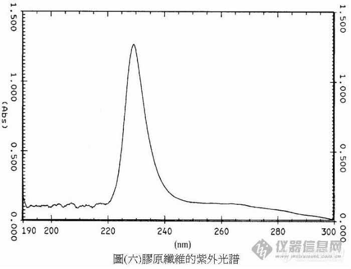
我们专题主要是研究胶原纤维,费伦有提过胶原纤维存在红外光传输的特征波段,不过在国内外研究极少提到有关细胞与细胞外基质的光传输,大都是提到有关化学反应的过程,现在是想找在胶原纤维的[color=#DC143C]UV与IR光谱里那一个波峰[/color]透过纤连蛋白(fibronectin)传输光讯号到细胞上的受体,之前是有找到有关细胞不含胞器(只剩肌动蛋白丝actin、整键蛋白integrin)也能移动,所以我们就假设细胞的移动可能是胶原纤维所操控,讲得有点多了,因为就只差这一点专题就完成了,可以请专家提出一些意见吗?谢谢[IMG]http://www.cella.cn/book/10/images/image006.jpg[/IMG][img]http://ng1.17img.cn/bbsfiles/images/2008/04/200804301729_87430_1025911_3.jpg[/img][img]http://ng1.17img.cn/bbsfiles/images/2008/04/200804301733_87431_1025911_3.jpg[/img]
[align=center][font='times new roman'][size=16px]超速离心法[/size][/font][font='times new roman'][size=16px]富集[/size][/font][font='times new roman'][size=16px]细胞分泌的外[/size][/font][font='times new roman'][size=16px]泌[/size][/font][font='times new roman'][size=16px]体[/size][/font][/align][align=left][font='times new roman'][size=16px]引言[/size][/font][/align]本章利用超速离心法富集细胞分泌的外泌体。采用透射电子显微镜,Western Blot实验,纳米颗粒示踪分析等表征了外泌体的形态结构及分布特征。最后,用增殖实验和划痕实验测试了富集得到的外泌体的生物完整性[font='宋体']。[/font][align=left][font='times new roman'][size=16px]细胞分泌的[/size][/font][font='times new roman'][size=16px]外[/size][/font][font='times new roman'][size=16px]泌[/size][/font][font='times new roman'][size=16px]体表征[/size][/font][/align][img]" style="max-width: 100% max-height: 100% [/img]利用透射电子显微镜对超速离心法得到的外泌体进行了表征。如图(a)所示,洗脱后的外泌体具有典型的杯状结构。纳米颗粒示踪分析技术显示富集的外泌体粒径分布较窄,平均粒径为115.3 nm(图 b)。Western blot方法验证了富集方法的有效性。如图(c)所示,经超速离心法富集后,典型的低丰度外泌体标记物HSC70、TSG101、CD63和CD9可得到有效检测。以上结果证明,基于超速离心法对外泌体的富集可行性。[align=center][font='黑体'][size=14px]图[/size][/font][font='黑体'][size=14px] (a)重悬液中外[/size][/font][font='黑体'][size=14px]泌[/size][/font][font='黑体'][size=14px]体的透射电子显微镜表征,(b)[/size][/font][font='黑体'][size=14px]纳米颗粒示踪分析技术[/size][/font][font='黑体'][size=14px]表征外[/size][/font][font='黑体'][size=14px]泌[/size][/font][font='黑体'][size=14px]体的尺寸分布,(c)[/size][/font][font='黑体'][size=14px]Western blot方法[/size][/font][font='黑体'][size=14px]表征[/size][/font][font='黑体'][size=14px]外[/size][/font][font='黑体'][size=14px]泌[/size][/font][font='黑体'][size=14px]体标记物HSC70、TSG101、CD63和CD9[/size][/font][font='黑体'][size=14px]蛋白条带[/size][/font][/align][align=left][font='times new roman'][size=16px][color=#000000] [/color][/size][/font][font='times new roman'][size=16px][color=#000000] [/color][/size][/font][font='times new roman'][size=16px]完整性验证[/size][/font][/align][img]" style="max-width: 100% max-height: 100% [/img]首先用细胞增殖用于评估超速离心法分离的外泌体的生物完整性。不同浓度(10[font='times new roman'][sup][size=16px]6[/size][/sup][/font]、10[font='times new roman'][sup][size=16px]8[/size][/sup][/font]、10[font='times new roman'][sup][size=16px]10[/size][/sup][/font]颗粒/孔)的H1299细胞外泌体分别与H1299细胞共培养24、48和72小时。如图(a)所示,H1299细胞来源的外泌体以剂量依赖性方式显著促进细胞增殖。此外,伤口愈合实验显示,H1299细胞来源的外泌体加速了伤口愈合速度,并以剂量和时间依赖的方式提高了伤口愈合质量(图 b)。上述结果表明,通过超速离心法富集的外泌体具有完整性,可以促进细胞增殖、分化和迁移。[align=center][font='黑体'][size=14px]图[/size][/font][font='黑体'][size=14px](a)H[/size][/font][font='黑体'][size=14px]1299[/size][/font][font='黑体'][size=14px]细胞来源的外[/size][/font][font='黑体'][size=14px]泌[/size][/font][font='黑体'][size=14px]体对细胞增殖的影响结果,(b)[/size][/font][font='黑体'][size=14px]H1299细胞来源的外[/size][/font][font='黑体'][size=14px]泌[/size][/font][font='黑体'][size=14px]体对细胞[/size][/font][font='黑体'][size=14px]划后伤口愈合[/size][/font][font='黑体'][size=14px]的影响结果[/size][/font][/align][align=left][font='times new roman'][size=16px]小结[/size][/font][/align]本章利用超速离心法富集了细胞来源的外泌体,并对富集得到的外泌体进行了一系列表征表征。通过透射电子显微镜和纳米颗粒示踪分析技术分析表明得到的外泌体具有典型的杯状结构和小的粒径分布,证明了富集方法的可行性。Western Blot结果表明,富集后得到的外泌体溶液经裂解后,特征蛋白HSC70、CD63、TSG101和CD81,其含量比富集前显著提高。利用细胞增殖实验和划痕实验证明超速离心法富集得到的外泌体具有良好的生物学活性,可应用于下游的实验研究。
[align=center][img=,600,216]https://img1.17img.cn/17img/images/202403/uepic/0115a8aa-1863-4da0-bf05-5b02661ffb4e.jpg[/img][/align]近年来,细胞外囊泡 (Extracellular vesicles,EV)的研究热度正在持续增长,与EV相关的文献数量呈指数级增长,已成为生命科学和生物医学研究领域内的一大热点话题。前不久,国际细胞外囊泡学会(ISEV)发布了最新版的细胞外囊泡研究指南[color=#00b050][b]《Minimal information for studies of extracellular vesicles(MISEV2023): From basic to advanced approaches》[/b][/color],在MISEV2014和MISEV2018版本基础上整合了来自ISEV专家工作组和1000多名研究人员的反馈意见,加强了研究设计和实验细节,并为新的应用领域提出了建议和指导。[color=#000000][b]MISEV2023重点对EV命名、样品收集和预处理、EV分离与浓缩、EV表征、EV研究技术方法、EV释放与摄取、EV功能研究、EV体内实验进行了介绍。[/b][/color](文末附全文链接)[align=center][color=#c0504d][b][size=20px]关于ISEV和MISEV简介[/size][/b][/color][/align]MISEV指南由国际外囊泡协会(ISEV)编制,ISEV是研究和使用细胞外囊泡的科学家和临床医生的主要专业协会,通过其年会、专题研讨会和其他会议、同行评审期刊、在线学习平台以及与其他学会的合作,吸引了世界各地的不同研究人员群体。因此,ISEV具有独特的优势,可以指导制定和传播关于最佳实践指南和科学考虑的专家共识。MISEV 2014是ISEV发表的第一篇EV研究指南,旨在为EV研究提供可靠的支撑,MISEV 2018对EV研究发展过程中的方法和手段进行了深入的且批判性的评估,其中大部分内容至今仍然有效。而MISEV 2023与之前的版本一样,为EV研究人员提供了简明扼要的建议和指导,对 MISEV2018 中提出的要点进行了完善,并增加了对新发展领域的建议和指导。其目的是帮助EV研究和应用领域的从业人员针对每个EV来源、类型、研究问题或应用展开最佳实践。[align=center][b][size=20px][color=#c0504d]关于EV命名[/color][/size][/b][/align]MISEV 2023保留了MISEV 2018的EV定义,但删除了2018年使用的“自然释放”的用词(新定义:EV是指从细胞中释放出来的颗粒,由脂质双层分隔,并且不能自行复制,即不包含功能性细胞核),以避免排除了通过细胞培养生产的EV。一般来说,ISEV建议使用通用术语“EV”和该术语的扩展,而不是使用具有误导性的术语,如与难以确定的生物发生途径相关的“exosomes(外泌体)”和“ectosomes(核外颗粒体)”。这两个术语是与假定的生物发生途径有关,需要谨慎使用且需要有强有力的证据。术语“exosomes(外泌体)”是指通过多泡体(MVB)释放的来自细胞内部的EV,而术语ectosomes(核外颗粒体,又称微囊泡Microvesicle、微粒Microparticle)是指细胞膜出芽形成的EV。由于目前大多数EV分离技术不能富集由不同机制产生的EV,且没有外泌体、核外颗粒体或其他EV亚型的通用分子标记。因此,ISEV不鼓励使用基于生物发生的术语,除非对此类EV群体进行了专门的分离和表征。相关术语及定义:[align=center][img=,600,639]https://img1.17img.cn/17img/images/202403/uepic/dce5a128-4ecf-4c0c-ae0c-31e02862f1d1.jpg[/img][/align][align=center][b][size=20px][color=#c0504d]EV的收集和预处理[/color][/size][/b][/align]样本采集、预处理、储存等因素可能会对EV数量和质量造成影响,MISEV2023对需要注意的一些因素给出了建议。对于不同样本都适用的因素,给出了普适建议,另外也针对细胞培养物(cell culture‐conditioned medium,CCM)、细菌、血液、尿液、脑脊液、唾液、滑液、乳汁、实体组织共计9类EV来源样本的采集及处理给出了具体建议。[b]1.血液[/b]血液是EV研究中最常见的生物体液样本,但血液样本面临供体变化、分析前处理、血液中血细胞、血小板、脂蛋白及其他蛋白成分的影响。基于此,MISEV2023对血液样本的收集与处理给出了以下建议:? 相较于其他样本,供体对血液及血液EV的影响较大,因此当收集血液样本时,需详细的记录和报告。? 静脉采血应使用管径较大的采血针,以最大限度减少血小板活化和溶血。为减少细菌和皮肤细胞污染、避免组织因子介导的血小板活化,弃去少量抽到的血液是一种有效的做法(例如,人类抽血时丢弃前面的2-3 mL)。? 选用与下游分析兼容的采血管和抗凝剂。? 采血后,应避免过度摇晃和低温,并尽快处理为血浆或血清,以减少血小板激活和EV释放。? 制备血浆或血清时,应选择能够有效去除血小板但不影响EV的方法。若使用离心法,吸取上清时应从上向下吸上清液,并在沉淀上方保留一定量的血浆或血清,以免干扰沉淀导致血小板释放。? 血液EV的主要污染物/共分离物包括血小板、脂蛋白、溶血产物以及大量可溶性/聚集蛋白,检测时需说明任一污染物。[b]2.尿液[/b]尿液是继血液之后第二大用于EV研究的生物体液样本,可以通过非侵入性的方式连续获得大量样本。尿液EV (uEV)研究的挑战源于uEV的来源细胞不同,以及受到液体摄入量、采样时间、饮食、运动、年龄、性别、药物以及健康状况的影响。基于此,MISEV2023对尿液样本的收集与处理给出了以下建议:? 应使用无细胞尿液/无细胞的尿液生物库。? 在适当情况下,报告uEV污染物/共分离成分(THP、白蛋白、其他过滤到尿液中的蛋白)的去除方法和去除效果。? 为实现标准化,收集uEV和非EV尿液(如肌酐、PSA等)数据,用于估计绝对或相对排泄率。[b]3.细胞培养物[/b]MISEV2018针对CCM中提出的建议仍然有效,包括但不限于描述培养基的组成和制备,记录生产细胞的特征、细胞培养条件、物理或化学刺激物处理(如果有)、CCM收获的频率和时间间隔及方法、EV分离之前CCM的储存处理。如果细胞来源不是已建立的细胞系,则应报告采集和预培养条件,如酶消化。? 如使用血清或其他添加剂,需说明来源和用量。如果使用的添加剂已经去除了EV,需说明去除方法并评估去除程度(包括稀释,通过离心的方法去除EV时稀释可能是必要的)。? 应将非条件(空白)培养基作为对照进行处理和定性,以评估培养基本身对EV检测的影响。[b]4.细菌[/b]细菌EV和细菌来源的多样性,很难就样品类型、预处理、分离、收集和表征给出普适性建议。MISEV2023建议在处理细菌样本时需要注意以下事项:? 除其他培养参数外,细菌培养物收获时需说明细菌生长阶段。? 尽量缩短EV分离/浓缩前的储存时间,尤其是在样本未经过滤的情况下。? 当细菌EV样本来自体内或环境,应考虑宿主EV和环境中非目标EV的影响。? LPS(脂多糖)和LTA(脂磷壁酸)可分别作为革兰氏阴性菌和革兰氏阳性菌的EV通用标志物,但在许多特定细菌物种中,该特定标志物仍然不可用。? 细菌EV的非囊泡共分离物可能包括毛、鞭毛、噬菌体和蛋白质、脂蛋白和核蛋白复合物。MISEV2023的建议旨在提高EV研究的严谨性、可重复性和透明度,帮助细胞外囊泡研究和应用领域的从业者根据EV来源、EV类型、研究内容、应用方向选择或制定最佳实践方案。[color=#191b1f]需要说明的是,[/color]MISEV2023的内容建立在MISEV2014和MISEV2018的基础之上,前两份指南中的指导建议很大程度上仍然有效,读者在参阅MISEV2023时应结合之前的文件。[color=#191b1f]下表列出了可供参考的文章:[/color][align=center][img=1.png,600,330]https://img1.17img.cn/17img/images/202403/uepic/1b5a57a4-f9c7-4abe-ac48-423a3c72de9f.jpg[/img][/align]参考:1.[i]权威发布!细胞外囊泡研究国际指南MISEV2023[/i] 2.[i]干货分享|外泌体研究红宝书—MISEV 2023解读(一)[/i] 3.[i]MISEV2023解读:全面认识细胞外囊泡[/i]附全文:[img]https://img1.17img.cn/17img/images/202101/pic/80056faa-b411-482e-9e52-14210fe10051.gif[/img][url=https://img1.17img.cn/17img/files/202403/attachment/07210f6f-ba10-4594-a029-01fcb39d3d64.pdf]J of Extracellular Vesicle - 2024 - Welsh - Minimal information for studies of extracellular vesicles MISEV2023 From.pdf[/url][来源:仪器信息网] 未经授权不得转载[align=right][/align]
[b][url=http://www.f-lab.cn/cell-analyzers/cell-stainer.html]细胞过滤筛Cell stainer[/url][/b]是[b]细胞过滤网,细胞筛网[/b],需要把细胞标记到芯片上。细胞过滤筛还具有夹持工具用于吸附细胞筛网,方便用户操作。[img=细胞过滤筛]http://www.f-lab.cn/Upload/cell-stainer.JPG[/img][img=细胞过滤筛]http://www.f-lab.cn/Upload/cell-label.JPG[/img]细胞过滤筛Cell stainer 细胞过滤筛Cell stainer 应用:cell labelling细胞过滤筛:[url]http://www.f-lab.cn/cell-analyzers/cell-stainer.html[/url]
[font='times new roman'][size=18px][color=#000000]显微镜下[/color][/size][/font][font='times new roman'][size=18px][color=#000000]顺铂处理[/color][/size][/font][font='times new roman'][size=18px][color=#000000]后细胞外[/color][/size][/font][font='times new roman'][size=18px][color=#000000]泌[/color][/size][/font][font='times new roman'][size=18px][color=#000000]体的分泌[/color][/size][/font][font='times new roman'][size=16px]为了[/size][/font][font='times new roman'][size=16px]进一步验证[/size][/font][font='times new roman'][size=16px]Tim4@ILI-01[/size][/font][font='times new roman'][size=16px]免疫亲和[/size][/font][font='times new roman'][size=16px]材料的捕获性能,[/size][/font][font='times new roman'][size=16px]采用[/size][/font][font='times new roman'][size=16px]不同浓度的[/size][/font][font='times new roman'][size=16px]顺铂处理[/size][/font][font='times new roman'][size=16px]H1299[/size][/font][font='times new roman'][size=16px]细胞来验证[/size][/font][font='times new roman'][size=16px]Tim4@ILI-01[/size][/font][font='times new roman'][size=16px]免疫亲和[/size][/font][font='times new roman'][size=16px]材料是否可以检测细胞培养上清液中外[/size][/font][font='times new roman'][size=16px]泌[/size][/font][font='times new roman'][size=16px]体分泌的变化。如图[/size][/font][font='times new roman'][size=16px]3-18 A[/size][/font][font='times new roman'][size=16px]所示,[/size][/font][font='times new roman'][size=16px]随着顺铂浓度[/size][/font][font='times new roman'][size=16px]的增加,细胞活力[/size][/font][font='times new roman'][size=16px]持续下降[/size][/font][font='times new roman'][size=16px],[/size][/font][font='times new roman'][size=16px]细胞分泌[/size][/font][font='times new roman'][size=16px]外[/size][/font][font='times new roman'][size=16px]泌[/size][/font][font='times new roman'][size=16px]体[/size][/font][font='times new roman'][size=16px]的[/size][/font][font='times new roman'][size=16px]浓度随着药物浓度的增加呈现先小幅增加后持续下降的趋势。外[/size][/font][font='times new roman'][size=16px]泌[/size][/font][font='times new roman'][size=16px]体浓度的下降[/size][/font][font='times new roman'][size=16px]主要[/size][/font][font='times new roman'][size=16px]是由大多数细胞活性降低[/size][/font][font='times new roman'][size=16px]所致[/size][/font][font='times new roman'][size=16px]。[/size][/font][font='times new roman'][size=16px][back=#ffff00]活细胞数量及其形态变化进一步证实了[/back][/size][/font][font='times new roman'][size=16px][back=#ffff00]顺铂仅[/back][/size][/font][font='times new roman'][size=16px][back=#ffff00]在特定浓度下调控癌细胞外[/back][/size][/font][font='times new roman'][size=16px][back=#ffff00]泌[/back][/size][/font][font='times new roman'][size=16px][back=#ffff00]体的分泌[/back][/size][/font][font='times new roman'][size=16px][back=#ffff00],[/back][/size][/font][font='times new roman'][size=16px]顺铂诱导[/size][/font][font='times new roman'][size=16px]细胞毒性存在浓度依赖性机制[/size][/font][font='times new roman'][size=16px](图[/size][/font][font='times new roman'][size=16px]3-18 B[/size][/font][font='times new roman'][size=16px])[/size][/font][font='times new roman'][size=16px]。为了进一步验证外[/size][/font][font='times new roman'][size=16px]泌[/size][/font][font='times new roman'][size=16px]体是否能提高细胞的化疗耐药能力,我们用不同浓度的顺铂[/size][/font][font='times new roman'][size=16px]([/size][/font][font='times new roman'][size=16px]0 [/size][/font][font='times new roman'][size=16px]μM[/size][/font][font='times new roman'][size=16px],[/size][/font][font='times new roman'][size=16px]1[/size][/font][font='times new roman'][size=16px] [/size][/font][font='times new roman'][size=16px]μM[/size][/font][font='times new roman'][size=16px]和[/size][/font][font='times new roman'][size=16px]10[/size][/font][font='times new roman'][size=16px] [/size][/font][font='times new roman'][size=16px]μM[/size][/font][font='times new roman'][size=16px])[/size][/font][font='times new roman'][size=16px]处理细胞,[/size][/font][font='times new roman'][size=16px]其分泌的[/size][/font][font='times new roman'][size=16px]外[/size][/font][font='times new roman'][size=16px]泌[/size][/font][font='times new roman'][size=16px]体孵育[/size][/font][font='times new roman'][size=16px]下一代[/size][/font][font='times new roman'][size=16px]细胞后,细胞[/size][/font][font='times new roman'][size=16px]在相同刺激条件下[/size][/font][font='times new roman'][size=16px]存活率较对照组明显提高,以[/size][/font][font='times new roman'][size=16px]10[/size][/font][font='times new roman'][size=16px] [/size][/font][font='times new roman'][size=16px]μM[/size][/font][font='times new roman'][size=16px]浓度效果最好[/size][/font][font='times new roman'][size=16px](图[/size][/font][font='times new roman'][size=16px]3-18 C[/size][/font][font='times new roman'][size=16px])[/size][/font][font='times new roman'][size=16px]。[/size][/font][font='times new roman'][size=16px]实验[/size][/font][font='times new roman'][size=16px]表明,[/size][/font][font='times new roman'][size=16px]H1299[/size][/font][font='times new roman'][size=16px]细胞暴露于特定浓度[/size][/font][font='times new roman'][size=16px]的顺铂后[/size][/font][font='times new roman'][size=16px],外[/size][/font][font='times new roman'][size=16px]泌[/size][/font][font='times new roman'][size=16px]体的分泌[/size][/font][font='times new roman'][size=16px]量增加,并且这些[/size][/font][font='times new roman'][size=16px]外[/size][/font][font='times new roman'][size=16px]泌[/size][/font][font='times new roman'][size=16px]体[/size][/font][font='times new roman'][size=16px]可以[/size][/font][font='times new roman'][size=16px]降低其他[/size][/font][font='times new roman'][size=16px]H1299[/size][/font][font='times new roman'][size=16px]细胞[/size][/font][font='times new roman'][size=16px]对顺铂的[/size][/font][font='times new roman'][size=16px]敏感性,增加了它们[/size][/font][font='times new roman'][size=16px]对顺铂的[/size][/font][font='times new roman'][size=16px]耐药性。以上研究表明,来自肺腺癌的外[/size][/font][font='times new roman'][size=16px]泌[/size][/font][font='times new roman'][size=16px]体在肺癌化疗耐药中起着重要作用[/size][/font][font='times new roman'][size=16px],[/size][/font][font='times new roman'][size=16px]这个结果与之前的[/size][/font][font='times new roman'][size=16px]报道[/size][/font][font='times new roman'][size=16px]一致。[/size][/font][table][tr][td][img]https://ng1.17img.cn/bbsfiles/images/2023/06/202306302213052787_7255_5389809_3.jpeg[/img][/td][/tr][/table][align=center][font='times new roman']图[/font][font='times new roman']3-18 [/font][font='times new roman']顺铂对[/font][font='times new roman']H1299[/font][font='times new roman']细胞外[/font][font='times new roman']泌[/font][font='times new roman']体分泌的影响:([/font][font='times new roman']A[/font][font='times new roman'])[/font][font='times new roman']H1299[/font][font='times new roman']细胞的剂量反应;([/font][font='times new roman']B[/font][font='times new roman'])不同浓度[/font][font='times new roman']顺铂[/font][font='times new roman']处理[/font][font='times new roman']H1299[/font][font='times new roman']细胞[/font][font='times new roman']3[/font][font='times new roman']天后的形态;([/font][font='times new roman']C[/font][font='times new roman'])外[/font][font='times new roman']泌[/font][font='times new roman']体作用后的[/font][font='times new roman']H1299[/font][font='times new roman']细胞[/font][font='times new roman']对顺铂的[/font][font='times new roman']剂量反应。[/font][font='times new roman'] [/font][font='times new roman']比例尺:[/font][font='times new roman']200 [/font][font='times new roman']μ[/font][font='times new roman']m[/font][/align]
【序号】:1【作者】:杨涛【题名】:不同层软骨细胞与骨髓间充质干细胞在体内外三维共培养时细胞外基质特点的实验研究【期刊】:大连医科大学【年、卷、期、起止页码】:2017【全文链接】:https://kns.cnki.net/kcms2/article/abstract?v=3uoqIhG8C447WN1SO36whLpCgh0R0Z-ifBI1L3ks338rpyhinzvy7Kt4RMGSMaxxH2WkQVeTlx_sUqdJWIk0PB_cP8FsBxdg&uniplatform=NZKPT
很多培养细胞的实验室对无菌的要求都有细微不同,关于紫外灯的照射时间大体上可以分为两派,一派开始前照射30分钟,一派是只要没有实验紫外灯就一直开着,直到下次实验开始前关闭,这两种哪种方式好呢?当然不能一概而论,当然从节能上看是第一种好,但是第一种能不能达到好的杀菌效果?那么一般来说是没有问题的,但是如果对于人数比较多的实验室,建议采用第二种方法,当然紫外不是避免污染的唯一方法,还要配合酒精擦拭和酒精灯消毒等。下面我们来看一般的消毒方法。实验进行前,超净台以紫外灯照射 30 分钟灭菌,以 75 % 酒精擦拭无菌操作台面,并开启超净工作台吹风5分钟以上分钟后,开始实验操作。每次操作只处理一株细胞株,以避免失误混淆或细胞间污染。实验完毕后,将实验物品带出工作台,以 75 % 酒精擦拭无菌操作台。无菌操作工作区域应保持清洁及宽敞,必要物品,例如支架、吸管等公用物品可以暂时放置,其它个人实验用品用完应立即拿出。实验用品以 75% 酒精擦拭后才带入无菌操作台内。实验操作应在中央无菌区域,一般勿在边缘区域操作。小心取用无菌之实验物品,避免造成污染。勿碰触吸管尖头部或是容器瓶口,亦不要在打开之容器正上方操作实验。容器打开后,以手夹住瓶盖并握住瓶身,倾斜约 45° 角取用,尽量勿将瓶盖盖口朝上放置桌面。注:由于大多数实验室酒精不是先用现配,由于酒精的挥发性较强,一般建议配置80%的酒精备用。
不知道哪位同学会使用紫外-可见分光光度计来检测细胞内EGCG和镉离子形成的螯合物呢?EGCG是绿茶中的一种物质,能够螯合金属离子。希望能指点一二;将不胜感激
偶欲通过[url=https://insevent.instrument.com.cn/t/Wp][color=#3333ff]原子吸收光谱[/color][/url]测定细胞钙含量,门外汉,遍寻不到相关的样品前处理方法,望大虾赐教!细胞钙以磷酸钙形式存在。需要灰化么——看资料说灰化是为了分解有机物,可是细胞样实在想象不出来如何进行灰化?酸化的时候,用多大浓度的酸比较合适,酸化时间如何掌握,低浓度的酸需要驱酸么?用什么容器盛装比较合适阿?普通的聚乙烯管子可以么?试验选用什么级别的水?试验室只有三蒸水,玻璃烧出来的。
【序号】:2【作者】:郜坤洁【题名】:人脐带脱细胞细胞外基质水凝胶的制备及相关性能研究【期刊】:南方医科大学【年、卷、期、起止页码】:2022【全文链接】:https://kns.cnki.net/kcms2/article/abstract?v=3uoqIhG8C447WN1SO36whLpCgh0R0Z-i16_wNaYct1rCckkTLVqOrRrxFeU2NGsFOH53JjrctrPrUUGId2crPEl-rpQXKB0w&uniplatform=NZKPT
多维流式细胞仪可同时进行多参数测量,在特定空间内对细胞群进行分析。若要实现该多维空间的合理使用,每个特定参数需提供额外信息来识别细胞群,并确保其动态范围能够最大限度地加以利用。本研究就白细胞的光信号散射情况进行了详细说明,从而促进了多维流式细胞分析的开展。细胞制备技术的提升对获得高分辨率光散射信号至关重要,可以实现粒细胞、单核细胞、颗粒状和非颗粒状淋巴球的完全分离。对搜集前向散射光的角度进行了改进,以提升白细胞的区分度。尽管正交光散射信号能够区分颗粒状和非颗粒状淋巴细胞,但仍无法利用线性或对数函数的形式将分辨率和动态范围显示出来。而在正交光散射信号中应用多项式函数,则可将白细胞全部以高分辨率显示出来。关联前向和正交光散射信号可实现高分辨率光散射与非线性显示的结合,使细胞群呈现等距分布状态。使用这种方式,可将外周血中性粒细胞、嗜酸细胞、嗜碱粒细胞、单核细胞、颗粒状和非颗粒状淋巴细胞等都显示出来,占据与正交和前向光散射相关的不同位置。出人意料的是,嗜碱粒细胞是处在了颗粒状淋巴和单核细胞附近而非中性和嗜酸性粒细胞。流式细胞术中的人体白细胞光散射特性主要应用于区分淋巴细胞、单核细胞和粒细胞。前向光散射信号与细胞的大小和折光率有关,而正交光散射信号则与细胞的粒度有关。一项对正交光散射信号更进一步的分析显示出了淋巴细胞成分的差异,即非颗粒状淋巴细胞的信号比颗粒状的要低。此外,该方法还显示了白血球的正交光散射信号在不同疾病状态下的变化情况。高分辨率光散射要在最佳角度收集散射参数,并对散射光的收集光路进行优化。改进细胞制备方法对最大限度地实现对细胞群的分离至关重要。改变制备流程可能导致细胞群分辨率的提高或降低。通过光散射,可从测量中排除受损细胞和无核细胞的干扰,从而提高细胞群的分辨率。正交光散射信号的动态范围不允许在相同线性尺度上同时观察淋巴细胞群和中性粒细胞。本研究提供了一种新方法,通过对正交光散射信号进行数字信号处理转换,实现了白细胞群在光散射显示中更加均衡的分布。这种转换提升了淋巴细胞分辨率,实现了细胞的可视化,而动态范围的确定对中性粒细胞的观察也十分重要。因此,重新对细胞群在多维空间进行定位可使细胞群在制备过程中实现完美分离。
[size=16px]细胞外小泡通过[/size][size=16px]PTEN[/size][size=16px]基因[/size][size=16px]途径[/size][size=16px]靶向治疗[/size][size=16px]HCC[/size][size=16px]PTEN[/size][size=16px]是一种抑癌基因,负向调控肿瘤的发生。有研究表明,肝癌细胞外[/size][size=16px]泌[/size][size=16px]体[/size][size=16px]miR-155[/size][size=16px]抑制邻旁或远端受体[/size][size=16px]HCC[/size][size=16px]细胞中[/size][size=16px]PTEN[/size][size=16px]表达,活化[/size][size=16px]PI3K/AKt[/size][size=16px]信号通路以促进[/size][size=16px]HCC[/size][size=16px]细胞的增殖[/size][font='times new roman'][sup][size=16px][[/size][/sup][/font][font='times new roman'][sup][size=16px]23][/size][/sup][/font][size=16px]。[/size][size=16px]Zhang[/size][size=16px]等[/size][font='times new roman'][sup][size=16px][[/size][/sup][/font][font='times new roman'][sup][size=16px]24][/size][/sup][/font][size=16px]研究发现,[/size][size=16px]HCC[/size][size=16px]患者血清中脂肪细胞来源外[/size][size=16px]泌[/size][size=16px]体[/size][size=16px]circDB[/size][size=16px]表达水平上调,通过与[/size][size=16px]HCC[/size][size=16px]细胞中[/size][size=16px]miR-34a[/size][size=16px]结合,上调[/size][size=16px]去泛素相关[/size][size=16px]分子[/size][size=16px]USP7[/size][size=16px]的表达,通过减少细胞增殖过程中的[/size][size=16px]DNA[/size][size=16px]损伤来促进肿瘤发生。[/size][size=16px]Mao[/size][size=16px]等[/size][font='times new roman'][sup][size=16px][[/size][/sup][/font][font='times new roman'][sup][size=16px]25][/size][/sup][/font][size=16px]在研究中提到了癌症相关成纤维细胞[/size][size=16px]([/size][size=16px]C[/size][size=16px]ancer associated fibroblasts[/size][size=16px],[/size][size=16px]CAFs[/size][size=16px])[/size][size=16px]调节[/size][size=16px]HCC[/size][size=16px]细胞增殖。转移性肝癌细胞来源[/size][size=16px]外[/size][size=16px]泌[/size][size=16px]体[/size][size=16px]中的[/size][size=16px]NID1[/size][size=16px]通过激活肺脏中的[/size][size=16px]CAFs[/size][size=16px],促进其旁分泌肿瘤坏死因子受体[/size][size=16px]1[/size][size=16px]([/size][size=16px]TNFR1[/size][size=16px])[/size][size=16px],进而诱导[/size][size=16px]HCC[/size][size=16px]细胞克隆的形成,表现出更加活跃的增殖能力。新血管生成不仅能为肿瘤提供大量的氧气和营养,促进肿瘤的生长,还作为转移细胞进入体循环的入口点,[/size][size=16px]介[/size][size=16px]导肿瘤侵袭转移能力的增强。[/size][size=16px]Lin [/size][size=16px]等[/size][font='times new roman'][sup][size=16px][[/size][/sup][/font][font='times new roman'][sup][size=16px]21][/size][/sup][/font][size=16px]研究发现[/size][size=16px]HCC[/size][size=16px]患者微血管密度与血浆[/size][size=16px]miR-210[/size][size=16px]表达水平具有明显相关性。研究发现,当肝癌细胞来源的外[/size][size=16px]泌[/size][size=16px]体[/size][size=16px]miR-210[/size][size=16px]与单层内皮细胞共孵育后,[/size][size=16px]HCC[/size][size=16px]细胞来源外[/size][size=16px]泌[/size][size=16px]体[/size][size=16px]miR-210[/size][size=16px]通过下调[/size][size=16px]SMAD4[/size][size=16px]和[/size][size=16px]STAT6[/size][size=16px]蛋白表达来促进毛细血管生成。肿瘤干细胞样[/size][size=16px]CD90+[/size][size=16px]肝癌细胞通过外[/size][size=16px]泌[/size][size=16px]体高水平表达[/size][size=16px]lncRNA H19[/size][size=16px],显著增加促血管生成因子[/size][size=16px]VEGF[/size][size=16px]和相应受体[/size][size=16px]VEGF-R1[/size][size=16px]的释放,也能上调促血管生成作用[/size][font='times new roman'][sup][size=16px][[/size][/sup][/font][font='times new roman'][sup][size=16px]26][/size][/sup][/font][size=16px]。[/size][size=16px]Shao[/size][size=16px]等[/size][font='times new roman'][sup][size=16px][[/size][/sup][/font][font='times new roman'][sup][size=16px]27][/size][/sup][/font][size=16px]研究发现,[/size][size=16px]HCC[/size][size=16px]细胞来源[/size][size=16px]外[/size][size=16px]泌[/size][size=16px]体[/size][size=16px]的[/size][size=16px]miR-584-5p[/size][size=16px]通过结合磷酸烯醇式丙酮酸激酶[/size][size=16px]1[/size][size=16px]([/size][size=16px]PCK1[/size][size=16px])[/size][size=16px]抑制其活性,诱导核因子[/size][size=16px]E2[/size][size=16px]相关因子[/size][size=16px]2[/size][size=16px]([/size][size=16px]Nrf2[/size][size=16px])[/size][size=16px]活化,上调血管内皮生长因子[/size][size=16px]A[/size][size=16px]([/size][size=16px]VEGFA[/size][size=16px])[/size][size=16px]表达,促进内皮细胞增殖,增强血管生成能力。[/size][size=16px]Wang[/size][size=16px]等[/size][font='times new roman'][sup][size=16px][[/size][/sup][/font][font='times new roman'][sup][size=16px]28][/size][/sup][/font][size=16px]认为,[/size][size=16px]HCC[/size][size=16px]细胞来源的外[/size][size=16px]泌[/size][size=16px]体中[/size][size=16px]miR-1290[/size][size=16px]富集,当其被血管内皮细胞内化后,能够抑制胞内[/size][size=16px]SMEK1[/size][size=16px]的表达,削弱其对[/size][size=16px]VEGFR2[/size][size=16px]磷酸化的抑制作用,从而诱导[/size][size=16px]VEGFR2[/size][size=16px]的活化和血管生成。除了非编码[/size][size=16px]RNA[/size][size=16px]介[/size][size=16px]导血管生成外,蛋白质分子也可参与其中。血管紧张素转运蛋白[/size][size=16px]([/size][size=16px]V[/size][size=16px]asorin[/size][size=16px],[/size][size=16px]VASN[/size][size=16px])[/size][size=16px]是一种[/size][size=16px]Ⅰ[/size][size=16px]型跨膜蛋白,在肿瘤发生和血管生成中发挥重要作用。[/size][size=16px]Huang[/size][size=16px]等[/size][font='times new roman'][sup][size=16px][[/size][/sup][/font][font='times new roman'][sup][size=16px]29][/size][/sup][/font][size=16px]研究发现,[/size][size=16px]HepG2[/size][size=16px]细胞外[/size][size=16px]泌[/size][size=16px]体高表达[/size][size=16px]VASN[/size][size=16px],被受体[/size][size=16px]HUVEC[/size][size=16px]内化摄取后,内皮细胞的增殖能力增强,大量的内皮细胞聚集有利于血管生成。[/size][size=16px]HCC[/size][size=16px]早期患者可进行肝切除术治疗,外[/size][size=16px]泌体一些[/size][size=16px]信号分子的表达与术后复发时间、总生存期长短、转移潜能等密切相关,可作为评估预后的指标。[/size][size=16px]Wang[/size][size=16px]等[/size][font='times new roman'][sup][size=16px][[/size][/sup][/font][font='times new roman'][sup][size=16px]30][/size][/sup][/font][size=16px]跟踪随访[/size][size=16px]183[/size][size=16px]例行肝细胞癌切除术后的丙型肝炎病毒相关肝细胞性肝癌患[/size][size=16px]者[/size][size=16px]10[/size][size=16px]年发现,这些患者血清外[/size][size=16px]泌[/size][size=16px]体中的分化拮抗非蛋白质编码[/size][size=16px]RNA[/size][size=16px]([/size][size=16px]D[/size][size=16px]ifferentiation antagonizing non-protein coding RNA[/size][size=16px],[/size][size=16px]DANCR[/size][size=16px])[/size][size=16px]的表达水平与[/size][size=16px]HCC[/size][size=16px]复发呈正相关,并且成为[/size][size=16px]HCC[/size][size=16px]复发和病死[/size][size=16px]率相关[/size][size=16px]的最具预测性的因素。另外一项研究发现,[/size][size=16px]HCC[/size][size=16px]患者外[/size][size=16px]泌[/size][size=16px]体[/size][size=16px]miR-103[/size][size=16px]表达与[/size][size=16px]HCC[/size][size=16px]的转移潜能和复发风险呈正相关,外[/size][size=16px]泌[/size][size=16px]体中[/size][size=16px]miR-103[/size][size=16px]水平[/size][size=16px]高表达[/size][size=16px]提示预后较差[/size][font='times new roman'][sup][size=16px][[/size][/sup][/font][font='times new roman'][sup][size=16px]28][/size][/sup][/font][size=16px]。[/size][size=16px]Jung[/size][size=16px]等[/size][font='times new roman'][sup][size=16px][[/size][/sup][/font][font='times new roman'][sup][size=16px]31][/size][/sup][/font][size=16px]检测并分析[/size][size=16px]14[/size][size=16px]例[/size][size=16px]HCC[/size][size=16px]患者[/size][size=16px]([/size][size=16px]其中[/size][size=16px]8[/size][size=16px]例患者[/size][size=16px]1[/size][size=16px]年内没有发生肿瘤转移,[/size][size=16px]6[/size][size=16px]例患者发生肿瘤转移[/size][size=16px])[/size][size=16px]血清外[/size][size=16px]泌[/size][size=16px]体的[/size][size=16px]miRNA[/size][size=16px]表达谱后发现,有[/size][size=16px]61[/size][size=16px]种[/size][size=16px]miRNA[/size][size=16px]在转移性[/size][size=16px]HCC[/size][size=16px]患者血清外[/size][size=16px]泌[/size][size=16px]体中表达显著上调,提示[/size][size=16px]HCC[/size][size=16px]的不良预后。外[/size][size=16px]泌体因[/size][size=16px]其具有低免疫原性、低毒性、可进行人工修饰,并且能够自由穿过生物屏障的特点,是非常理想的药物输送载体。外[/size][size=16px]泌体既[/size][size=16px]可以包裹[/size][size=16px]HCC[/size][size=16px]化疗药物,通过外层脂质膜保护作用使其不容易被降解,延长其在机体内作用时间,也可以包裹提高[/size][size=16px]HCC[/size][size=16px]细胞对化疗药物敏感度的[/size][size=16px]miRNA[/size][size=16px]等,利于化疗药物在机体内发挥抑瘤作用。[/size][size=16px]Zhang[/size][size=16px]等[/size][font='times new roman'][sup][size=16px][[/size][/sup][/font][font='times new roman'][sup][size=16px]32][/size][/sup][/font][size=16px]将阿霉素或索拉菲尼装载至红细胞来源的[/size][size=16px]外[/size][size=16px]泌[/size][size=16px]体[/size][size=16px]中,该载药[/size][size=16px]外[/size][size=16px]泌[/size][size=16px]体[/size][size=16px]可明显抑制小鼠原位肝癌细胞的生长,并且其对肝癌的抑制作用强于传统化疗药物给药方式及剂量所诱导的肝癌抑制作用。[/size][size=16px]miR-122[/size][size=16px]表达下调可激活[/size][size=16px]IGF-1R[/size][size=16px]进而活化[/size][size=16px]RAS/RAF/ERK[/size][size=16px]信号转导途径,[/size][size=16px]介[/size][size=16px]导[/size][size=16px]HCC[/size][size=16px]细胞对索拉菲尼耐药性的形成,过表达[/size][size=16px]miR-122[/size][size=16px]时可使得[/size][size=16px]HCC[/size][size=16px]细胞对索拉菲尼敏感而诱导细胞凋亡[/size][font='times new roman'][sup][size=16px][[/size][/sup][/font][font='times new roman'][sup][size=16px]33][/size][/sup][/font][size=16px]。[/size][size=16px]Lou [/size][size=16px]等[/size][font='times new roman'][sup][size=16px][[/size][/sup][/font][font='times new roman'][sup][size=16px]34][/size][/sup][/font][size=16px]在研究中用编码[/size][size=16px]miR-122[/size][size=16px]的质粒转染脂肪间充质干细胞[/size][size=16px]([/size][size=16px]AMSC[/size][size=16px])[/size][size=16px]后,产生的外[/size][size=16px]泌[/size][size=16px]体可通过分泌[/size][size=16px]miR-122[/size][size=16px]增加[/size][size=16px]HCC[/size][size=16px]细胞对索拉菲尼的敏感度。此外,外[/size][size=16px]泌[/size][size=16px]体还能够通过包裹抑制肿瘤细胞生长和侵袭的[/size][size=16px]miRNA[/size][size=16px],使其转移到[/size][size=16px]HCC[/size][size=16px]细胞后逆转其恶性表型。人肝成纤维细胞[/size][size=16px]miR-335-5p[/size][size=16px]表达上调可抑制邻旁[/size][size=16px]HCC[/size][size=16px]细胞增殖,进而抑制肿瘤生成,[/size][size=16px]CAFs[/size][size=16px]外[/size][size=16px]泌[/size][size=16px]体分泌的[/size][size=16px]miR-320a[/size][size=16px]可作为一种抗肿瘤[/size][size=16px]miRNA[/size][size=16px],与其下游靶点[/size][size=16px]PBX3[/size][size=16px]结合,抑制肝癌细胞的增殖、迁移[/size][font='times new roman'][sup][size=16px][[/size][/sup][/font][font='times new roman'][sup][size=16px]35-36][/size][/sup][/font][size=16px]。因此,选择合适的外[/size][size=16px]泌[/size][size=16px]体装载这些[/size][size=16px] miRNA[/size][size=16px],为[/size][size=16px]HCC[/size][size=16px]的靶[/size][size=16px]向治疗[/size][size=16px]提供了新的途径。[/size]
生物质谱技术在细胞生物学中的应用桑志红 王红霞 综述 概 论 蛋白分离与显色 蛋白质鉴定 数据库查寻 灵敏度 具体示例 展望未来(相关文献)摘 要 基因组计划的飞速发展使我们提早进入"后基因组时代",而质谱技术的重要进展使得通过酶解、质量分析、序列分析及其数据库检索对蛋白质进行高通量快速鉴定的技术方法应运而生,并成为"后基因组时代"的关键核心技术。这种技术的应用范围已经从细胞,组织以及整个有机体中蛋白质的表达到蛋白质翻译后修饰等等方面。本文简要综述生物质谱技术在细胞生物学等学科中的应用。 过去的十年经历并见证了生命科学革命性的变化. 大规模基因组测序技术的问世使人类基因组计划最终目标的实现比预期一再提前。与此同时,近几年间已有10余种模式生物的基因组序列测定告罄,3年内还将有40种左右生物的基因组全序列问世。因此大多数人同意我们现在已经提早进入"后基因组时代"(post-genome era), 目前我们所面临的挑战是如何破解基因组计划已获得的大量序列信息并加以应用。这个问题的关键是基因的生物学功能不能只通过对核酸一级结构(序列)的检测来确定。研判一个未知基因的功能、与其他基因产物及其亚细胞结构之间的功能联系, 最终都必须通过在蛋白水平对基因产物的研究才能确定。蛋白质组这个名词是近几年才提出来的,它用来描述一个细胞的全部蛋白质,而在蛋白水平上进行大规模的研究引出了新的术语蛋白质组学。蛋白质表达图谱是依靠蛋白质显示技术和精确定量技术对细胞或组织中蛋白质表达总况进行比较(2), 这个领域最近已有综述(11)。细胞图谱蛋白质组学是指应用生物质谱技术鉴定蛋白质及其相互作用并确定在亚细胞中的定位。本文的目的是 简要综述生物质谱技术在细胞生物学领域中越来越多的应用,并为该领域正考虑应用这种技术的研究者提供一些的有用的信息。 作为一个新的研究领域,蛋白质组学发展的关键是近年来质谱技术的革新。这种革新极大地促进了质谱技术在生命科学研究中的应用。质谱现在可以作为将各种蛋白质与序列数据库联系起来的桥梁。生物质谱根据质量数和所载电荷数不同的多肽片断在磁场中产生不同轨道而以质荷比(m/z)方式来分离它们。80年代末,随着两种崭新的尤其适合蛋白质研究的软电离方式ESI(电喷雾电离)和MALDI(基质辅助激光解吸附电离)的出现,质谱成为现代蛋白质科学中最重要和不可缺少的组成部分。 生物质谱最强大的应用功能之一是能够鉴定蛋白质复合物的组成成分(19)。细胞中一些最重要的生命过程都是通过多蛋白质复合体来执行和调节的,但由于蛋白质鉴定的困难,大多数上述蛋白复合体都是未知的。生物质谱灵敏度的不断提高显著地促进了对具有生物学功能和治疗潜力的蛋白复合体的鉴定,例如,NF-k B信号通路,CD95(FAS/APO-1)介导的细胞死亡途径,和核受体介导的转录信号传导过程中形成的蛋白复合体。在某些情况下,复合物可通过常规蛋白纯化的方法进行纯化,如剪接体复合(25),酵母纺锤体复合物(29)及VHL肿瘤抑制复合物(18)。然而,更常见的是,复合物中的组成成分通过一步免疫沉淀或免疫亲和步骤后就可纯化,这种方法甚至可以用于鉴定那些用常规蛋白纯化和鉴定技术所不及的一些过渡态或不稳定的复合物。因为生物质谱技术的介入,现在已经不再需要通过抗体进行免疫印迹实验,而是通过生物质谱技术对免疫沉淀获得的蛋白复合体组分直接进行蛋白序列分析。以酵母P24复合物鉴定为例,应用上游表位标签策略有可能不需制备抗目标蛋白的抗体就能对蛋白质复合物进行鉴定(13)。这种方法(36)对于基因组序列已完全清楚和遗传稳定的生物(如芽殖酵母,啤酒酵母)尤为简便, 例如对RENT复合物和促有丝分裂后期复合物(49)。因为这种方法的成功应用,使人们对上游表位标签策略-蛋白纯化-生物质谱分析的方法兴趣倍增。值得一提的是,将能被特殊蛋白酶切除的连接子(接头)掺入表位标签是尤为有利的(如下所述)。 上述方法是通过识别蛋白复合物中相互作用和配对的各组分而达到对蛋白鉴定的目的,另一种策略则是通过对分离纯化的细胞器蛋白组成进行鉴定而在亚细胞水平对蛋白质定位. 用这种方法确定蛋白质位置, 对评价蛋白质潜在的功能将是大有帮助的. 应用这种方法,我们称为细胞器蛋白质组学, 已经发现正常工作状态下的细胞器含有比我们以前所知道的数量多得多的蛋白种类。然而实际上,由于质谱极高的分辨率和灵敏度,纯化后的细胞器组分即使只有微量的混杂,也能被质谱分辨并误认为是细胞器的组成部分。因此,如何充分的纯化以保证至少绝大多数被鉴定的蛋白质都来自同一种细胞器,成为制约上述工作的瓶颈。 蛋白质翻译后修饰也是蛋白鉴定工作中的一个重要方面。根据DNA序列信息并不能可靠预测或推导出蛋白质翻译后的修饰。而质谱技术已经被证明对研究蛋白质翻译后的修饰(例如磷酸化和糖基化)是极为有用的,特别是对序列已知的蛋白的鉴定。例如:Betts等人(1a)用这种方法成功地鉴定了从小鼠大脑中分离的神经纤维蛋白体内磷酸化位点。同样方法, Wong等人(45)确定了钙联蛋白质(calnexin)C未端的磷酸化位点。在糖基化的例子中, Carr等人(5)采用液相色谱与质谱联用技术选择性地鉴定了糖蛋白中N- 和O-联接的寡糖. 稍后, 本文将会通过对E-选择素中糖基化位点的鉴定来进一步说明这种方法.
产品简介:保存液快速对脱落上皮细胞、腺细胞、白细胞等进行很好的保存和固定,保持标本采集时的原始细胞形态,防止细胞在保存过程中发生变形、自溶等。并通过制片使细胞均匀涂布在载玻片上制成薄层细胞涂片。染色后细胞结构在显徵镜下清晰易辨,同时把血液、粘液和炎症细胞减少到最底程度,从而易发现和确认异常细胞。更有利于从细胞的形态变化判定细胞的病变程度,使判定结果更加准确可靠,提高异常细胞的检出率,大大提高宫颈癌筛查方法的特异性和诊断的准确率。·产品性能特点::红细胞处理能力强:无需另加裂解液,既可将全部红细胞彻底清除,同时完美保存有诊断价值的各种有核细胞形态,从而对于临床上重度宫颈糜烂病人(或大量血细胞标本)能轻松一次性处理干净·消化分解黏液能力强:充分消化粘黏液,去除标本中普遍存在的黏液等干扰成份,释放具有诊断价值的细胞,保留有价值的诊断背景,有效提高检出率,检测结果准确。·细胞形态:核结构完整,其中核膜、核仁、核染色质颗粒及分布清晰可见,胞浆的嗜染性正常,有利于鉴别细胞的类别及来源。 细胞萃取:采用梯度离心分离萃取及红细胞处理专利技术和黏液消化技术多合一去除液基细胞学标本中的血液、黏液等干扰成份,富集提取细胞及诊断成份。 ·兼容性强:保存的细胞同时可做免疫细胞化学、HPV-DNA和衣原体等病原微生物的分子生物学检测,无需多次采样的烦恼。·应用广泛:细胞保存液临床运用非常广泛,除了运用宫颈细胞学检查外,还有胸腹积液、尿液、滑膜液、支气管冲洗液、脑脊液、针吸穿刺细胞及痰液标本细胞检测。·保存时间长:细胞在保存液中保存30天形态不变,真正保持细胞原始形态,更接近本身的组织学结构,更有利于恶性病变与良性反应性改变的鉴别诊断。·保存液细胞包裹技术,可以使细胞均匀悬浮,保证操作者在涂片标本时的随机性,任意取样涂片都具有代表性。http://ng1.17img.cn/bbsfiles/images/2011/06/201106231241_301155_2324710_3.jpg
广州中山大学研究发现天然病毒M1可杀灭癌细胞中新网广州10月13日电(许青青 蔡珊珊) 记者13日从广州中山大学获悉,该校中山医学院颜光美教授课题组研究发现天然病毒M1能选择性地感染并杀伤包括肝癌、结直肠癌、膀胱癌、黑色素瘤在内的多种体外培养的癌细胞,而对正常细胞无毒副作用。全球癌症发病率呈现快速增长态势,现有的治疗手段远远未能满足临床需求。颜光美教授课题组发现,M1病毒是一种从中国海南岛分离得到的天然病毒,能选择性地感染并杀伤包括肝癌、结直肠癌、膀胱癌、黑色素瘤在内的多种体外培养的癌细胞,而对正常细胞无毒副作用。整体动物实验表明,经尾静脉注射的M1病毒能显著富集在肿瘤组织并抑制肿瘤生长,正常器官则不受影响。除细胞水平及动物实验之外,课题组还使用临床标本离体活组织培养模型进一步证实了上述新型溶瘤病毒的有效性和特异性。据悉,该研究成果对阐明新型天然溶瘤病毒M1选择性杀伤肿瘤细胞的机制和研发新型靶向抗肿瘤药物都具有重要意义。相关资料:癌细胞增殖方式癌细胞是一种变异的细胞,是产生癌症的病源,癌细胞与正常细胞不同,有无限生长、转化和转移三大特点,也因此难以消灭。癌细胞由“叛变”的正常细胞衍生而来,经过很多年才长成肿瘤。“叛变”细胞脱离正轨,自行设定增殖速度,累积到10亿个以上我们才会察觉。癌细胞的增殖速度用倍增时间计算,1个变2个,2个变4个,以此类推。1912年8月13日,法国医生发现癌细胞。
细胞在体外进行培养,失去了机体的调节和控制。因此,除满足营养的要求外,还必须使细胞生存环境尽量接近活体的环境。外环境的培养条件如温度、渗透压、酸碱度等均能影响细胞的生长。 一、温度 一般哺乳类及禽类细胞体外培养的适宜温度是37~38℃。温度过高或过低都会影响到细胞的生长。细胞耐受低温的能力比抗热的能力强,在低温下,细胞的代谢活力及核分裂降低。温度不低于0℃时,虽影响细胞代谢,但并无伤害作用;把细胞置于25~35℃时,细胞仍能生存和生长,但速度减缓;放在40℃数小时后,再置回37℃培养细胞仍能继续生长。但如果在40℃下暴露时间太长,对细胞生长不利,甚至变圆脱落于瓶壁。若温度过低,在降到冰点以下时,细胞因胞外水和胞质结冰而受损死亡。但若向培养液中加入甘油或二甲亚砜等保护剂,封入安瓿中后,置于液氮中,可起保护作用,此时细胞可耐受-70℃以下温度,能长期储存,解冻后细胞复苏,仍能继续生长增殖,细胞生物性状不受任何影响。此为保存细胞的主要手段。 高温对细胞培养不利。细胞在39~40℃培养1小时,能受到一定损伤,但仍有可能恢复,但不能忍受温度再升高2℃,持续数小时,即在41~42℃中培养1小时,细胞损伤严重,温度至43℃以上时细胞多数被杀死。高温主要引起酶的灭活、类脂质破坏,核分裂的破坏,产生凝固酶使细胞发生凝固,另外使蛋白质变性。因此,体外培养细胞时一定要避免高温。 二、渗透压 细胞在高渗溶液或低渗溶液中,可以立即发生皱缩或肿胀、破裂。所以,渗透压是体外培养细胞的重要条件之一。哺乳动物和其他动物组织细胞体外培养的渗透压的维持主要与NaCl有关,但不能忽视其他电介质渗透压的关系。渗透压与单位体积溶媒内溶质的分子数和离子数成正比。为此,按一定比例控制培养液中离子平衡,维持正常渗透压是很重要的。这不仅是为了维持细胞张力,而且是为了调节细胞的代谢。因为细胞外离子输送和离子浓度改变着其他营养物质的输送(如氨基酸、蔗糖等),直接影响细胞基本合成系统。 理想的渗透压因细胞的类型及种族而异,人血浆渗透压为290mmol/L,被视为是体外培养人类细胞的理想渗透压。哺乳类动物细胞的渗透压一般为290~300mmol/L。人胚肺成纤维细胞为250~325mmol/L,鼠则为310mmol/L左右。在实际应用中,260~320mmol/L的渗透压可适于大多数细胞。
德国和以色列研究人员能够利用实验室器皿里的少量细胞培育出老鼠精子。他们认为,利用从睾丸里提出来的“生殖细胞”培育精子的这项技术,最终将能在人类身上产生作用 新浪科技讯 北京时间1月5日消息,科学家已经在体外培育精子方面取得新突破,这一重大发现将能帮助不育男性生育属于自己的孩子,而不是利用捐献精子。 德国和以色列研究人员能够利用实验室器皿里的少量细胞培育出老鼠精子。他们认为,利用从睾丸里提出来的“生殖细胞”培育精子的这项技术,最终将能在人类身上产生作用。该科研组现在正在“尽快”在人类身上重现这一结果。德国明斯特大学的斯特凡-斯库拉德教授领导的这个科研组,是世界上首个能利用生殖细胞培育精子的研究组。生殖细胞是睾丸里负责产生精子的细胞。 科学家在生殖细胞被称作果胶体的一种特殊化合物环绕的环境下培育精子,这种环境与睾丸里的环境非常类似。以色列班固利恩大学的穆罕默德-胡雷赫尔教授也在培养精子,他说:“我认为,我们将能通过从睾丸里提取包含生殖细胞的组织,在实验室环境下刺激精子产生,最终培育出男性精子。”有关这项精子试验的发现,已经出现在《自然》杂志上。获得这一发现的科学家现在已经开始试验在体外培育人类精子。 英国国家医疗服务体系(NHS)负责人、男性不育顾问斯蒂芬-戈登大加称赞这一突破。他说:“这是一项出人意料的新发现,它将彻底变革不育治疗,令每一位男性都能有自己的亲骨肉。不育男性都想有自己的孩子,但是目前他们的愿望还无法变成现实。但是通过老鼠试验获得的重大发现,使这种情况有可能变成现实。”英国顶级不育专家、爱丁堡大学的理查德-沙普教授希望参与这项试验,他说:“这是向成功培育出人类精子迈出的重要一步。” 过去50多年间,男性不育问题变得越来越严重,男性的精子数大量减少。导致这种情况的部分原因是污染和塑料包装里含有雌激素等环境因素。泌尿科医生戈登说:“即使借助最新的显微外科技术,仍有数千名男性无法生育亲骨肉,只能依靠精子捐献。”胡雷赫尔表示,他的科研组目前正在“尽快”在人类身上再现老鼠试验取得的成功,以便帮助不育男性。“我们已经采用了相同试验,就如我们在实验室里利用老鼠做试验一样,我们采用人类细胞,不过目前还未取得成功。我们相信,只要它在老鼠等哺乳动物身上有作用,它就对人类也有效。我们正在利用大量不同化合物进行研究,以便获得生殖细胞,培育精子。我们认为它有可能变成现实,而且或许很快就会梦想成真。” 这项研究成果发表在本月的《亚洲男性学》杂志上。胡雷赫尔说:“我们能产生可以繁育后代的精子,我们可以利用它们繁育小老鼠。这些精子显然很健康,而且也不存在遗传受损问题。我们花了多年时间才达到这个阶段,因此产生人类精子的技术不会在一夜间出现,但是老鼠试验取得成功后,我们已经开始人类研究。”为了加快制造人类精子的方法取得成功的步伐,胡雷赫尔的科研组打算开始与爱丁堡大学的理查德-沙普进行合作。 沙普说:“这项研究显示,在体外培养人类精子并非不可能。生殖细胞需要合适环境。这是让它们以为它们仍在睾丸里的关键。”他认为,一种新颖方法或许能让它变成现实。他建议把活老鼠作为制造人类精子的“寄主”。他说:“你要做的就是提取一些含生殖细胞的人类睾丸组织,然后把它们放置在老鼠的皮肤下,利用老鼠‘卵化’这些细胞。然后取出培育出来的精子,用来治疗不育。但是我们首先需要证明萃取出来的精子不包含老鼠细胞,如果我们希望利用该技术,我想我们就可以做到这些。” 戈登也在私营新生命诊所治疗不育,他说:“有几十亿人投入到女性不孕的研究中来,但是人们对男性不育的研究兴趣较小。有很多不育男性希望这项研究取得成功,因为这样他们以后就不用依靠捐献精子要小孩了。” 用在实验室里培育出来的人类精子治疗不育之前,需要获得相关部门的许可。但是沙普等研究人员认为,这个障碍将会被攻克。他说:“我们需要证明的主要问题,是精子不存在遗传损伤,并与睾丸产生的精子一样。用来进行女性不孕治疗的卵子和晶胚,也接受了类似检测。”
[align=center][font='times new roman'][size=21px]肿瘤细胞分泌的外[/size][/font][font='times new roman'][size=21px]泌[/size][/font][font='times new roman'][size=21px]体在机体内的作用[/size][/font][/align][font='times new roman'][size=16px]摘要[/size][/font][font='times new roman'][size=16px] [/size][/font][font='times new roman'][size=16px]肿瘤细胞通过产生,释放和利用外[/size][/font][font='times new roman'][size=16px]泌[/size][/font][font='times new roman'][size=16px]体来促进肿瘤发生发展。肿瘤来源的外[/size][/font][font='times new roman'][size=16px]泌[/size][/font][font='times new roman'][size=16px]体在肿瘤中的作用机制以成为目前的研究热点。外[/size][/font][font='times new roman'][size=16px]泌[/size][/font][font='times new roman'][size=16px]体作为信息载体,可将遗传信息从肿瘤细胞传递到位于近处或远处的正常或其他异常细胞。所有体液中[/size][/font][font='times new roman'][size=16px]均[/size][/font][font='times new roman'][size=16px]发现了肿瘤来源的外[/size][/font][font='times new roman'][size=16px]泌[/size][/font][font='times new roman'][size=16px]体,与靶细胞接触后,[/size][/font][font='times new roman'][size=16px]外[/size][/font][font='times new roman'][size=16px]泌[/size][/font][font='times new roman'][size=16px]体[/size][/font][font='times new roman'][size=16px]会改变受体[/size][/font][font='times new roman'][size=16px]细胞[/size][/font][font='times new roman'][size=16px]的表型和功能属性,[/size][/font][font='times new roman'][size=16px]起到促进[/size][/font][font='times new roman'][size=16px]血管生成,血栓形成,免疫抑制,肿瘤转移和耐药的作用。[/size][/font][font='times new roman'][size=16px]外[/size][/font][font='times new roman'][size=16px]泌[/size][/font][font='times new roman'][size=16px]体[/size][/font][font='times new roman'][size=16px]在抑制宿主抗肿瘤反应和[/size][/font][font='times new roman'][size=16px]介导[/size][/font][font='times new roman'][size=16px]耐药中发挥重要作用。肿瘤来源的外[/size][/font][font='times new roman'][size=16px]泌[/size][/font][font='times new roman'][size=16px]体可能会干扰癌症患者的免疫治疗。它们在癌症进展以及癌症治疗中的生物学作用表明肿瘤来源的外[/size][/font][font='times new roman'][size=16px]泌[/size][/font][font='times new roman'][size=16px]体是致癌转化的关键组成部分。[/size][/font][font='times new roman'][size=16px]关键词[/size][/font][font='times new roman'][size=16px] [/size][/font][font='times new roman'][size=16px]外[/size][/font][font='times new roman'][size=16px]泌[/size][/font][font='times new roman'][size=16px]体;肿瘤转移;耐药;免疫抑制;血栓形成[/size][/font][font='times new roman'][size=16px]多细胞生物[/size][/font][font='times new roman'][size=16px]体[/size][/font][font='times new roman'][size=16px]中相邻细胞之间的通讯通常包括细胞内物质的交换和共享,这些[/size][/font][font='times new roman'][size=16px]过程[/size][/font][font='times new roman'][size=16px]是通过细胞间连接、突触或通过吞噬作用形成的,都需要细胞接触并且在短距离内进行。相反,外[/size][/font][font='times new roman'][size=16px]泌体代表[/size][/font][font='times new roman'][size=16px]了信息传递的独特形式,既可以在短距离传递,也可以在长距离下进行信息交流。肿瘤来源的外[/size][/font][font='times new roman'][size=16px]泌[/size][/font][font='times new roman'][size=16px]体可以将信号从肿瘤细胞转移到远端组织和器官。[/size][/font][font='times new roman'][size=16px]外[/size][/font][font='times new roman'][size=16px]泌[/size][/font][font='times new roman'][size=16px]体[/size][/font][font='times new roman'][size=16px]存在于机体循环中,并可以随时进入身体的各个部位。它们带有能够与内皮细胞接触并促进外[/size][/font][font='times new roman'][size=16px]泌[/size][/font][font='times new roman'][size=16px]体进入血管和组织的表面成分[/size][/font][font='times new roman'][size=16px][[/size][/font][font='times new roman'][size=16px]1,2[/size][/font][font='times new roman'][size=16px]][/size][/font][font='times new roman'][size=16px]。但是肿瘤来源的外[/size][/font][font='times new roman'][size=16px]泌[/size][/font][font='times new roman'][size=16px]体仅占患者血浆中总外[/size][/font][font='times new roman'][size=16px]泌[/size][/font][font='times new roman'][size=16px]体的[/size][/font][font='times new roman'][size=16px]一[/size][/font][font='times new roman'][size=16px]小部分,且该部分的含量可根据肿瘤进展而改变。[/size][/font]1. [font='times new roman'][size=16px]外[/size][/font][font='times new roman'][size=16px]泌[/size][/font][font='times new roman'][size=16px]体在肿瘤转移中的作用[/size][/font][font='times new roman'][size=16px]肿瘤[/size][/font][font='times new roman'][size=16px]细胞[/size][/font][font='times new roman'][size=16px]的转移过程[/size][/font][font='times new roman'][size=16px]开始[/size][/font][font='times new roman'][size=16px]于肿瘤细胞经历[/size][/font][font='times new roman'][size=16px]了[/size][/font][font='times new roman'][size=16px]上皮间质转化([/size][/font][font='times new roman'][size=16px]Epithelial-to-mesenchymal transition[/size][/font][font='times new roman'][size=16px],[/size][/font][font='times new roman'][size=16px]EMT[/size][/font][font='times new roman'][size=16px])。肿瘤细胞[/size][/font][font='times new roman'][size=16px]获得[/size][/font][font='times new roman'][size=16px]迁移[/size][/font][font='times new roman'][size=16px]能力,并[/size][/font][font='times new roman'][size=16px]进入血液[/size][/font][font='times new roman'][size=16px]和[/size][/font][font='times new roman'][size=16px]淋巴系统[/size][/font][font='times new roman'][size=16px],逐渐[/size][/font][font='times new roman'][size=16px]转移[/size][/font][font='times new roman'][size=16px]到其他组织[/size][/font][font='times new roman'][size=16px]。[/size][/font][font='times new roman'][size=16px]这些[/size][/font][font='times new roman'][size=16px]肿瘤细胞产生具有独特分子特征的外[/size][/font][font='times new roman'][size=16px]泌[/size][/font][font='times new roman'][size=16px]体,[/size][/font][font='times new roman'][size=16px]主要包含[/size][/font][font='times new roman'][size=16px]EMT[/size][/font][font='times new roman'][size=16px]相关的蛋白质与迁移和侵袭所需的分子,[/size][/font][font='times new roman'][size=16px]如[/size][/font][font='times new roman'][size=16px]前列腺癌释放的外[/size][/font][font='times new roman'][size=16px]泌[/size][/font][font='times new roman'][size=16px]体中的α[/size][/font][font='times new roman'][size=16px]v[/size][/font][font='times new roman'][size=16px]β[/size][/font][font='times new roman'][size=16px]6[/size][/font][font='times new roman'][size=16px]整联蛋白[/size][/font][font='times new roman'][size=16px][3][/size][/font][font='times new roman'][size=16px],白血病[/size][/font][font='times new roman'][size=16px]或[/size][/font][font='times new roman'][size=16px]乳腺癌衍生的外[/size][/font][font='times new roman'][size=16px]泌[/size][/font][font='times new roman'][size=16px]体的[/size][/font][font='times new roman'][size=16px]Wnt[/size][/font][font='times new roman'][size=16px]通路[/size][/font][font='times new roman'][size=16px]成分[/size][/font][font='times new roman'][size=16px][4,5][/size][/font][font='times new roman'][size=16px],以及胃肠道间质瘤([/size][/font][font='times new roman'][size=16px]GIST[/size][/font][font='times new roman'][size=16px])产生的外[/size][/font][font='times new roman'][size=16px]泌[/size][/font][font='times new roman'][size=16px]体中的[/size][/font][font='times new roman'][size=16px]KIT [/size][/font][font='times new roman'][size=16px][6][/size][/font][font='times new roman'][size=16px]。这些外[/size][/font][font='times new roman'][size=16px]泌[/size][/font][font='times new roman'][size=16px]体中缺氧诱导因子[/size][/font][font='times new roman'][size=16px]([/size][/font][font='times new roman'][size=16px]HIF[/size][/font][font='times new roman'][size=16px])[/size][/font][font='times new roman'][size=16px]的含量增加,[/size][/font][font='times new roman'][size=16px]促炎因子[/size][/font][font='times new roman'][size=16px]的含量也增加[/size][/font][font='times new roman'][size=16px][7][/size][/font][font='times new roman'][size=16px]。准备迁移的肿瘤细胞产生的外[/size][/font][font='times new roman'][size=16px]泌[/size][/font][font='times new roman'][size=16px]体可与机体的血管、基质成分和免疫细胞相互作用,完成转移前的准备[/size][/font][font='times new roman'][size=16px][8][/size][/font][font='times new roman'][size=16px]。黑色素瘤来源的外[/size][/font][font='times new roman'][size=16px]泌[/size][/font][font='times new roman'][size=16px]体显示在前哨淋巴结中积累,刺激血管生成,重塑细胞外基质并诱导黑色素瘤细胞富集到淋巴结中[/size][/font][font='times new roman'][size=16px][9][/size][/font][font='times new roman'][size=16px]。[/size][/font][font='times new roman'][size=16px]Peinado[/size][/font][font='times new roman'][size=16px]研究团队证明了,从高度转移的鼠类黑[/size][/font][font='times new roman'][size=16px]色素瘤细胞衍生的外[/size][/font][font='times new roman'][size=16px]泌[/size][/font][font='times new roman'][size=16px]体能够将骨髓重编程为转移前的生理状态。现在已有研究支持肿瘤细胞分泌的外[/size][/font][font='times new roman'][size=16px]泌[/size][/font][font='times new roman'][size=16px]体与高度侵袭性的黑色素瘤细胞的发展有关,与空白对照组相比,实验组小鼠先前外[/size][/font][font='times new roman'][size=16px]泌[/size][/font][font='times new roman'][size=16px]体进行过注射处理,其体内黑色素瘤细胞的增殖和转移速率明显提高[/size][/font][font='times new roman'][size=16px][10][/size][/font][font='times new roman'][size=16px]。在许多有关鼠类和人体肿瘤来源的外[/size][/font][font='times new roman'][size=16px]泌[/size][/font][font='times new roman'][size=16px]体的近期研究中,已证明这些外[/size][/font][font='times new roman'][size=16px]泌[/size][/font][font='times new roman'][size=16px]体还携带微小[/size][/font][font='times new roman'][size=16px]RNA[/size][/font][font='times new roman'][size=16px]分子,将其转移至正常细胞并诱导其遗传和蛋白质谱发生变化,从而有利于转移形成[/size][/font][font='times new roman'][size=16px][11,12][/size][/font][font='times new roman'][size=16px]。肿瘤来源的外[/size][/font][font='times new roman'][size=16px]泌[/size][/font][font='times new roman'][size=16px]体已被证明带有[/size][/font][font='times new roman'][size=16px]CD39[/size][/font][font='times new roman'][size=16px]和[/size][/font][font='times new roman'][size=16px]CD73[/size][/font][font='times new roman'][size=16px],它们是催化腺苷产生的外核苷酸酶[/size][/font][font='times new roman'][size=16px][13][/size][/font][font='times new roman'][size=16px]。腺苷在机体内可[/size][/font][font='times new roman'][size=16px]介[/size][/font][font='times new roman'][size=16px]导免疫抑制,发挥促进血管生成并驱动细胞基质重塑的重要作用,所有这些功能都促进肿瘤细胞迁移及其进入淋巴结。肿瘤外[/size][/font][font='times new roman'][size=16px]泌体支持[/size][/font][font='times new roman'][size=16px]转移的能力可以通过腺苷参与不同类别的分子途径[/size][/font][font='times new roman'][size=16px][14][/size][/font][font='times new roman'][size=16px]。[/size][/font]2. [font='times new roman'][size=16px]外[/size][/font][font='times new roman'][size=16px]泌[/size][/font][font='times new roman'][size=16px]体在肿瘤耐药性中的作用[/size][/font][font='times new roman'][size=16px]肿瘤对放射和化学药物的抵抗作用是肿瘤患者临床治疗中面对的严重问题,至今尚未得到解决。值得注意的是有研究指出肿瘤分泌的外[/size][/font][font='times new roman'][size=16px]泌[/size][/font][font='times new roman'][size=16px]体在肿瘤的耐药性中起重要作用;肿瘤来源的外[/size][/font][font='times new roman'][size=16px]泌[/size][/font][font='times new roman'][size=16px]体通过多种机制促进耐药性的发展,例如肿瘤细胞可以将化学治疗性药物(例如顺铂)浓缩并通过外[/size][/font][font='times new roman'][size=16px]泌[/size][/font][font='times new roman'][size=16px]体从细胞质中去除[/size][/font][font='times new roman'][size=16px][15][/size][/font][font='times new roman'][size=16px];此外肿瘤细胞还可以简单地将化疗药物包装到外[/size][/font][font='times new roman'][size=16px]泌[/size][/font][font='times new roman'][size=16px]体中以保护自己免受细胞毒性作用。耐药性肿瘤细胞可以通过外[/size][/font][font='times new roman'][size=16px]泌[/size][/font][font='times new roman'][size=16px]体将抗性传递给敏感细胞,从而产生新的耐药性肿瘤细胞株。例如,已显示某些[/size][/font][font='times new roman'][size=16px]RNA[/size][/font][font='times new roman'][size=16px]([/size][/font][font='times new roman'][size=16px]miR-100[/size][/font][font='times new roman'][size=16px],[/size][/font][font='times new roman'][size=16px]miR-222[/size][/font][font='times new roman'][size=16px],[/size][/font][font='times new roman'][size=16px]miR-30a[/size][/font][font='times new roman'][size=16px]和[/size][/font][font='times new roman'][size=16px]miR-17[/size][/font][font='times new roman'][size=16px])在外[/size][/font][font='times new roman'][size=16px]泌[/size][/font][font='times new roman'][size=16px]体中从抗阿霉素和多[/size][/font][font='times new roman'][size=16px]西他赛[/size][/font][font='times new roman'][size=16px]的乳腺癌耐药细胞系转移至敏感细胞[/size][/font][font='times new roman'][size=16px]系[/size][/font][font='times new roman'][size=16px]赋予抗药性[/size][/font][font='times new roman'][size=16px][16][/size][/font][font='times new roman'][size=16px]。有研究报道,在乳腺癌中,由[/size][/font][font='times new roman'][size=16px]HER2[/size][/font][font='times new roman'][size=16px]+[/size][/font][font='times new roman'][size=16px]细胞系或[/size][/font][font='times new roman'][size=16px]HER2[/size][/font][font='times new roman'][size=16px]+[/size][/font][font='times new roman'][size=16px]的肿瘤患者[/size][/font][font='times new roman'][size=16px]产生的[/size][/font][font='times new roman'][size=16px]携带[/size][/font][font='times new roman'][size=16px]HER2[/size][/font][font='times new roman'][size=16px]的外[/size][/font][font='times new roman'][size=16px]泌[/size][/font][font='times new roman'][size=16px]体[/size][/font][font='times new roman'][size=16px]可以[/size][/font][font='times new roman'][size=16px]清除特异性抗肿瘤药[/size][/font][font='times new roman'][size=16px]物曲妥珠单[/size][/font][font='times new roman'][size=16px]抗[/size][/font][font='times new roman'][size=16px][17,18][/size][/font][font='times new roman'][size=16px]。多[/size][/font][font='times new roman'][size=16px]西他赛[/size][/font][font='times new roman'][size=16px]耐药性已在前列腺癌中进行了研究,发现其抗药性是通过多药耐药蛋白[/size][/font][font='times new roman'][size=16px]-1[/size][/font][font='times new roman'][size=16px]([/size][/font][font='times new roman'][size=16px]MDR-1 / P-[/size][/font][font='times new roman'][size=16px]gp[/size][/font][font='times new roman'][size=16px])的外[/size][/font][font='times new roman'][size=16px]泌体转移[/size][/font][font='times new roman'][size=16px]而赋予的,多药耐药蛋白[/size][/font][font='times new roman'][size=16px]-1[/size][/font][font='times new roman'][size=16px]是一种[/size][/font][font='times new roman'][size=16px]P-[/size][/font][font='times new roman'][size=16px]糖蛋白转运蛋白,通常在耐药肿瘤中过表达[/size][/font][font='times new roman'][size=16px][19][/size][/font][font='times new roman'][size=16px]。[/size][/font][font='times new roman'][size=16px]顺铂耐药[/size][/font][font='times new roman'][size=16px]的卵巢癌产生的外[/size][/font][font='times new roman'][size=16px]泌[/size][/font][font='times new roman'][size=16px]体中富含其他转运蛋白,例如[/size][/font][font='times new roman'][size=16px]MDR-2[/size][/font][font='times new roman'][size=16px],[/size][/font][font='times new roman'][size=16px]ATP-7A[/size][/font][font='times new roman'][size=16px]和[/size][/font][font='times new roman'][size=16px]ATP-7B [/size][/font][font='times new roman'][size=16px][15][/size][/font][font='times new roman'][size=16px]。最近的研究表明,耐药性部分归因于外[/size][/font][font='times new roman'][size=16px]泌[/size][/font][font='times new roman'][size=16px]体中的[/size][/font][font='times new roman'][size=16px]miRNA[/size][/font][font='times new roman'][size=16px]从耐药性癌细胞向敏感性癌细胞的细胞间转移[/size][/font][font='times new roman'][size=16px][16][/size][/font][font='times new roman'][size=16px]。黑色素瘤动物模型的体内研究表明,质子泵抑制剂([/size][/font][font='times new roman'][size=16px]PPI[/size][/font][font='times new roman'][size=16px])和低[/size][/font][font='times new roman'][size=16px]pH[/size][/font][font='times new roman'][size=16px]剂的联合使用可有效降低外[/size][/font][font='times new roman'][size=16px]泌[/size][/font][font='times new roman'][size=16px]体[/size][/font][font='times new roman'][size=16px]对顺铂的[/size][/font][font='times new roman'][size=16px]耐药性[/size][/font][font='times new roman'][size=16px][20][/size][/font][font='times new roman'][size=16px]。尽管现有的研究表明外[/size][/font][font='times new roman'][size=16px]泌[/size][/font][font='times new roman'][size=16px]体与肿瘤的耐药[/size][/font][font='times new roman'][size=16px]性转移有关,但更详细的分子和遗传学分析对于确认上述研究并确定该过程中的潜在机制是十分重要的。[/size][/font]3. [font='times new roman'][size=16px]外[/size][/font][font='times new roman'][size=16px]泌[/size][/font][font='times new roman'][size=16px]体对宿主免疫功能的影响[/size][/font][font='times new roman'][size=16px]肿瘤来源的外[/size][/font][font='times new roman'][size=16px]泌[/size][/font][font='times new roman'][size=16px]体[/size][/font][font='times new roman'][size=16px]不仅仅在[/size][/font][font='times new roman'][size=16px]肿瘤微环境[/size][/font][font='times new roman'][size=16px]起[/size][/font][font='times new roman'][size=16px]免疫抑制或免疫刺激作用,与循环[/size][/font][font='times new roman'][size=16px]系统[/size][/font][font='times new roman'][size=16px]以及各种淋巴器官中的免疫细胞也[/size][/font][font='times new roman'][size=16px]可以[/size][/font][font='times new roman'][size=16px]相互[/size][/font][font='times new roman'][size=16px]作用[/size][/font][font='times new roman'][size=16px]。[/size][/font][font='times new roman'][size=16px]例如,[/size][/font][font='times new roman'][size=16px]白血病胚泡衍生的外[/size][/font][font='times new roman'][size=16px]泌[/size][/font][font='times new roman'][size=16px]体在血浆中[/size][/font][font='times new roman'][size=16px]聚集[/size][/font][font='times new roman'][size=16px]并直接与免疫细胞作用[/size][/font][font='times new roman'][size=16px][21][/size][/font][font='times new roman'][size=16px]。在肿瘤存在的情况下,外[/size][/font][font='times new roman'][size=16px]泌[/size][/font][font='times new roman'][size=16px]体与周围免疫细胞的相互作用会导致免疫抑制[/size][/font][font='times new roman'][size=16px][22][/size][/font][font='times new roman'][size=16px]。[/size][/font][font='times new roman'][size=16px]实验性小鼠模型的体内研究表明,注射肿瘤来源的外[/size][/font][font='times new roman'][size=16px]泌[/size][/font][font='times new roman'][size=16px]体后,外周免疫细胞的功能发生改变,这些改变[/size][/font][font='times new roman'][size=16px]导致[/size][/font][font='times new roman'][size=16px]肿瘤生长和更短的生长周期[/size][/font][font='times new roman'][size=16px][23][/size][/font][font='times new roman'][size=16px]。将离体分离的人[/size][/font][font='times new roman'][size=16px]T[/size][/font][font='times new roman'][size=16px]细胞、[/size][/font][font='times new roman'][size=16px]B[/size][/font][font='times new roman'][size=16px]细胞或[/size][/font][font='times new roman'][size=16px]NK[/size][/font][font='times new roman'][size=16px]细胞与肿瘤来源的外[/size][/font][font='times new roman'][size=16px]泌[/size][/font][font='times new roman'][size=16px]体共同孵育,导致[/size][/font][font='times new roman'][size=16px]其[/size][/font][font='times new roman'][size=16px]介[/size][/font][font='times new roman'][size=16px]导的抗肿瘤功能部分或完全丧失,其机制与上述中外[/size][/font][font='times new roman'][size=16px]泌[/size][/font][font='times new roman'][size=16px]体的机制相同。癌症患者血液和淋巴器官中常见免疫抑制因子,并且似乎与血浆中外[/size][/font][font='times new roman'][size=16px]泌[/size][/font][font='times new roman'][size=16px]体的水平相关。循环肿瘤源性外[/size][/font][font='times new roman'][size=16px]泌[/size][/font][font='times new roman'][size=16px]体的分子和遗传特征部分反映了在肿瘤细胞中发现的分子和遗传特征,这些特征正在作为鉴定癌症进展的非侵入性生物标志物的潜在方法被广泛研究[/size][/font][font='times new roman'][size=16px][24][/size][/font][font='times new roman'][size=16px]。[/size][/font][font='times new roman'][size=16px]肿瘤源性外[/size][/font][font='times new roman'][size=16px]泌[/size][/font][font='times new roman'][size=16px]体的免疫抑制机制之一是癌症患者循环中活化的[/size][/font][font='times new roman'][size=16px]CD8[/size][/font][font='times new roman'][size=16px]+[/size][/font][font='times new roman'][size=16px] T[/size][/font][font='times new roman'][size=16px]效应细胞的凋亡。癌症患者循环中几乎所有的[/size][/font][font='times new roman'][size=16px]CD8[/size][/font][font='times new roman'][size=16px]+[/size][/font][font='times new roman'][size=16px] T[/size][/font][font='times new roman'][size=16px]淋巴细胞都表达表面[/size][/font][font='times new roman'][size=16px]CD95[/size][/font][font='times new roman'][size=16px],同时有许多表达[/size][/font][font='times new roman'][size=16px]PD-1 [/size][/font][font='times new roman'][size=16px][25][/size][/font][font='times new roman'][size=16px]。因此,它们分别受到携带[/size][/font][font='times new roman'][size=16px]FasL[/size][/font][font='times new roman'][size=16px]膜形式[/size][/font][font='times new roman'][size=16px]的外[/size][/font][font='times new roman'][size=16px]泌[/size][/font][font='times new roman'][size=16px]体或携带[/size][/font][font='times new roman'][size=16px]PD-L1[/size][/font][font='times new roman'][size=16px]的外[/size][/font][font='times new roman'][size=16px]泌[/size][/font][font='times new roman'][size=16px]体的凋亡影响。这些凋亡诱导分子在外[/size][/font][font='times new roman'][size=16px]泌[/size][/font][font='times new roman'][size=16px]体中的表达水平与癌症患者循环中对细胞凋亡敏感的活化[/size][/font][font='times new roman'][size=16px]CD8[/size][/font][font='times new roman'][size=16px]+[/size][/font][font='times new roman'][size=16px] T[/size][/font][font='times new roman'][size=16px]细胞的频率相关。此外,循环中的[/size][/font][font='times new roman'][size=16px]CD8[/size][/font][font='times new roman'][size=16px]+[/size][/font][font='times new roman'][size=16px] T[/size][/font][font='times new roman'][size=16px]细胞的“自发凋亡”与疾病分期和预后之间存在显着相关性[/size][/font][font='times new roman'][size=16px][26][/size][/font][font='times new roman'][size=16px]。[/size][/font][font='times new roman'][size=16px]TEX[/size][/font][font='times new roman'][size=16px]介[/size][/font][font='times new roman'][size=16px]导的导致活化[/size][/font][font='times new roman'][size=16px]CD8[/size][/font][font='times new roman'][size=16px]+[/size][/font][font='times new roman'][size=16px] T[/size][/font][font='times new roman'][size=16px]细胞凋亡的信号与靶细胞中的早期膜变化([/size][/font][font='times new roman'][size=16px]即膜联蛋白[/size][/font][font='times new roman'][size=16px]V[/size][/font][font='times new roman'][size=16px]结合)、半[/size][/font][font='times new roman'][size=16px]胱天冬酶[/size][/font][font='times new roman'][size=16px]3[/size][/font][font='times new roman'][size=16px]裂解、线粒体细胞色素[/size][/font][font='times new roman'][size=16px]C[/size][/font][font='times new roman'][size=16px]释放、线粒体膜电位([/size][/font][font='times new roman'][size=16px]MMP[/size][/font][font='times new roman'][size=16px])的损失以及最后的[/size][/font][font='times new roman'][size=16px]DNA[/size][/font][font='times new roman'][size=16px]片段[/size][/font][font='times new roman'][size=16px]化有关[/size][/font][font='times new roman'][size=16px][27][/size][/font][font='times new roman'][size=16px]。[/size][/font][font='times new roman'][size=16px] PI3K / AKT[/size][/font][font='times new roman'][size=16px]途径成为活化的[/size][/font][font='times new roman'][size=16px]CD8[/size][/font][font='times new roman'][size=16px]+[/size][/font][font='times new roman'][size=16px] T[/size][/font][font='times new roman'][size=16px]细胞中肿瘤来源的外[/size][/font][font='times new roman'][size=16px]泌[/size][/font][font='times new roman'][size=16px]体的主要靶标。有研究发现调节[/size][/font][font='times new roman'][size=16px]PI3K-AKT[/size][/font][font='times new roman'][size=16px]信号的[/size][/font][font='times new roman'][size=16px]PTEN[/size][/font][font='times new roman'][size=16px]是外[/size][/font][font='times new roman'][size=16px]泌[/size][/font][font='times new roman'][size=16px]体的组成成分,并[/size][/font][font='times new roman'][size=16px]介[/size][/font][font='times new roman'][size=16px]导受体细胞中的磷酸酶活性。将活化的[/size][/font][font='times new roman'][size=16px]CD8[/size][/font][font='times new roman'][size=16px]+[/size][/font][font='times new roman'][size=16px] T[/size][/font][font='times new roman'][size=16px]细胞与肿瘤来源的外[/size][/font][font='times new roman'][size=16px]泌[/size][/font][font='times new roman'][size=16px]体共同孵育会导致严重的时间依赖性[/size][/font][font='times new roman'][size=16px]AKT[/size][/font][font='times new roman'][size=16px]去磷酸化,并同时下调抗凋亡蛋白[/size][/font][font='times new roman'][size=16px]Bcl-2[/size][/font][font='times new roman'][size=16px],[/size][/font][font='times new roman'][size=16px]Bcl-xL[/size][/font][font='times new roman'][size=16px]和[/size][/font][font='times new roman'][size=16px]MCl-1[/size][/font][font='times new roman'][size=16px]的表达水平,同时,肿瘤细胞分泌的外[/size][/font][font='times new roman'][size=16px]泌[/size][/font][font='times new roman'][size=16px]体会上调促凋亡蛋白[/size][/font][font='times new roman'][size=16px]Bax[/size][/font][font='times new roman'][size=16px][28][/size][/font][font='times new roman'][size=16px]。这些研究表明,肿瘤来源的外[/size][/font][font='times new roman'][size=16px]泌[/size][/font][font='times new roman'][size=16px]体通过参与外在和[/size][/font][font='times new roman'][size=16px]内在的凋亡途径来诱导活化的[/size][/font][font='times new roman'][size=16px]CD8[/size][/font][font='times new roman'][size=16px]+[/size][/font][font='times new roman'][size=16px] T[/size][/font][font='times new roman'][size=16px]细胞凋亡[/size][/font][font='times new roman'][size=16px][22][/size][/font][font='times new roman'][size=16px]。体外数据与癌症患者循环[/size][/font][font='times new roman'][size=16px]T[/size][/font][font='times new roman'][size=16px]细胞中促凋亡或抗凋亡家族成员表达的类似变化的报道一致[/size][/font][font='times new roman'][size=16px][7][/size][/font][font='times new roman'][size=16px]。[/size][/font][font='times new roman'][size=16px]肿瘤来源的外[/size][/font][font='times new roman'][size=16px]泌[/size][/font][font='times new roman'][size=16px]体可能会激活宿主的免疫系统。由于发现了肿瘤相关抗原([/size][/font][font='times new roman'][size=16px]TAA[/size][/font][font='times new roman'][size=16px])、[/size][/font][font='times new roman'][size=16px]MHC[/size][/font][font='times new roman'][size=16px]分子、伴侣蛋白(例如热休克蛋白[/size][/font][font='times new roman'][size=16px]HSP-70[/size][/font][font='times new roman'][size=16px]和[/size][/font][font='times new roman'][size=16px]HSP-90[/size][/font][font='times new roman'][size=16px])等,因此,研究人员对肿瘤衍生的外[/size][/font][font='times new roman'][size=16px]泌[/size][/font][font='times new roman'][size=16px]体的免疫刺激作用进行了详细的研究。实际上,肿瘤细胞释放并被免疫系统内化的外[/size][/font][font='times new roman'][size=16px]泌[/size][/font][font='times new roman'][size=16px]体是开发抗肿瘤疫苗中[/size][/font][font='times new roman'][size=16px]TAA[/size][/font][font='times new roman'][size=16px]的良好来源[/size][/font][font='times new roman'][size=16px][29,30][/size][/font][font='times new roman'][size=16px]。有研究报道在鼠类肿瘤模型中进行的疫苗接种研究证实,使用肿瘤衍生的外[/size][/font][font='times new roman'][size=16px]泌[/size][/font][font='times new roman'][size=16px]体进行有效的疫苗接种可诱导小鼠产生强大的抗肿瘤免疫力和肿瘤排斥反应[/size][/font][font='times new roman'][size=16px][31][/size][/font][font='times new roman'][size=16px]。基于这些报告,在人类临床试验中,肿瘤来源的外[/size][/font][font='times new roman'][size=16px]泌[/size][/font][font='times new roman'][size=16px]体分别被认为是疫苗佐剂和治疗性疫苗的设计的免疫激活剂和免疫原性抗原的贡献者。[/size][/font]4. [font='times new roman'][size=16px]外[/size][/font][font='times new roman'][size=16px]泌[/size][/font][font='times new roman'][size=16px]体在血栓形成过程中的作用[/size][/font][font='times new roman'][size=16px]晚期恶性肿瘤患者可能会产生足以危及生命的血栓。有研究指出,携带转移因子([/size][/font][font='times new roman'][size=16px]Tf[/size][/font][font='times new roman'][size=16px])的肿瘤来源外[/size][/font][font='times new roman'][size=16px]泌[/size][/font][font='times new roman'][size=16px]体可以诱导癌症相关的血栓形成[/size][/font][font='times new roman'][size=16px][32][/size][/font][font='times new roman'][size=16px]。[/size][/font][font='times new roman'][size=16px] [/size][/font][font='times new roman'][size=16px]Tf[/size][/font][font='times new roman'][size=16px]也被称为凝血因子,其在患者体内的过表达与肿瘤的进展和转移密切相关[/size][/font][font='times new roman'][size=16px][33][/size][/font][font='times new roman'][size=16px]。当癌细胞发生[/size][/font][font='times new roman'][size=16px]EMT[/size][/font][font='times new roman'][size=16px]过程时,它们开始释放含有高水平[/size][/font][font='times new roman'][size=16px]Tf[/size][/font][font='times new roman'][size=16px]的外[/size][/font][font='times new roman'][size=16px]泌[/size][/font][font='times new roman'][size=16px]体。这些富含[/size][/font][font='times new roman'][size=16px]Tf[/size][/font][font='times new roman'][size=16px]的外[/size][/font][font='times new roman'][size=16px]泌[/size][/font][font='times new roman'][size=16px]体可以被内皮细胞内化,并诱导其快速转化为促凝血表型。癌症患者血浆中存在大量促凝囊泡是一种不良预后因素[/size][/font][font='times new roman'][size=16px][32][/size][/font][font='times new roman'][size=16px]。但是,目前尚不清楚外[/size][/font][font='times new roman'][size=16px]泌体转移[/size][/font][font='times new roman'][size=16px]Tf[/size][/font][font='times new roman'][size=16px]及其促血栓作用如何促进癌症进展和转移形成。[/size][/font][font='times new roman'][size=16px]总结[/size][/font][font='times new roman'][size=16px]在过去的[/size][/font][font='times new roman'][size=16px]10[/size][/font][font='times new roman'][size=16px]年里,外[/size][/font][font='times new roman'][size=16px]泌[/size][/font][font='times new roman'][size=16px]体作为细胞间传递信息的载体而被熟知。虽然信息传递可能是外[/size][/font][font='times new roman'][size=16px]泌[/size][/font][font='times new roman'][size=16px]体的主要生物学作用,但这种囊泡通讯机制似乎超越了几乎所有的细胞功能,并调节所有正常和异常细胞的分子和遗传特征。肿瘤来源的外[/size][/font][font='times new roman'][size=16px]泌[/size][/font][font='times new roman'][size=16px]体引起了人们的兴趣,因为人们认为它们不仅能将肿瘤的信息传递给附近或远处的正常细胞,而且还能改变这些靶细胞的表型和功能,从而促进肿瘤的进展。在[/size][/font][font='times new roman'][size=16px]TME[/size][/font][font='times new roman'][size=16px]中,这些外[/size][/font][font='times new roman'][size=16px]泌[/size][/font][font='times new roman'][size=16px]体直接或间接有助于肿瘤的发生发展。在癌症中,循环外[/size][/font][font='times new roman'][size=16px]泌[/size][/font][font='times new roman'][size=16px]体的负荷和功能与健康供体不同。肿瘤来源的外[/size][/font][font='times new roman'][size=16px]泌[/size][/font][font='times new roman'][size=16px]体是血浆内容物的重要组成部分,它们的分子和基因图谱在疾病或治疗过程中发生变化,并且部分反映了母体肿瘤细胞的特征。此外,通过自分泌或旁分泌信号,肿瘤来源的外[/size][/font][font='times new roman'][size=16px]泌[/size][/font][font='times new roman'][size=16px]体调节肿瘤生长,驱动新生血管形成、细胞分化、迁移和生存,并协调转移性肿瘤扩散。肿[/size][/font][font='times new roman'][size=16px]瘤来源的外[/size][/font][font='times new roman'][size=16px]泌体似乎[/size][/font][font='times new roman'][size=16px]在整个癌变过程中发挥作用,并被肿瘤细胞设定为促进癌变的过程。它们还能抑制抗肿瘤免疫反应。此外,它们还可以将癌基因和致癌蛋白或其转录本从肿瘤细胞中转移到正常细胞。有趣的是,正常造血或组织细胞产生的外[/size][/font][font='times new roman'][size=16px]泌[/size][/font][font='times new roman'][size=16px]体可以[/size][/font][font='times new roman'][size=16px]介[/size][/font][font='times new roman'][size=16px]导抗肿瘤反应并维持体内平衡。区分好的和坏的外[/size][/font][font='times new roman'][size=16px]泌体成为[/size][/font][font='times new roman'][size=16px]未来沉默肿瘤来源的外[/size][/font][font='times new roman'][size=16px]泌[/size][/font][font='times new roman'][size=16px]体疗法的主要挑战。肿瘤来源的外[/size][/font][font='times new roman'][size=16px]泌[/size][/font][font='times new roman'][size=16px]体作为治疗靶点或癌症生物标志物进入这个领域之前,还需要进行更多的基础和临床工作。[/size][/font][align=center][font='times new roman'][size=21px][color=#000000]参考文献[/color][/size][/font][/align][1] [font='times new roman']Skog J, W[/font][font='times new roman']ü[/font][font='times new roman']rdinger[/font][font='times new roman'] T, Van Rijn S, et al. Glioblastoma [/font][font='times new roman']microvesicles[/font][font='times new roman'] transport RNA and proteins that promote [/font][font='times new roman']tumour[/font][font='times new roman'] growth and provide diagnostic biomarkers[/font][font='times new roman'][J].[/font][font='times new roman'] Nature Cell Biology, 2008, 10(12):1470-1476.[/font][2] [font='times new roman']Al-[/font][font='times new roman']Nedawi[/font][font='times new roman'] K, Meehan B, [/font][font='times new roman']Kerbel[/font][font='times new roman'] R S, et al. Endothelial expression of autocrine VEGF upon the uptake of tumor-derived [/font][font='times new roman']microvesicles[/font][font='times new roman'] containing oncogenic EGFR[J].[/font][font='times new roman'] [/font][font='times new roman']Proceedings of the National Academy of Sciences,[/font][font='times new roman'] [/font][font='times new roman']2009, 106(10):3794-3799.[/font][3] [font='times new roman']Bretz[/font][font='times new roman'] N P, [/font][font='times new roman']Ridinger[/font][font='times new roman'] J, Rupp A K, et al. Body fluid exosomes promote secretion of inflammatory cytokines in monocytic cells via Toll-like receptor signaling[J]. The Journal of biological chemistry, 2013, 288(51):36691.[/font][4] [font='times new roman']Chalmin[/font][font='times new roman'] F, [/font][font='times new roman']Ladoire[/font][font='times new roman'] S, Grégoire M, et al. Membrane-associated Hsp72 from tumor-derived exosomes mediates STAT3-dependent immunosuppressive function of mouse and human myeloid-derived suppressor cells[J]. Journal of Clinical Investigation, 2010, 120(2):457-471.[/font][5] [font='times new roman']Gross J C, Chaudhary V, [/font][font='times new roman']Bartscherer[/font][font='times new roman'] K, et al. Active [/font][font='times new roman']Wnt[/font][font='times new roman'] proteins are secreted on exosomes[J]. Nature Cell Biology, 2012, 14(10):1036-[/font][font='times new roman']10[/font][font='times new roman']45.[/font][6] [font='times new roman']Yang C, Kim S H, Bianco N R, et al. Tumor-Derived Exosomes Confer Antigen-Specific Immunosuppression in a Murine Delayed-Type Hypersensitivity Model[J]. [/font][font='times new roman']PLoS[/font][font='times new roman'] ONE, 2011, 6(8):1-11.[/font][7] [font='times new roman']Hoffmann T K, [/font][font='times new roman']Dworacki[/font][font='times new roman'] G, [/font][font='times new roman']Tsukihiro[/font][font='times new roman'] T, et al.[/font][font='times new roman'] Spontaneous Apoptosis of Circulating T Lymphocytes in Patients with Head and Neck Cancer and Its Clinical Importance[J]. Clinical Cancer Research, 2002, 8(8):2553-2562.[/font][8] [font='times new roman']Zhang H G, Grizzle W E. Exosomes: a novel pathway of local and distant intercellular communication that facilitates the growth and metastasis of neoplastic lesions[J]. The American Journal of Pathology, 2014, [/font][font='times new roman']184( 1[/font][font='times new roman']):28-41.[/font][9] [font='times new roman']Hood J L, San R S, Wickline S A. Exosomes Released by Melanoma Cells Prepare Sentinel Lymph Nodes for Tumor Metastasis[J]. Cancer Research, 2011, 71(11):3792-3801.[/font][10] [font='times new roman']Peinado[/font][font='times new roman'] H, [/font][font='times new roman']Aleckovic[/font][font='times new roman'] M, [/font][font='times new roman']Lavotshkin[/font][font='times new roman'] S, et al. Melanoma exosomes educate bone marrow progenitor cells toward a pro-metastatic phenotype through MET[J]. Nature Medicine, 2012, 18(6):883.[/font][11] [font='times new roman']Yu S, Liu C, [/font][font='times new roman']Su[/font][font='times new roman'] K, et al. Tumor Exosomes Inhibit Differentiation of Bone Marrow Dendritic Cells[J]. Journal of Immunology, 2007, 178(11):6867-6875.[/font][12] [font='times new roman']Altevogt[/font][font='times new roman'] P, [/font][font='times new roman']Bretz[/font][font='times new roman'] N P, [/font][font='times new roman']Ridinger[/font][font='times new roman'] J, et al. Novel insights into exosome-induced, tumor-associated inflammation and immunomodulation[J]. Seminars in Cancer Biology, 2014, 28:51-57.[/font][13] [font='times new roman']Schuler P[/font][font='times new roman'] [/font][font='times new roman']J, [/font][font='times new roman']Saze[/font][font='times new roman'] Z, Hong C[/font][font='times new roman'] [/font][font='times new roman']S, et al. [/font][font='times new roman']Human CD4+CD39+ regulatory T cells produce adenosine upon co-expression of surface CD73 or contact with CD73+ exosomes or CD73+ cells[J]. Clinical & Experimental Immunology, 2014, [/font][font='times new roman']177[/font][font='times new roman'](2)[/font][font='times new roman']:531[/font][font='times new roman']-5[/font][font='times new roman']43.[/font][14] [font='times new roman']Muller-[/font][font='times new roman']Haegele[/font][font='times new roman'] S, Muller L, Whiteside T L. Immunoregulatory activity of adenosine and its role in human cancer progression[J]. Expert Review of Clinical Immunology, 2014, 10(7):897.[/font][15] [font='times new roman']Safaei[/font][font='times new roman'] R, Larson B[/font][font='times new roman'] [/font][font='times new roman']J, Cheng T[/font][font='times new roman'] [/font][font='times new roman']C, et al. Abnormal lysosomal trafficking and enhanced [/font][font='times new roman']exosomal[/font][font='times new roman'] export of cisplatin in drug-resistant human ovarian carcinoma cells[J].[/font][font='times new roman'] Molecular Cancer Therapeutics, 2005, 4(10):1595-1604.[/font][16] [font='times new roman']Mrizak[/font][font='times new roman'] D, Martin N, [/font][font='times new roman']Barjon[/font][font='times new roman'] C,[/font][font='times new roman'] [/font][font='times new roman']et al. Effect of nasopharyngeal carcinoma-derived exosomes on human regulatory T cells[J]. [/font][font='times new roman']Journal of the National Cancer Institute,[/font][font='times new roman'] 2015[/font][font='times new roman'], [/font][font='times new roman']107(12):363.[/font][17] [font='times new roman']Ciravolo[/font][font='times new roman'] V, Huber V, [/font][font='times new roman']Ghedini[/font][font='times new roman'] G[/font][font='times new roman'] [/font][font='times new roman']C, et al.[/font][font='times new roman'] Potential role of HER2-overexpressing exosomes in countering trastuzumab-based therapy[J]. Journal of Cellular Physiology, 2012, 227(2):658-667.[/font][18] [font='times new roman']Amorim M, Fernandes G, Oliveira P, et al. The overexpression of a single oncogene (ERBB2/HER2) alters the proteomic landscape of extracellular [/font][font='times new roman']vesicles[/font][font='times new roman'].[/font][font='times new roman'][[/font][font='times new roman']J]. Proteomics, 2014, 14(12)[/font][font='times new roman']:1472-1479[/font][font='times new roman'].[/font][19] [font='times new roman']Claire C, Sweta R, O’Brien Keith, et al. Docetaxel-Resistance in Prostate Cancer: Evaluating Associated Phenotypic Changes and Potential for Resistance Transfer via Exosomes[J]. [/font][font='times new roman']Plos[/font][font='times new roman'] One, 2012, 7(12[/font][font='times new roman']):e[/font][font='times new roman']50999-.[/font][20] [font='times new roman']Federici C, Petrucci F, [/font][font='times new roman']Caimi[/font][font='times new roman'] S, et al. Exosome Release and Low pH Belong to a Framework of Resistance of Human Melanoma Cells to Cisplatin[J]. [/font][font='times new roman']Plos[/font][font='times new roman'] One, 2014, [/font][font='times new roman']9(2[/font][font='times new roman']):e[/font][font='times new roman']88193[/font][font='times new roman'].[/font][21] [font='times new roman']Szczepanski[/font][font='times new roman'] M[/font][font='times new roman'] [/font][font='times new roman']J, [/font][font='times new roman']Szajnik[/font][font='times new roman'] M, Welsh A,[/font][font='times new roman'] et al[/font][font='times new roman']. Blast-derived [/font][font='times new roman']microvesicles[/font][font='times new roman'] in sera from patients [/font][font='times new roman']with acute myeloid leukemia suppress natural killer cell function via [/font][font='times new roman']membraneassociated[/font][font='times new roman'] transforming growth factor-beta1[J]. [/font][font='times new roman']Haematologica[/font][font='times new roman'], [/font][font='times new roman']2011[/font][font='times new roman'], [/font][font='times new roman']96[/font][font='times new roman'](9)[/font][font='times new roman']:1302[/font][font='times new roman']-130[/font][font='times new roman']9.[/font][22] [font='times new roman']Whiteside T[/font][font='times new roman'] [/font][font='times new roman']L. Immune modulation of T-cell and NK (natural killer) cell activities by TEXs ([/font][font='times new roman']tumour[/font][font='times new roman']-derived [/font][font='times new roman']exosomes)[[/font][font='times new roman']J]. Biochemical Society Transactions, 2013, 41(1):245-251.[/font][23] [font='times new roman']Curtale[/font][font='times new roman'] G, [/font][font='times new roman']Citarella[/font][font='times new roman'] F, [/font][font='times new roman']Carissimi[/font][font='times new roman'] C, et al. An emerging player in the adaptive immune response: microRNA-146a is a modulator of IL-2 expression and [/font][font='times new roman']activationinduced[/font][font='times new roman'] cell death in T lymphocytes[J]. Blood[/font][font='times new roman'], [/font][font='times new roman']2010[/font][font='times new roman'],[/font][font='times new roman'] 115[/font][font='times new roman'](2)[/font][font='times new roman']:265[/font][font='times new roman']-2[/font][font='times new roman']73.[/font][24] [font='times new roman']Dinarello[/font][font='times new roman'] C A. Interleukin-1 and interleukin-1 [/font][font='times new roman']antagonism[/font][font='times new roman'].[/font][font='times new roman'][[/font][font='times new roman']J].[/font][font='times new roman'] Blood, 1991, 77(8):1627.[/font][25] [font='times new roman']Schuler P[/font][font='times new roman'] [/font][font='times new roman']J, Schilling B, [/font][font='times new roman']Harasymczuk[/font][font='times new roman'] M, et al. Phenotypic and functional characteristics of CD4+ CD39+ FOXP3+ and CD4+ CD39+ FOXP3neg T-cell subsets in cancer patients[J]. [/font][font='times new roman']European Journal of Immunology,[/font][font='times new roman'] 2012[/font][font='times new roman'],[/font][font='times new roman'] 42[/font][font='times new roman'](7)[/font][font='times new roman']:187[/font][font='times new roman']6-18[/font][font='times new roman']85.[/font][26] [font='times new roman']Kim J[/font][font='times new roman'] [/font][font='times new roman']W, [/font][font='times new roman']Wieckowski[/font][font='times new roman'] E, Taylor D[/font][font='times new roman'] [/font][font='times new roman']D[/font][font='times new roman'], [/font][font='times new roman']et al[/font][font='times new roman']. [/font][font='times new roman']Fas[/font][font='times new roman'] ligand-positive membranous vesicles isolated from sera of patients with oral cancer induce apoptosis of activated T lymphocytes[J]. [/font][font='times new roman']Clinical Cancer Research,[/font][font='times new roman'] 2005[/font][font='times new roman'],[/font][font='times new roman'] 11[/font][font='times new roman'](3)[/font][font='times new roman']:1010[/font][font='times new roman']-10[/font][font='times new roman']20[/font][font='times new roman'].[/font][27] [font='times new roman']Czystowska[/font][font='times new roman'] [/font][font='times new roman']M, Han J, [/font][font='times new roman']Szczepanski[/font][font='times new roman'] M J, et al. IRX-2, a novel immunotherapeutic, protects human T cells from tumor-induced cell death[J]. Cell Death & Differentiation, 2009, 16(5):708-718.[/font][28] [font='times new roman']Czystowska[/font][font='times new roman'] M, [/font][font='times new roman']Szczepanski[/font][font='times new roman'] M J, [/font][font='times new roman']Szajnik[/font][font='times new roman'] M, et al. Mechanisms of T-cell protection from death by IRX-2: a new immunotherapeutic[J]. Cancer Immunology Immunotherapy, 2011, 60(4):495-506.[/font][29] [font='times new roman']Li W, Kong L[/font][font='times new roman'] [/font][font='times new roman']B, Li J[/font][font='times new roman'] [/font][font='times new roman']T, et al. MiR-568 inhibits the activation and function of CD4(+) T cells and Treg cells by targeting NFAT5[J]. International Immunology[/font][font='times new roman'],[/font][font='times new roman'] 2014[/font][font='times new roman'],[/font][font='times new roman'] 26(5):269–[/font][font='times new roman']2[/font][font='times new roman']81.[/font][30] [font='times new roman']Gracias D T, [/font][font='times new roman']Katsikis[/font][font='times new roman'] P D. MicroRNAs: key components of immune regulation[J]. [/font][font='times new roman']Advances in experimental medicine and biology,[/font][font='times new roman'] 2011[/font][font='times new roman'],[/font][font='times new roman'] 780:15[/font][font='times new roman']-[/font][font='times new roman']26[/font][font='times new roman'].[/font][31] [font='times new roman']Baxevanis[/font][font='times new roman'] C[/font][font='times new roman'] [/font][font='times new roman']N, [/font][font='times new roman']Anastasopoulou[/font][font='times new roman'] E[/font][font='times new roman'] [/font][font='times new roman']A, [/font][font='times new roman']Voutsas[/font][font='times new roman'] I[/font][font='times new roman'] [/font][font='times new roman']F, [/font][font='times new roman']et al[/font][font='times new roman']. Immune biomarkers: how well do they serve prognosis in human [/font][font='times new roman']cancers?[[/font][font='times new roman']J]. Expert review of molecular diagnostics[/font][font='times new roman'],[/font][font='times new roman'] 2015[/font][font='times new roman'],[/font][font='times new roman'] 15[/font][font='times new roman'](1)[/font][font='times new roman']:49–59.[/font][32] [font='times new roman']Chowdhury F, Williams A, Johnson P. Validation and comparison of two multiplex technologies, Luminex and Mesoscale Discovery, for human cytokine profiling[J]. Journal of Immunological Methods, 2009, 340(1):55-64.[/font][33] [font='times new roman']Dai R, Ahmed S A. MicroRNA, a new paradigm for understanding immunoregulation, inflammation, and autoimmune diseases[J]. Translational Research, 2011, 157(4):163-179.[/font]
[b][url=http://www.f-lab.cn/cell-analyzers/cell-filtration.html]一次性细胞过滤器[/url][/b]是[b]一次性细胞过滤筛[/b]基础上升级而来的[b]细胞微过滤[/b]系统,采用[b]细胞过滤微筛[/b]结构过滤细胞,细胞过滤微筛包含了150000个直径为5微米+/-0.2微米的微孔,过滤面积高达8x8mm,满足40ml容积的过滤。[b]一次性细胞过滤[/b]器应用细胞技术CTC细胞技术罕见细胞探测单细胞分析一次性细胞过滤筛规格外部尺寸:57x24mm 或 75x24mm 材料:HIPS微筛芯片:10x10mm 微孔直径:5微米(其它规格可提供)微孔间距:14微米微孔数量:150000个滤膜材料:1微米厚氮化硅负压:0-250mBar 一次过滤容积:最大40mL[img=一次性细胞过滤器]http://www.f-lab.cn/Upload/cell-filtration.JPG[/img] [img=一次性细胞过滤器]http://www.f-lab.cn/Upload/filtration-disposable.JPG[/img] [img=一次性细胞过滤器]http://www.f-lab.cn/Upload/filtration-tube.JPG[/img] 高级一次性细胞过滤器 一次性细胞过滤筛 简易一次性细胞过滤筛/器[img=一次性细胞过滤器]http://www.f-lab.cn/Upload/cell-label.JPG[/img][img=一次性细胞过滤器]http://www.f-lab.cn/Upload/cell-filtration.JPG[/img]一次性细胞过滤器:[url]http://www.f-lab.cn/cell-analyzers/cell-filtration.html[/url]
[align=center][font='宋体'][size=18px][color=#000000]超速离心法收集[/color][/size][/font][font='宋体'][size=18px][color=#000000]肿瘤细胞[/color][/size][/font][font='宋体'][size=18px][color=#000000]外泌体[/color][/size][/font][/align][font='times new roman'][size=16px]将之前冻存于[/size][/font][font='times new roman'][size=16px]-80[/size][/font][font='times new roman'][size=16px]℃冰箱中的人肺腺癌细胞[/size][/font][font='times new roman'][size=16px]H1299[/size][/font][font='times new roman'][size=16px],人胃癌细胞[/size][/font][font='times new roman'][size=16px]MGC-803[/size][/font][font='times new roman'][size=16px]和人乳腺癌细胞[/size][/font][font='times new roman'][size=16px]MDA-MB-231[/size][/font][font='times new roman'][size=16px]放入复苏盒,[/size][/font][font='times new roman'][size=16px]37[/size][/font][font='times new roman'][size=16px]℃水浴锅融化,[/size][/font][font='times new roman'][size=16px][color=#000000]1[/color][/size][/font][font='times new roman'][size=16px][color=#000000]5[/color][/size][/font][font='times new roman'][size=16px][color=#000000]00 rpm[/color][/size][/font][font='times new roman'][size=16px][color=#000000]离心[/color][/size][/font][font='times new roman'][size=16px][color=#000000]3[/color][/size][/font][font='times new roman'][size=16px][color=#000000] min[/color][/size][/font][font='times new roman'][size=16px][color=#000000]。在超净操作台中,丢弃上清液,迅速加入[/color][/size][/font][font='times new roman'][size=16px][color=#000000]4[/color][/size][/font][font='times new roman'][size=16px][color=#000000] [/color][/size][/font][font='times new roman'][size=16px][color=#000000]mL[/color][/size][/font][font='times new roman'][size=16px][color=#000000]新鲜培养基,将细胞悬液混匀[/color][/size][/font][font='times new roman'][size=16px][color=#000000]后[/color][/size][/font][font='times new roman'][size=16px][color=#000000]转移到[/color][/size][/font][font='times new roman'][size=16px][color=#000000]25 cm[/color][/size][/font][font='times new roman'][size=16px][color=#000000]2[/color][/size][/font][font='times new roman'][size=16px][color=#000000]的培养瓶中。倒置显微镜观察后放入[/color][/size][/font][font='times new roman'][size=16px][color=#000000]37[/color][/size][/font][font='times new roman'][size=16px][color=#000000]℃[/color][/size][/font][font='times new roman'][size=16px][color=#000000],[/color][/size][/font][font='times new roman'][size=16px][color=#000000]5% CO[/color][/size][/font][font='times new roman'][size=10px][color=#000000]2 [/color][/size][/font][font='times new roman'][size=16px][color=#000000]恒温培养箱中培养。[/color][/size][/font][align=left][font='times new roman'][size=18px][color=#000000]细胞培养与传代[/color][/size][/font][/align][font='times new roman'][size=16px][color=#000000]将[/color][/size][/font][font='times new roman'][size=16px][color=#000000]H1299[/color][/size][/font][font='times new roman'][size=16px][color=#000000]重悬于含有[/color][/size][/font][font='times new roman'][size=16px][color=#000000]10[/color][/size][/font][font='times new roman'][size=16px][color=#000000]%[/color][/size][/font][font='times new roman'][size=16px][color=#000000]([/color][/size][/font][font='times new roman'][size=16px][color=#000000]v/v[/color][/size][/font][font='times new roman'][size=16px][color=#000000])[/color][/size][/font][font='times new roman'][size=16px][color=#000000]FBS[/color][/size][/font][font='times new roman'][size=16px][color=#000000]的[/color][/size][/font][font='times new roman'][size=16px][color=#000000]RPMI-1640[/color][/size][/font][font='times new roman'][size=16px][color=#000000]培养基中,[/color][/size][/font][font='times new roman'][size=16px][color=#000000]在[/color][/size][/font][font='times new roman'][size=16px][color=#000000]恒温[/color][/size][/font][font='times new roman'][size=16px][color=#000000]培养箱中培养。当达到[/color][/size][/font][font='times new roman'][size=16px][color=#000000]90[/color][/size][/font][font='times new roman'][size=16px][color=#000000]%[/color][/size][/font][font='times new roman'][size=16px][color=#000000]汇合时,去除旧培养基,加入[/color][/size][/font][font='times new roman'][size=16px][color=#000000]PBS[/color][/size][/font][font='times new roman'][size=16px][color=#000000]缓冲液清洗两遍以去除部分死细胞和细胞碎片。弃上清后,加入[/color][/size][/font][font='times new roman'][size=16px][color=#000000]0.25%[/color][/size][/font][font='times new roman'][size=16px][color=#000000]胰蛋白酶[/color][/size][/font][font='times new roman'][size=16px][color=#000000]-EDTA[/color][/size][/font][font='times new roman'][size=16px][color=#000000]溶液[/color][/size][/font][font='times new roman'][size=16px][color=#000000]1[/color][/size][/font][font='times new roman'][size=16px][color=#000000] [/color][/size][/font][font='times new roman'][size=16px][color=#000000]mL[/color][/size][/font][font='times new roman'][size=16px][color=#000000],在[/color][/size][/font][font='times new roman'][size=16px][color=#000000]37[/color][/size][/font][font='times new roman'][size=16px][color=#000000]℃条件下消化细胞[/color][/size][/font][font='times new roman'][size=16px][color=#000000]2[/color][/size][/font][font='times new roman'][size=16px][color=#000000] [/color][/size][/font][font='times new roman'][size=16px][color=#000000]min[/color][/size][/font][font='times new roman'][size=16px][color=#000000]。通过倒置显微镜观察到细胞之间相互分离后,迅速加入[/color][/size][/font][font='times new roman'][size=16px][color=#000000]2[/color][/size][/font][font='times new roman'][size=16px][color=#000000] [/color][/size][/font][font='times new roman'][size=16px][color=#000000]mL[/color][/size][/font][font='times new roman'][size=16px][color=#000000]新鲜培养基,轻轻吹打使培养皿上的细胞脱落[/color][/size][/font][font='times new roman'][size=16px][color=#000000],[/color][/size][/font][font='times new roman'][size=16px][color=#000000]1500 rpm[/color][/size][/font][font='times new roman'][size=16px][color=#000000]离心[/color][/size][/font][font='times new roman'][size=16px][color=#000000]3[/color][/size][/font][font='times new roman'][size=16px][color=#000000] min[/color][/size][/font][font='times new roman'][size=16px][color=#000000]。去除上清液[/color][/size][/font][font='times new roman'][size=16px][color=#000000],[/color][/size][/font][font='times new roman'][size=16px][color=#000000]抽取适量细胞悬液加入新的培养皿中,补充适量培养基进去吹打混匀。放入培养箱中继续传代培养。[/color][/size][/font][font='times new roman'][size=16px][color=#000000]MGC-803[/color][/size][/font][font='times new roman'][size=16px][color=#000000]和[/color][/size][/font][font='times new roman'][size=16px][color=#000000]MDA-MB-231[/color][/size][/font][font='times new roman'][size=16px][color=#000000]在含[/color][/size][/font][font='times new roman'][size=16px][color=#000000]10[/color][/size][/font][font='times new roman'][size=16px][color=#000000]%[/color][/size][/font][font='times new roman'][size=16px][color=#000000]([/color][/size][/font][font='times new roman'][size=16px][color=#000000]v/v[/color][/size][/font][font='times new roman'][size=16px][color=#000000])[/color][/size][/font][font='times new roman'][size=16px][color=#000000]FBS[/color][/size][/font][font='times new roman'][size=16px][color=#000000]的[/color][/size][/font][font='times new roman'][size=16px][color=#000000]DMEM[/color][/size][/font][font='times new roman'][size=16px][color=#000000]培养基中生长。其他操作与[/color][/size][/font][font='times new roman'][size=16px][color=#000000]H1299[/color][/size][/font][font='times new roman'][size=16px][color=#000000]细胞[/color][/size][/font][font='times new roman'][size=16px][color=#000000]相同。[/color][/size][/font][align=left][font='calibri'][size=21px]细胞冻存[/size][/font][/align][font='times new roman'][size=16px]配置冻存液[/size][/font][font='times new roman'][size=16px]FBS:DMSO=9:1[/size][/font][font='times new roman'][size=16px],吹打混匀。取培养至覆盖培养皿表面[/size][/font][font='times new roman'][size=16px]9[/size][/font][font='times new roman'][size=16px]0[/size][/font][font='times new roman'][size=16px]%[/size][/font][font='times new roman'][size=16px]的细胞,同传代步骤处理,离心后去除上清液,加入适量的冻存液后吹打均匀,转移至冻存管中密封,填写冻存信息,将冻存管放于冻存盒后转移至[/size][/font][font='times new roman'][size=16px]-[/size][/font][font='times new roman'][size=16px]80[/size][/font][font='times new roman'][size=16px]℃冰箱中备用。[/size][/font][align=left][font='times new roman'][size=18px][color=#000000]培养基收集[/color][/size][/font][/align][font='times new roman'][size=16px]细胞在无外泌体的培养基中生长至覆盖培养皿表面[/size][/font][font='times new roman'][size=16px]9[/size][/font][font='times new roman'][size=16px]0[/size][/font][font='times new roman'][size=16px]%[/size][/font][font='times new roman'][size=16px]时,收集细胞培养上清。将上述培养基进行[/size][/font][font='times new roman'][size=16px]300 g[/size][/font][font='times new roman'][size=16px],[/size][/font][font='times new roman'][size=16px]10 min[/size][/font][font='times new roman'][size=16px]的离心,以去除死细胞;收集的上清液继续进行[/size][/font][font='times new roman'][size=16px]2000 g[/size][/font][font='times new roman'][size=16px],[/size][/font][font='times new roman'][size=16px]10 min[/size][/font][font='times new roman'][size=16px]的离心,用来去除细胞碎片;最后将上清液[/size][/font][font='times new roman'][size=16px]10000 g[/size][/font][font='times new roman'][size=16px]离心[/size][/font][font='times new roman'][size=16px]30 min[/size][/font][font='times new roman'][size=16px],并用[/size][/font][font='times new roman'][size=16px]0.22 [/size][/font][font='times new roman'][size=16px]μ[/size][/font][font='times new roman'][size=16px]m[/size][/font][font='times new roman'][size=16px][color=#000000]过滤器去除较大的细胞外囊泡,收集得到的溶液即为包含外泌体的培养基样品。[/color][/size][/font][font='times new roman'][size=18px][color=#000000]收集外泌体[/color][/size][/font][font='times new roman'][size=16px]首先,将培养基放入离心管进行严格配平,对称的两只离心管要精确到小数点后[/size][/font][font='times new roman'][size=16px]4[/size][/font][font='times new roman'][size=16px]位保持一致。将转子放入离心机时,要保证转子完全平衡。抽真空,慢慢将速度上升到[/size][/font][font='times new roman'][size=16px]100000 g[/size][/font][font='times new roman'][size=16px]进行[/size][/font][font='times new roman'][size=16px]70 min[/size][/font][font='times new roman'][size=16px]的离心。离心后弃上清加入[/size][/font][font='times new roman'][size=16px]PBS[/size][/font][font='times new roman'][size=16px]缓冲液,进行第二次[/size][/font][font='times new roman'][size=16px]100000 g[/size][/font][font='times new roman'][size=16px],[/size][/font][font='times new roman'][size=16px]70 min[/size][/font][font='times new roman'][size=16px]的离心。弃去上清液,将产物重悬于[/size][/font][font='times new roman'][size=16px]100 [/size][/font][font='times new roman'][size=16px]μ[/size][/font][font='times new roman'][size=16px]L PBS[/size][/font][font='times new roman'][size=16px]缓冲液中,放入[/size][/font][font='times new roman'][size=16px]-80[/size][/font][font='times new roman'][size=16px]℃冰箱中保存。[/size][/font][align=left][font='times new roman'][size=18px][color=#000000]外泌体的透射电镜表征[/color][/size][/font][/align][font='times new roman'][size=16px]首先取[/size][/font][font='times new roman'][size=16px]10 [/size][/font][font='times new roman'][size=16px]μ[/size][/font][font='times new roman'][size=16px]L[/size][/font][font='times new roman'][size=16px]外泌体溶液与等量的[/size][/font][font='times new roman'][size=16px]4%[/size][/font][font='times new roman'][size=16px]多聚甲醛混匀滴加到铜网上,过夜风干将其固定在铜栅格上。采用[/size][/font][font='times new roman'][size=16px]20 [/size][/font][font='times new roman'][size=16px]μ[/size][/font][font='times new roman'][size=16px]L 2%[/size][/font][font='times new roman'][size=16px]磷钨酸溶液负染[/size][/font][font='times new roman'][size=16px]3-5 min[/size][/font][font='times new roman'][size=16px],用滤纸吸取多余的染料,可在[/size][/font][font='times new roman'][size=16px]100 kV[/size][/font][font='times new roman'][size=16px]的透射电镜下观察外泌体的形貌。[/size][/font][font='times new roman'][size=16px]首先采用[/size][/font][font='times new roman'][size=16px]TEM[/size][/font][font='times new roman'][size=16px]观察了所捕获外泌体的形貌。如图所示,可从[/size][/font][font='times new roman'][size=16px]TEM[/size][/font][font='times new roman'][size=16px]图片中清楚的看到膜泡结构,所捕获外泌体具有典型的圆杯状膜结构,与之前报告的外泌体形貌一致。[/size][/font][table][tr][td][align=center][img]https://ng1.17img.cn/bbsfiles/images/2021/08/202108012213366216_1219_5111497_3.jpeg[/img][/align][/td][/tr][/table][align=center][font='times new roman']图[/font][font='times new roman'] [/font][font='times new roman']1 [/font][font='times new roman']透射电镜表征外泌体的形貌。标准尺:[/font][font='times new roman']500 nm[/font][/align][font='times new roman'][size=16px]进一步通过[/size][/font][font='times new roman'][size=16px]NTA[/size][/font][font='times new roman'][size=16px]确定了捕获外泌体的大小和分布。如图[/size][/font][font='times new roman'][size=16px]2[/size][/font][font='times new roman'][size=16px]所示,所捕获外泌体平均大小为[/size][/font][font='times new roman'][size=16px]110.1 [/size][/font][font='times new roman'][size=16px]±[/size][/font][font='times new roman'][size=16px] 1.0 nm[/size][/font][font='times new roman'][size=16px]。此外,从图中可看出所捕获外泌体呈单峰分布,表明捕获的外泌体具有高纯度。[/size][/font][table][tr][td][align=center][img]https://ng1.17img.cn/bbsfiles/images/2021/08/202108012213368577_5681_5111497_3.jpeg[/img][/align][/td][/tr][/table][align=center][font='times new roman']图[/font][font='times new roman'] [/font][font='times new roman']2[/font][font='times new roman'] NTA[/font][font='times new roman']表征外泌体的尺寸[/font][/align][align=center][/align][font='times new roman'][size=16px]将细胞培养液、富集后的外泌体洗脱液、富集后的上清液和洗涤缓冲液(第一次洗涤和第二次洗涤)用[/size][/font][font='times new roman'][size=16px]SDS-PAGE[/size][/font][font='times new roman'][size=16px]进行分离。如图[/size][/font][font='times new roman'][size=16px]3[/size][/font][font='times new roman'][size=16px]所示,通过对比其他泳道,[/size][/font][font='times new roman'][size=16px]Tim4@ILI-01[/size][/font][font='times new roman'][size=16px]免疫亲和材料分离的外泌体样品中[/size][/font][font='times new roman'][size=16px]70 kDa[/size][/font][font='times new roman'][size=16px]处的大量污染蛋白含量明显降低。[/size][/font][table][tr][td][align=center][img]https://ng1.17img.cn/bbsfiles/images/2021/08/202108012213370540_8358_5111497_3.jpeg[/img][/align][/td][/tr][/table][align=center][font='times new roman']图[/font][font='times new roman'] [/font][font='times new roman']3[/font][font='times new roman'] SDS-PAGE[/font][font='times new roman']分析从[/font][font='times new roman']4 mL[/font][font='times new roman']培养基中分离出的外泌体蛋白质(考马斯亮蓝染色)[/font][/align][font='times new roman'][size=16px]对分离得到的外泌体进行表征。通过[/size][/font][font='times new roman'][size=16px]TEM[/size][/font][font='times new roman'][size=16px]和[/size][/font][font='times new roman'][size=16px]NTA[/size][/font][font='times new roman'][size=16px]分析表明分离得到的外泌体的形貌和大小与文献报道一致。[/size][/font][font='times new roman'][size=16px]Western blotting[/size][/font][font='times new roman'][size=16px]实验结果表明,经富集后的细胞培养基中的外泌体标志性蛋白质[/size][/font][font='times new roman'][size=16px]HS[/size][/font][font='times new roman'][size=16px]P[/size][/font][font='times new roman'][size=16px]70[/size][/font][font='times new roman'][size=16px]、[/size][/font][font='times new roman'][size=16px]CD[/size][/font][font='times new roman'][size=16px]63[/size][/font][font='times new roman'][size=16px]、[/size][/font][font='times new roman'][size=16px]TSG101[/size][/font][font='times new roman'][size=16px]和[/size][/font][font='times new roman'][size=16px]CD81[/size][/font][font='times new roman'][size=16px]的含量比富集前显著提高。[/size][/font]
液氮槽vaporphase长期储存。-20℃不可超过1小时,以防止胞内冰晶过大,造成细胞大量死亡,亦可跳过此步骤直接放入-80℃冰箱中,惟存活率稍微降低一些。(2)程序降温:利用已设定程序的等速降温机以-1~-3℃/分钟之速度由室温降至(-80℃以下)-120℃,再放在液氮槽vaporphase长期储存。适用于悬浮型细胞与hybridoma之保存。3、步骤:(1)冷冻前24-48小时更换半量或全量培养基,使细胞处于指数生长期。(2)配制冷冻保存溶液(使用前配制):另取一离心管,加入培养基、血清,逐滴加入二甲基亚砜(DMSO)至20%浓度,即制成双倍的冻存液,置于室温下待用。(3)离心收集培养之细胞,用加血清的培养基重悬起细胞,取少量细胞悬浮液(约0.1ml)计数细胞浓度及冻前存活率。(4)取与细胞悬液等量的冻存液,缓慢逐滴加入细胞悬液,并晃动试管,制成细胞冻存悬液(DMSO最后浓度为5~10%),使细胞浓度为1~5×106cells/ml,混合均匀,分装于已标示完全之冷冻保存管中,1~2ml/vial,并取少量细胞悬浮液作污染检测。严密封口后,注明细胞名称、代数、日期。然后进行冻存。4、注意事项:(1)欲冷冻保存之细胞应在生长良好(logphase)且存活率高之状态,约为80~90%致密度。冷冻前检测细胞是否仍保有其特有性质,例如hybridoma应在冷冻保存前一至二日测试是否有抗体之产生。(2)细胞在液氮中可长期冻存无限时间,而不会影响细胞活力;在-70度可保存数月。(3)注意冷冻保护剂之品质。DMSO应为试剂级等级,无菌且无色(以0.22micron FGLP Telflon过滤或是直接购买无菌产品,如Sigma D-2650),以5~10ml小体积分装,4℃避光保存,勿作多次解冻。Glycerol亦应为试剂级等级,以高压蒸汽灭菌后避光保存。在开启后一年内使用,因长期储存后对细胞会有毒性。本方法中先制备双倍冻存液,可避免DMSO直接加入时释放的热量对细胞的损伤。缓慢逐滴加入细胞悬液是使细胞逐步适应高渗,可降低细胞受损。DMSO可能引起部分白血病细胞株的分化,可换用10%甘油冻存。(4)冷冻保存之细胞浓度:①normal human fibroblast:1~3×10⁶cells/ml②hybridoma:1~3×106cells/ml,细胞浓度不要太高,某些hybridoma会因冷冻浓度太高而在解冻24小时后死去。③adherent tumor lines:5~7×10⁶,依细胞种类而异。Adenocarcinoma解冻后须较高之浓度,而HeLa只需1~3×10⁶cells/ml④other suspensions:5~10×106cells/ml,human lymphocyte须至少5×10⁶cells/ml。(5)冷冻保护剂浓度为5或10%DMSO,若是不确定细胞之冷冻条件,在做冷冻保存之同时,亦应作一个backup culture,以防止冷冻失败。(6)冻存可用10%~90%的血清,一般高浓度血清有助于维护细胞活力,此处介绍20%终浓度有利于细胞悬浮而少沉积(4度时),复苏存活率在80%~90%以上,对原代培养细胞,以90%血清冻存更为有效。二、冷冻细胞活化1、冷冻细胞之活化原则为快速解冻,以避免冰晶重新结晶而对细胞造成伤害,导致细胞之死亡。2、细胞活化后,约需数日,或继代一至二代,其细胞生长或特性表现才会恢复正常(例如产生单株抗体或是其它蛋白质)。3、材料37℃恒温水槽、新鲜培养基、无菌吸管/离心管/培养瓶、液氮或干冰容器4、步骤:(1)操作人员应戴防护面罩及手套,防止冷冻管可能爆裂之伤害。(2)自液氮或干冰容器中取出冷冻管,检查盖子是否旋紧,由于热胀冷缩过程,此时盖子易松掉。(3)将新鲜培养基置于37℃水槽中回温,回温后喷以70%酒精并擦拭之,移入无菌操作台内。(4)取出冷冻管,立即放入37℃水槽中快速解冻,轻摇冷冻管使其在1分钟内全部融化,以70%酒精擦拭保存管外部,移入无菌操作台内。(5)取出解冻之细胞悬浮液,缓缓加入有培养基之培养容器内(稀释比例为1:10~1:15),混合均匀,放入CO2培养箱培养。取0.1ml解冻细胞悬浮液作存活测试。(6)解冻后是否立即去除冷冻保护剂(例如DMSO或glycerol),依细胞种类而异,一般而言,大都不需要立即去除冷冻保护剂。惟若要立即去除,则将解冻之细胞悬浮液加入含有5-10ml培养基之离心管内,离心1000rpm,5分钟,移去上清液,加入新鲜培养基,混合均匀,放入CO2培养箱培养。(7)若不需立即去除冷冻保存剂,则在解冻培养后隔日更换培养基。三、细胞计数与存活测试1、原理:(1)计算细胞数目可用血球计数盘或是Coultercounter粒子计数器自动计数。(2)血球计数盘一般有二个chambers,每个chamber中细刻9个1mm2大正方形,其中4个角落之正方形再细刻16个小格,深度均为0.1mm。当chamber上方盖上盖玻片后,每个大正方形之体积为1mm2×0.1mm=1.0x10-4ml。使用时,计数每个大正方形内之细胞数目,乘以稀释倍数,再乘以104,即为每ml中之细胞数目。(3)存活测试之步骤为dyeexclusion,利用染料会渗入死细胞中而呈色,而活细胞因细胞膜完整,染料无法渗入而不会呈色。一般使用蓝色之trypan blue染料,如果细胞不易吸收trypan blue,则用红色之Erythrosin bluish。计算细胞活率:活细胞数/(活细胞数+死细胞数)×100%。计数应在台盼兰染色后数分钟内完成,随时间延长,部分活细胞也开始摄取染料;因为台盼兰对蛋白质有很强的亲和力,用不含血清的稀释液,可以使染色计数更为准确。2、材料:0.4%w/v trypan blue(GibcoBRL15250-061);Erythosin bluish stain;取0.1gram Erythrosin bluish(SigmaE-9259)及0.05gram preservative methyl paraben(SigmaH-3647)溶于100mlCa++/Mg++freesaline;血球计数盘及盖玻片(Hemocytometerandcoverslip);计数器(counter);低倍倒立显微镜;粒子计数器(Coultercounter,CoulterElectronics)。白细胞稀释液(4%乙酸溶液)。3、步骤:(1)取50μl细胞悬浮液与50μl trypan blue(orErythrosinbluish)等体积混合均匀于1.5ml小离心管中。(2)取少许混合液(约15μl)自血球计数盘chamber上方凹槽加入,盖上盖玻片,于100倍倒立显微镜下观察,活细胞不染色,死细胞则为蓝色(或红色-Erythrosin bluish)。(3)计数四个大方格之细胞总数,再除4,乘以稀释倍数(至少乘以2,因与trypanblue等体积混合),最后乘以104,即为每ml中细胞悬浮液之细胞数。若细胞位于线上,只计上线与右线之细胞(或计下线与左线之细胞)。注:4大格细胞总数×稀释倍数×104/4=细胞数/ml;每一大格的体积=0.1cm×0.1cm×0.01cm=10-4ml计数板计数时,最适浓度为5~10×105细胞/ml,此范围外计数误差偏大。高浓度细胞悬液,可取出部分作稀释或连续稀释后计数。5、范例:T75 monolayer culture制成10ml细胞悬浮液,取0.1ml溶液与0.1ml trypan blue混合均匀于试管中,取少许混合液加入血球计数盘,计数四大方格内之细胞数目。活细胞数/方格:55,62,49,59;死细胞数/方格:5,3,4,6;细胞总数=243平均细胞数/方格=60.75;稀释倍数=2;细胞数/ml:60.75×104×2(稀释倍数)=1.22×106;细胞数/flask(10ml):1.22×106×10ml=12.2×106存活率:225/243﹦92.6%
作者:丁香园网友Docofsoul《每日科学》2011年9月1日报道——由瑞士联邦理工学院(ETH)Yaakov Benenson教授与麻省理工Ron Weiss教授率领的研究小组成功地将生物“计算机”诊断网络导入人类细胞。该网络有识别某些肿瘤细胞的能力,利用五种肿瘤特异性分子因子的逻辑组合,进而触发肿瘤细胞毁灭过程。http://img1.jiansuo.net/cms/upload/userfiles/image/2011/09/04/1315042501_small.jpg细胞微机布线图:所有五种因子必须处于相应的正确状态,由此触发细胞死亡(图片来源:y Benenson Y. 教授 R. Weis教授)开发活体细胞内运作的生物电脑,是ETH苏黎世分院合成生物学教授Yaakov (Kobi) Benenson孜孜以求的目标,其职业生涯的大部分时间都倾注于此。他想建立既能侦测细胞生存状况、又能在细胞异常时对相应信息进行处理以提供合适的治疗响应的生物微机。目前,通过与麻省理工教授Ron Weiss以及团队成员(包括博士后学者Zhen Xie 与 Liliana Wroblewska、博士生Laura Prochazka)合作,他向这一目标迈出了重大一步。这一研究成果已发表于《Science》(见本文所附参考文献),论文介绍了一种多基因合成“电路”;此电路负责鉴别正常细胞与肿瘤细胞、继而进一步摧毁肿瘤细胞。其工作方式是:对细胞内五种肿瘤特异性分子因子及其出现频率进行抽样与综合;只有当所有这些因子在细胞内同时出现时,该电路才会作出正识别响应。这种方式使得侦测肿瘤的准确率非常高。研究者希望这一成果能够为高特异性抗癌治疗奠定基础。对肿瘤细胞的选择性破坏本研究对实验室培养的两种类型人类细胞进行了基因网络测试:海拉细胞(子宫颈癌细胞)与正常细胞。当基因生物微机被导入这两种不同的细胞类型时,只有海拉细胞被摧毁,而正常细胞则安然无恙。当然,取得这一结果需要做大量的基础工作。首先必须找出海拉细胞特有的分子组合。Benenson及其他小组成员在属于小RNA分子(MicroRNA或miRNA)这一类化合物的分子中找,终于确认其中一个miRNA组合(或者说“可识别属性”)只有海拉细胞才有,其它健康细胞类型内则不存在。发现这种可识别属性是一项颇具挑战性的任务。人体内既存在250种不同的健康细胞类型,此外也存在为数众多的肿瘤细胞的变异型(其中数百种可作实验室培养)。但miRNA多样性则更是不让须眉花样繁多,人类细胞中已得以描述的即达500到1000不同种类。Benenson指出:“每种健康或病损细胞类型都有其不同的miRNA分子处于开放或关闭状态。”可识别肿瘤属性中的五种因子确立一种miRNA“可识别属性”与发现一组症状以可靠诊断一种疾病有所不同。教授说:“一种症状,比如说发热吧,不可能由此概括出一种疾病。医生获得的信息越多,其诊断才越可靠。” 一年半前他从哈佛大学到ETH后,研究小组找到了几种因子,可由此可靠地将海拉细胞从所有其它健康细胞中鉴别出;结果表明,仅仅五种特定miRNA的组合(其中某些以高水平出现,某些则以极低水平出现)就足以将海拉细胞从其混迹的健康细胞中揪出来。与微机运作相似的网络Benenson介绍说:“这些miRNA因子在细胞内进行逻辑代数运算;该生物微机运用诸如‘与’与‘非’等逻辑操作将这些因子进行组合,并且,当全部因子的整体运算结果为逻辑‘真’值时,只产生所需要的结果——那就是细胞死亡。” 确实,研究者已经能够显示该网络在活体细胞内可以非常稳定地运作,可正确组合所有细胞内因子并给出正确的诊断。Benenson认为,这一成果代表该领域的一项重大成就。动物模型与基因疗法该研究小组想在下一步在合适的动物模型上测试该细胞计算方法,以期在未来创建诊断与治疗工具。这听起来可能象科幻小说,但Benenson相信其可行性;不过,仍有不少棘手的问题需要解决。比如,如何有效、安全地将外源基因导入细胞?这种DNA递送在目前情况下颇具挑战性。尤其是,该方法需要将外源基因暂时而不是永久导入细胞。现有的病毒导入法或化学导入法均未充分开发,需要进一步完善。Benenson说:“为人类提供一种功能完善的治疗方法还非常遥远。不过这一工作是重要的第一步,显示了单一细胞水平上这样一种选择性诊断方法具有可行性。”参考文献:1. Z. Xie, L. Wroblewska, L. Prochazka, R. Weiss, Y. Benenson. Multi-Input RNAi-Based Logic Circuit for Identification of Specific Cancer Cells. Science, 2011; 333 (6047): 1307 DOI: 10.1126/science.1205527
科技日报 2013年10月19日 星期六 科技日报讯 如果你问一个生物学家,某个细胞下一步会做什么?他可能先要问你该细胞的电压、氧化性、pH值、渗透性、葡萄糖浓度等等,然后才可能据此预测它是正要发起一个动作电位,还是要进入有丝分裂,抑或正在走向凋亡。但如果你能轻松地得到亚细胞范围的温度曲线图,比如每个线粒体、中心粒甚至内质网区的温度,就像母亲给孩子量体温那么容易,情况又会完全不一样。 据物理学家组织网10月16日报道,日本京都大学科学家最近将绿色荧光蛋白和沙门氏菌体内感受热量的一种蛋白融合在一起,制造出一种能检测细胞内部不同细胞器温度波动的基因编码“温度计”,并将细胞器温度变化与细胞内部功能联系在一起,有助于人们进一步理解细胞行为。相关论文发表在最近的《自然·方法学》杂志上。 制做这种新型“温度计”的关键,是一种已知的名为TlpA的蛋白,这种蛋白由沙门氏菌制造,其正常作用是作为一种自动调节抑制器,感知温度以控制转录,能在37℃左右进行迅速可逆的结构转录。研究人员把绿色荧光蛋白(GFP)的荧光片段与TlpA融合,使GFP的荧光光谱随温度变化,最后再把融合蛋白加入到能瞄准线粒体、内质网或细胞质膜蛋白的序列中。 这种以蛋白质为基础的新型热传感器还能通过基因编码,直接瞄准不同的细胞器,比如线粒体,同时测量膜蛋白和产生的能量,并在温度变化与细胞器的内部功能之间建立联系。在本实验中,研究人员能探测到褐脂肪线粒体的生热作用,并把温度与线粒体膜蛋白、三磷酸腺苷(ATP)生产联系在一起。 利用这种序列,他们能同时绘制出“感温”GFP随线粒体膜蛋白电压指示器JC-1的染色图。他们发现,在温度高的地方,电压也相应较高。他们还用另一种基因编码传感器(ATeam26)结合荧光共振成像(FRET)检测ATP,再次证实了这种相关性。ATP主要是在氧化磷酸化过程中由一种电化学泵产生的,反映了线粒体的质子变化曲线,与JC-1所指示的类似。 研究人员指出,这一技术充分发挥作用的最佳地方是脑细胞。它能更好地处理温度变化,不仅在轴突的内外,而且能在神经胶质细胞内部。胶质细胞包裹着髓磷脂,所以携带了脉冲能量的很大一部分,有助于人们更好地理解神经信号的传输。但这还有争议,脉冲神经元热动力学主要还是由实验驱动,而并非不太精确的外在温度传感器。(常丽君)
[b]求助上面的各位有哪些方法可以[color=#ff9d7d]检测细胞的吞噬能力[/color],如果用流式细胞仪分析有没有具体的实验步骤还有像这篇文献“Low intensity pulsed ultrasound accelerates macrophage phagocytosis by apathway that requires actin polymerization, Rho, and Src/MAPKs activity”中提到的测吞噬率的方法,有没有具体的中文实验步骤供参考,谢谢了。[/b]
白细胞与红细胞在此重新定向。白细胞(WBC)和红细胞(RBC)是血液中的重要组成部分,在生命体延续发展和生物治疗中具有不同的功能。红细胞,又称红血球,含有一种蛋白质称作血红蛋白。当血红蛋白从肺部吸收氧气时,血液呈红色。随着血液流经全身,血红蛋白向人体组织释放氧气。红细胞的生命周期为4个月,其形如圆盘,中间下凹,边缘较厚,呈圆饼状。白细胞,又称白血球,具有更加复杂的功能。白细胞构成了人体抵抗感染的一种防御机制。有多种不同类型的白细胞,其生命周期和功能各不相同。白细胞还能够产生一种特殊的蛋白质,称作抗体,能够识别并吞噬入侵人体的外来异物。 红细胞白细胞物理特征红细胞呈双凹圆盘状,无核。尺寸大约为6-8 μm。白细胞呈不规则性,但有一个核和外缓冲层。生命周期120天。几天,但在健康人体中可存活数天至数年不等。类型:血液中只有一种红细胞在血液中存在许多类型的白细胞,其功能各不相同:嗜中性粒细胞、T淋巴细胞、B淋巴细胞(巨噬细胞)、嗜酸性粒细胞、嗜碱性粒细胞。循环系统:心血管系统。心血管和淋巴系统总计红细胞700:1白细胞男性每立方毫米460-6200万个;女性每立方毫米4200-5400万个。每立方毫米4000 – 11000个功能:向身体的不同部位提供氧气,并负责运送二氧化碳和其它废物。产生抗体,对感染形成免疫力,有些具有噬菌功能。血液中含量:
7月6日,Cell Stem Cell杂志报道,来源于男性和女性的人类诱导多能干细胞,在表观遗传稳定性和癌基因的表达方面均有较大的差异。 虽然人类诱导多能干细胞(hiPSCs)在再生医学中具有巨大潜力,他们的表观遗传变异性表明,有些hiPSCs细胞系可能不适合人类治疗。目前对hiPSCs进行质量评估的基准很有限。 本研究表明,X染色体失活标记可以用来将表观遗传学上独特的hiPSCs和表型上独特的hiPSCs区分开来。XIST(X-inactive specific transcript)是一个X染色体上的胎盘哺乳动物的X染色体失活过程中发挥主要效应的RNA基因。Xist表达的缺失与X-连锁癌基因的表达上调、细胞在体外加速增长,在体内较差的分化密切相关。 在X染色体失活潜力的差异可导致女性hiPSC细胞系在表观遗传学上的差异,而男性hiPSC细胞系一般彼此相似,并且不过度表达癌基因。 生理水平的氧气含量和组蛋白去乙酰化酶(HDAC)抑制剂均不能促进女性hiPSC细胞系的培养。 在X染色体失活潜力的差异可导致女性hiPSC细胞系在表观遗传学上的差异,而男性hiPSC细胞系一般彼此相似,并且不过度表达癌基因。推荐关注:磷酸化特异性ELISA试剂盒 反义寡核苷酸类 生理水平的氧气含量和组蛋白去乙酰化酶(HDAC)抑制剂均不能促进女性hiPSC细胞系的培养。 据此,研究者得出这样的结论:在培养条件下,女性hiPSCs的表观遗传稳定性比男性的较差;Xist的丢失可能导致质量不理想的干细胞系。


 我要推广仪器
 下载APP
下载APP