氟在化学世界中具有重要地位。氟在所有原子中电负性最高、极化率最低。同时,氟是所有非惰性气体和非氢元素中半径最小的元素。通常,氟的引入使得有机化合物和无机化合物产生独特的物理性能、化学性能和生物性能。地壳中氟元素的丰度排在第13位,是自然界中含量最丰富的卤素。当前,氟已应用于制药、催化、生物、农业和材料等领域。在无机氧化物体系中,氟和氧的离子半径相似,具有较好的可替代性。因此,利用氟替代氧/羟基成为增强氧化物/羟基氧化物物化性质的有效途径之一。尽管氟化策略已在无机氧化物/羟基氧化物结构和性能改性中受到重视,但反应产物的结构分析仍是化学表征的难题。由于氟和氧对X射线和电子束的散射能力相近,致使准确区分和鉴别这两类元素变得困难。更复杂的是,X射线和电子束几乎不和氢原子相互作用,故X射线和电子束方法难以区分氟和羟基。因此,氟化产物中氟和氧/羟基的准确区分是确定取代位点、研究氟化反应规律以及明晰反应路径等课题的研究基础。[color=#ff0000]近日,中国科学院新疆理化技术研究所潘世烈团队与内蒙古医科大学教授额尔[/color][color=#ff0000]敦[/color][color=#ff0000]、台湾大学教授Hayashi [/color][color=#ff0000]Michitoshi[/color][color=#ff0000]、日本静冈大学教授Tetsuo Sasaki、日本神户大学教授Keisuke Tominaga,以水溶液中硼酸的氟化反应为研究对象,发展了基于高分辨率太赫兹光谱的结构解析方法。[/color]在本研究中,我们展示了太赫兹(THz)光谱为应对这一挑战提供的强大工具。该团队利用这一方法测定了反应产物中功能基元上氟和羟基的位点。结果表明,该反应体系中氟原子只出现在BO[font=等线][sub][size=13px]2[/size][/sub][/font]F[font=等线][sub][size=13px]2[/size][/sub][/font]阴离子功能基元上。在结构测定的基础上,该研究推导了水溶液中硼酸的氟化机理,提出了两步氟化历程。第一步是氟离子和硼酸分子B(OH)[font=等线][sub][size=13px]3[/size][/sub][/font]形成配位共价键,促使硼的电子轨道经历从sp[font=等线][sup][size=13px]2[/size][/sup][/font]到sp[font=等线][sup][size=13px]3[/size][/sup][/font]的转变,形成B(OH)[font=等线][sub][size=13px]3[/size][/sub][/font]F中间体。第二步是氟化剂产生的酸性环境使该中间体上的一个OH质子化,形成OH[font=等线][sub][size=13px]2[/size][/sub][/font][font=等线][sup][size=13px]+[/size][/sup][/font]优势离去基团。进而,氟离子通过亲核取代路径取代OH[font=等线][sub][size=13px]2[/size][/sub][/font][font=等线][sup][size=13px]+[/size][/sup][/font]基团,完成第二步氟化。基于高分辨率太赫兹光谱的结构分析方法,适应于含氟/氧、铍/硼、碳/氮等X射线难以识别元素对的结构体系以及用于研究其他羟基氧化物/氧化物氟化反应机理。[align=center][img=,500,256]https://img1.17img.cn/17img/images/202403/uepic/9cc47a87-9e7a-44a3-a144-71e69f2e9a0d.jpg[/img][/align][align=center]水溶液中硼酸的氟化路径示意图[/align]该方法为无机氟化学晶体结构基元精确解析和反应理论研究提供了新途径,而这一过程以前由于结构不明确而受到阻碍。在太赫兹光谱学的启发下,这项工作标志着我们在深入了解氧化物/氢氧化物氟化过程中的精确结构和反应机制方面又向前迈进了一步。。相关研究成果发表在《德国应用化学》上。新疆理化所为第一完成单位。研究工作得到科学技术部、国家自然科学基金委员会、中国科学院和新疆维吾尔自治区等的支持。[align=center][img=,500,205]https://img1.17img.cn/17img/images/202403/uepic/6715a417-4887-42ca-a47c-044234041f99.jpg[/img][/align][来源:仪器信息网] 未经授权不得转载[align=right][/align]
各位论坛的老师你们好,学生刚接触此方向,请教个有关超高分辨率傅里叶光谱仪的问题:超高分辨率傅里叶红外光谱仪的意义在哪里?具体实现起来有哪些技术难度不能被攻克?
近日,德国IBIDI公司成功开发出一款超高分辨率生物显微镜。该公司宣称基于新型随机光学重建显微技术“(d)STORM”,利用该公司独创的特殊塑料底板“μ-Slides”可实现超高分辨率观察活体细胞。 STED,SIM,(F)PALM 和(d)STORM等新型光学显微技术可有效避免衍射极限,获得纳米级水平的超高分辨率成像。这些超高分辨率显示技术可应用到生物实验研究,观察了解组织细胞分子结构。IBIDI公司采用了创新性的含有亲水性膜涂层的塑料材质底板“μ-Slides”替代传统玻璃底板,首次实现了“活体细胞”超高分辨率观察。这种被成为“ibi-Treat”的亲水性膜涂层性能可以与标准的细胞培养瓶和培养皿相媲美。 IBIDI公司相关研发工作受到了德国联邦教研部《生命科学领域光学技术—基本细胞功能》项目的资助。
[url=http://www.f-lab.cn/microscopes-system/picotweezers.html][b]高分辨率光镊系统[/b][/url]采用了德国picotweezers技术的细胞单分子力学捕获系统,是全球领先的超高分辨率激光光镊系统,是进口光镊品牌中具有超低光镊价格Optical Tweezers产品.[b]高分辨率光镊系统[/b]不仅具有光镊功能,还提供微视图像计算能力,非常方便单细胞生物力学分析.[b]高分辨率光镊系统通[/b]常与德国蔡司Axiovert、AxioA1或D1型显微镜配套使用,配备1W或5W的红外光纤激光器,提供激光捕获力高达400pN~2nN范围。高分辨率光镊系统配备压电定位位移台,在XYZ三轴三个方向具有200μm分辨率的扫描能力.[b]高分辨率光镊系统[/b]还具有视频分析功能,至少2.5nm的横向和轴向分辨率,其图像拍摄速率为200帧/秒,X、Y、Z互相成像速度为400赫兹,可对生物大分子进行0.1PN作用力分辨率的实时分析。[img=高分辨率光镊系统]http://www.f-lab.cn/Upload/ionovation-explorer.jpg[/img] [b]高分辨率光镊系统特色[/b]定量分析,在三维方向实现0.1 PN分辨率的生物为微力分析最大光阱捕获力可在1 W光纤激光器下达到400 PN通过光镊实现对捕获对象精度为纳米级别的操控 [b][b]高分辨率光镊系统[/b]应用[/b]单分子与活细胞的操控和分析 弹性模量分析、微流控分析 分子相互作用、纳米孔分析 [color=#666666][color=#000000]高分辨率光镊系统:[url]http://www.f-lab.cn/microscopes-system/picotweezers.html[/url][/color][/color]
上个月末,通用电气医疗集团(GE Healthcare)签署了一项协议,收购细胞成像产品制造商Applied Precision,具体收购金额不详。随着这次收购行动,GE Healthcare有望进入快速增长的细胞成像领域。 总部位于华盛顿西雅图郊外的Applied Precision开发并制造高分辨率以及超高分辨率的显微镜仪器,让研究人员能够以其他类型显微镜无法实现的规模来研究细胞过程。 一般显微镜所拥有的分辨率能让研究人员观察到200 nm及以上的物体。因此,对于大小在10 nm左右的胰岛素,一般的显微镜是无法看到的。然而,有了超高分辨率显微镜,研究人员就能看到。电镜的分辨率与超高分辨率显微镜相似,但它们不能活体观察细胞,而后者能做到。 GE Healthcare负责细胞技术的总经理Amr Abid向国外媒体透露,通过在此水平研究细胞功能,研究人员能够对功能异常细胞的机制有了更深入的了解。他举了一些例子,比如利用超高分辨率显微镜来研究HIV病毒如何穿透细胞,这为新药开发提供了信息。 几个世纪以来,科学家们都是利用光学显微镜对肉眼无法看到的结构进行观测,目前光学显微镜已经成为了实验室必备的实验器材之一,但是随着研究的深入,光学显微镜的分辨率已经无法达到科学家们的要求了。2008年,《Nature》杂志将超高分辨率显微技术评为年度技术。 Abid估计,如今整个显微镜市场大概在20亿-30亿美元。其中,超高分辨率显微镜占了约20%。Applied Precision和徕卡(Leica)是硬件方面的行业领先者,他们各自的市场份额大约为30%-35%。 GE目前不提供超高分辨率显微镜,也不曾开发它们。Applied Precision的产品是对GE细胞分析产品线的很好补充。GE也在探索一些方法,将其现有的细胞研究技术与Applied Precision的仪器捆绑起来。 目前,GE在细胞成像方面的旗舰产品是2009年上市的IN Cell平台。IN Cell Analyzer平台提供了一整套从自动化图像获取到数据的定量和深度分析以及可视化的强大工具,来协助整个高内涵分析过程。前不久,GE推出了最新版本的分析平台——IN Cell 6000。 据Abid透露,由于Applied Precision在高分辨率以及超高分辨率显微镜方面声名卓著,故GE打算保留其名称。该公司还计划保留全部130名员工,并在技术上继续投资。 GE还打算加大力度提高Applied Precision在亚太地区(如中国、印度和日本)的知名度,对于超高分辨率显微镜而言,这些区域是一个增长点,然而,Applied Precision目前的份额还很有限。
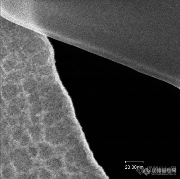
超高分辨率场发射扫描电镜,日本电子,欢迎有需要求的测试,量大优惠大[img]https://ng1.17img.cn/bbsfiles/images/2024/03/202403041546441431_3168_4087606_3.png[/img]
[em02] 由国家环境分析测试中心承建的二垩英实验室的主要设备,高分辨率质谱已经吊装完毕拉!一台将近1.4吨重的东西被顺利的吊上了三楼的实验室,真是不容易呀![em02]
2015年8月,台式电镜的领导者韩国COXEM(库赛姆)继2014年发布的EM-30 台式扫描电之后,今年再推出EM-30 Plus系列超高分辨率台式扫描电镜,将台式电镜的分辨率提高到了优于5nm分辨率的极限,可以与传统大型扫描电镜相媲美;同时设备配置了二次电子及背散射电子探测器,将台式电镜可对样品进行表面形貌分析、元素衬度分析以及二者相结合分析的技术发挥到了极致。COXEM(库赛姆)EM-30 Plus产品技术亮点:l 超高分辨率(5.0nm@30kV SE);l 更大的放大倍数x 150,000;l 更加简单易用的导航模式,更加快速的操作模式;l 同时刻进行二次电子像和背散射电子像收集;l 更加先进的低真空模式;l 全新升级的分析软件,舒适通用;l 从1kV-30kV同标准电镜相同的全加速电压量程;1、业界最高的分辨率COXEM(库赛姆)EM-30 Plus系列高分辨率台式(桌面式)扫描电镜打破了传统台式扫描电镜采用BSD探测器成像的局限性,利用创新的双聚光镜成像技术,采用大型扫描电镜成像方式,使用二次电子探测器作为基础成像单元,从而可以获得更高的分辨率(5nm),图像表面信息更丰富细腻,是真正意义上的高分辨率台式扫描电镜。2、同时刻进行二次电子像和背散射电子像收集 COXEM(库赛姆)EM-30 Plus系列台式电镜标配二次电电子(SE)+可伸缩式背散射电子(BSE)探测器,可以同时刻进行二次电子像和背散射电子像收集,既能实现对样品形貌衬度分析、元素衬度进行分析,又能实现将二者相结合进行分析,打破了传统台式电镜只能得到样品单一衬度像的瓶颈。COXEM(库赛姆)EM-30 Plus标配的可伸缩式背散射电子(BSE)可以非常有效的保护探测器不受损伤,更加体现了该款设备贴心的设计。同时该系列台式电镜也可选配EDS探测器(能谱仪),快速实现对材料微区化学成分进行定性及定量分析,是一款高信价比的微观分析设备。3、更加简单易用的导航模式,更加快速的操作模式 全新NanoStation™ 3.0软件系统,具有更加简单易用的导航模式,您只需要3步,首先对样品进行导航,然后设置成像条件。接下来对样品感兴趣的区域进行优化并自动采集图像。最后将结果可视化,而这些操作只需通过点击鼠标就可实现,为您高倍下快速寻找观测目标这个实际问题提供了完美的解决方案。
交叉极化为何可以提高分辨率?它的原理是什么?
本人目前正准备一项目,欲改良,开发设计一高分辨率的XRF仪器,能够实现对湿沉积物样品的直接测试,并且能够附加X射线照相\颜色反射率\密度等设备.急需要精通XRF研发技术的人员共同合作,有志者请与本人联系,共同探讨.可直接与本人联系,邮件地址:yangqh@gig.ac.cn
高分辨率质谱有什么优势?只是相邻的峰之间分离度高了吗?
[color=#444444]大家有没有遇到过 高分辨率质谱出现比分子离子峰的分子量还大的峰?[/color][color=#444444]这三个峰每相邻都差大概28,算了一下明显不是2M峰,【M+1】为479.9159, 3个大峰是683.5448, 711.5760, 739.6082.[/color][color=#444444]结构式一个喹啉环再连上一个 I 和1个1,2-二硫环戊4-烯-3-硫酮的片段。[/color][color=#444444]具体结构就不画啦![/color][color=#444444]谢谢大家啦[/color]
哪位大侠可以帮我解释一下,FFT补零对频率分辨率有影响吗,可以提高分辨率吗?
现在的科学技术发展迅速,也不知道现在最高分辨率的天平是多少?
失眠,无聊上网,看到最近石棉的帖子很多,顺便来转发一张石棉的图片。据说分辨率超高的哦,不清楚大家可以下载了再看图片![img]http://ng1.17img.cn/bbsfiles/images/2008/11/200811250212_120268_1602045_3.jpg[/img]
场发射透射电镜与高分辨率透射电镜有什么区别? 是不是场发射电镜的照片更清晰?急盼各位老师指点!!!!!!!!
请问api4000 [url=https://insevent.instrument.com.cn/t/Yp][color=#3333ff]液质联用仪[/color][/url]是高分辨率质谱吗
美国科学家称,利用世界上最先进的高分辨率光学显微镜,他们观察到了H2AX蛋白质在细胞核内的团状分布情况,以及DNA受损后它们如何移动到所需地方对基因进行“急救”或修复。 目前,有许多生物过程都是无法用视觉观察到的,原因是高分辨率电子显微镜常常因样品制备问题出现偏差,而光学显微镜虽然容易制备且能观察活细胞,但其分辨率却比较低。然而,通过对光波进行适当的操作,生物科学家扩展了光学显微镜的能力,成功地研制出4Pi显微镜,并通过它观察到了细胞的成分,其中包括细胞核的内部结构。 在新出版的美国《国家科学院学报》上,美国杰克逊实验室分子生物物理学所研究人员乔尔格• 毕瓦斯多夫及其合作者联合发表文章介绍说,借助4Pi光学显微镜,他们观察到了DNA双螺旋结构断裂情况下细胞的反应,并发现了DNA双螺旋结构断裂(即遗传物质严重受损)后引发的细胞内H2AX蛋白质一系列验证和修复损伤动作。如果细胞成分在修复过程中出现缺陷,则存在着发生癌症和免疫问题的危险,因此细胞内的反应十分重要。 H2AX是一种组蛋白。作为结构蛋白质,它们能缠绕在受损的DNA上,同时它们具有基因管理和基因修复的功能。H2AX在DNA受损后能快速做出反应,转变成γ-H2AX,这对协调发信号和修复等极其重要。 利用选择性着色技术和4Pi显微镜,毕瓦斯多夫还观察到H2AX组蛋白成团状均匀地分布在细胞核内。他认为,这种团状结构或许决定了DNA发生断裂时,γ-H2AX进行对应扩散的边界。 毕瓦斯多夫说:“H2AX团状分布也许为迅速和有效地应对DNA受损提供了平台。下一步,我们将分析H2AX团的位置及与其他细胞核成分的关系。”
[color=#444444]西服碱做高分辨率质谱用什么离子源[/color]
最开始调tune时,低、中、高分辨率状态都良好。而后做样时,开始的一个空白和前两个标准的峰和结果也都正常,但之后标准低分辨率元素的峰和结果都显示不了,中高分辨率正常。重新调tune,发现低分辨率已经扫不出来峰,或者呈现几条很乱的杂线,中高分辨率依旧正常。后用中高分辨率分别扫了li-in-u等元素,能出现较好的峰。请各位大神帮忙找找原因。十分感谢!
请问:耶拿-PQ 9000高分辨率电感耦合等离子体光谱仪怎么样,有用过的吗?故障率高吗,都表现在哪些方面?
光谱仪色散率和分辨率的不足,导致某些共存元素谱线之间重叠,采用高分辨率的分光系统,是否就意味着可以完全消除这类光谱干扰?现在光谱仪如ICP-OES在分辨率上都是如何做的?大家结合实际测试经验谈谈?
海洋光学推出了新款小型近红外光谱仪NIRQuest512-1.9 。这款高分辨率近红外光谱仪NIRQuest512-1.9的响应范围可达1100-1900纳米,从粮食生产和化学处理的变化监测到为半导体装配和医疗进行激光特征分析,该光谱仪可应用于各种领域。http://halmapr.com/news/halmacn/files/2012/09/nirquest512_1_9_blog.jpg
[color=#444444]求助!我最近测试了太赫兹时域光谱,只得到了时间和电场强度的数据,请问如何处理成折射光谱和吸收光谱的数据?[/color]
分子级高分辨率的激光扫描共焦显微镜和结构照明显微镜是在细胞生物学和其他相关领域强有力的研究工具,但是它们高昂的价格也使很多潜在用户望而却步。波士顿大学的科学家最近开发出一种显微新技术 (HiLo Microscopy),能够将普通的广域荧光显微镜变成可与激光扫描共焦显微镜和结构照明显微镜相媲美的高分辨率生物显微镜。这一技术包括一个简单的可以在均衡光源和结构光源之间自由转换的显微镜附件和一套功能强大的图像处理软件。该软件仅通过处理在均衡光源和结构光源条件下拍摄的两张分辨率不同的照片就可以得到全分辨率的三维图像。这一技术可用于任何现有的广域荧光显微镜,而成本大大低于激光扫描共焦显微镜和结构照明显微镜。由于成像机理简单,该技术的成像速度是常用的生物显微技术中最快的,而且操作简便,不受样本移动的影响。波士顿大学目前正在积极寻求企业合作,争取早日将这一突破性的技术推向市场。

http://ng1.17img.cn/bbsfiles/images/2012/09/201209301057_393843_1641557_3.jpg海洋光学高分辨率近红外光 谱仪
[align=center][font='times new roman'][size=16px][b]超高分辨[/b][/size][/font][font='times new roman'][size=16px][b]显微镜及其在生物医学领域的应用[/b][/size][/font][/align][align=center][font='times new roman'][size=14px]刘皎[/size][/font][font='times new roman'][sup][size=14px]1[/size][/sup][/font][font='times new roman'][size=14px],[/size][/font][font='times new roman'][sup][size=14px] [/size][/sup][/font][font='times new roman'][size=14px]吴晶[/size][/font][font='times new roman'][sup][size=14px]1[/size][/sup][/font][/align][align=center]1. [font='times new roman']北京大学医药卫生分析中心,北京,[/font][font='times new roman']100191[/font][/align][font='times new roman'][b]摘要[/b][/font][font='times new roman'][b] [/b][/font][font='times new roman']超高分辨显微镜([/font][font='times new roman']Super-Resolution Microscopy[/font][font='times new roman'])作为一类强大的科学工具,可以突破传统光学显微镜的分辨极限,实现对微小结构的高分辨率成像,已经在生物医学领域引起了广泛的关注和应用。本文将探讨超高分辨显微镜的不同类型和原理,介绍[/font][font='times new roman']其[/font][font='times new roman']在生物医学领域的应用[/font][font='times new roman']及展望其未来发展[/font][font='times new roman']。[/font][font='times new roman'][b]Abstract[/b][/font][font='times new roman']Super Resolution Microscopy[/font][font='times new roman'], as a powerful scientific tool, can break through the resolution limit of traditional optical microscopes and achieve high-resolution imaging of small structures. It has attracted widespread attention and application in the biomedical field. This article will explore the different types and principles of Super Resolution Microscopy, introduce their applications in the biomedical field, and look forward to their future development[/font][font='times new roman'].[/font][font='times new roman'][b]关键词[/b][/font][font='times new roman']超高分辨[/font][font='times new roman']显微镜,[/font][font='times new roman']成像技术[/font][font='times new roman'],应用[/font][font='times new roman'][b]1 [/b][/font][font='times new roman'][b]引言[/b][/font][font='times new roman']显微镜的产生和发展对于生命科学研究的进步有至关重要的作用[/font][font='times new roman'],它将微观世界呈现在大家面前,包括微生物的存在、组织细胞结构及生理病理活动等。显微镜技术的不断革新将成像分辨率不断提高,但相当长一段时间内光学成像无法突破一个极限值,即[/font][font='times new roman']xy[/font][font='times new roman']轴横向分辨率约[/font][font='times new roman']200nm[/font][font='times new roman'],[/font][font='times new roman']z[/font][font='times new roman']轴纵向分辨率约[/font][font='times new roman']500nm[/font][font='times new roman'],因此小于这个尺寸的生命活动和结构[/font][font='times new roman'],如病毒、亚细胞结构等,[/font][font='times new roman']是无法清楚地观察到的[/font][font='times new roman']。[/font][font='times new roman']聚焦点的光强会根据点扩散函数([/font][font='times new roman']point spread functio[/font][font='times new roman']n[/font][font='times new roman'],[/font][font='times new roman']PSF[/font][font='times new roman'])而展开[/font][font='times new roman'],[/font][font='times new roman']对于圆形孔径,[/font][font='times new roman']PSF[/font][font='times new roman']呈现为艾里斑([/font][font='times new roman']Airy disk[/font][font='times new roman'])的模式[/font][font='times new roman']。[/font][font='times new roman']激光扫描共聚焦显微镜([/font][font='times new roman']Confocal Laser Scanning Microscopy, CLSM[/font][font='times new roman'])的分辨率取决于[/font][font='times new roman']PSF[/font][font='times new roman']的大小,如果焦点很小,则每个像素[/font][font='times new roman']点[/font][font='times new roman']获取到的信息也很小,从而得到清晰锐利的图像;反之,则结果图像变得模糊。因此,[/font][font='times new roman']CLSM[/font][font='times new roman']成像的[/font][font='times new roman']主要挑战在于实现越来越小的[/font][font='times new roman']PSF[/font][font='times new roman']以获得更好的分辨率。德国物理学家恩斯特[/font][font='times new roman'][/font][font='times new roman']阿贝([/font][font='times new roman']Ernst Abbe[/font][font='times new roman'],[/font][font='times new roman']1840-1905[/font][font='times new roman']年)在[/font][font='times new roman']19[/font][font='times new roman']世纪[/font][font='times new roman']70[/font][font='times new roman']年代首次[/font][font='times new roman']提出阿贝衍射极限,即[/font][font='times new roman']由于衍射效应,[/font][font='times new roman']PSF[/font][font='times new roman']大[/font][font='times new roman']小与[/font][font='times new roman']λ/NA[/font][font='times new roman']成正比([/font][font='times new roman']d=0.61λ/NA[/font][font='times new roman']),其中[/font][font='times new roman']λ[/font][font='times new roman']是光的波长,[/font][font='times new roman']NA[/font][font='times new roman']是物镜最重要的参数[/font][font='times new roman']——[/font][font='times new roman']数值孔径[/font][font='times new roman']。由于可见光波长范围在[/font][font='times new roman']400-760nm[/font][font='times new roman']之间,[/font][font='times new roman']NA[/font][font='times new roman']值最大在[/font][font='times new roman']1.7[/font][font='times new roman']左右,所以分辨率极限在[/font][font='times new roman']200nm[/font][font='times new roman']左右。随着物理学和测量技术的进步,突破衍射极限的显微镜不断涌现,目前公认的超高分辨显微镜主要有三类,包括[/font][font='times new roman']结构照明显微镜([/font][font='times new roman']Structured Illumination Microscopy[/font][font='times new roman'],[/font][font='times new roman']SIM[/font][font='times new roman'])[/font][font='times new roman'],受激发射减耗显微镜([/font][font='times new roman']Stimulated Emission Depletion Microscopy[/font][font='times new roman'],[/font][font='times new roman']STED[/font][font='times new roman']),和[/font][font='times new roman']单分子定位显微镜。单分子定位显微镜包括光敏定位显微镜([/font][font='times new roman']Photoactivation Localization Microscopy[/font][font='times new roman'],[/font][font='times new roman']PALM[/font][font='times new roman'])和随机光学重建显微镜([/font][font='times new roman']Stochastic Optical Reconstruction Microscopy[/font][font='times new roman'],[/font][font='times new roman']STORM[/font][font='times new roman'])[/font][font='times new roman']。[/font][font='times new roman']2014[/font][font='times new roman']年三位科学家[/font][font='times new roman']史蒂芬[/font][font='times new roman'][/font][font='times new roman']霍尔([/font][font='times new roman']Stefan W. Hell[/font][font='times new roman'])[/font][font='times new roman']、埃里克[/font][font='times new roman'][/font][font='times new roman']贝兹([/font][font='times new roman']Eric Betzig[/font][font='times new roman'])和威廉[/font][font='times new roman'][/font][font='times new roman']莫纳([/font][font='times new roman']William E. Moerner[/font][font='times new roman'])因他们在超[/font][font='times new roman']高[/font][font='times new roman']分辨显微镜技术领域的贡献而获得了诺贝尔化学奖。[/font][font='times new roman'][b]2 [/b][/font][font='times new roman'][b]不同类型的超高分辨显微镜[/b][/font][font='times new roman'][b]2.1[/b][/font][font='times new roman'][b] [/b][/font][font='times new roman'][b]结构照明显微镜([/b][/font][font='times new roman'][b]Structured Illumination Microscopy[/b][/font][font='times new roman'][b],[/b][/font][font='times new roman'][b]SIM[/b][/font][font='times new roman'][b])[/b][/font][font='times new roman']SIM[/font][font='times new roman']本质是利用两束激发光在样品上进行干涉,产生明暗交替的莫尔条纹,高空间频率的莫尔条纹会放大激发条纹与样品空间频率不一致的结构,从而将样品中的高频信息整合入收集到的图像中。[/font][font='times new roman']通过投射特殊的光照明模式如格点或条纹光栅,以一定的模式照射样品,引入空间频率信息,采集多个图像并经过复杂的数据处理之后,重建高分辨率图像。由于每个图像都采用不同的结构照明模式,包含了不同的信息,合并后的图像能够展示出比传统显微镜更多的细节[/font][font='times new roman']。[/font][font='times new roman']相比于其他超高分辨成像技术,[/font][font='times new roman']SIM[/font][font='times new roman']最大的优势就是宽场[/font][font='times new roman']成像,速度快,基本可以达到实时观察。[/font][font='times new roman']SIM[/font][font='times new roman']技术的前身可以追溯到[/font][font='times new roman']20[/font][font='times new roman']世纪[/font][font='times new roman']70[/font][font='times new roman']年代初。当时,光学学家特奥多尔[/font][font='times new roman'][/font][font='times new roman']赫普恩([/font][font='times new roman']Theodor [/font][font='times new roman']H?upl[/font][font='times new roman'])首次提出了使用周期性光栅照明来提高显微镜分辨率的想法。这奠定了[/font][font='times new roman']SIM[/font][font='times new roman']技术的基础,尽管当时还没有实际的[/font][font='times new roman']SIM[/font][font='times new roman']显微镜。[/font][font='times new roman']21[/font][font='times new roman']世纪初期,史蒂芬[/font][font='times new roman'][/font][font='times new roman']霍尔([/font][font='times new roman']Stefan W. Hell[/font][font='times new roman'])和埃里克[/font][font='times new roman'][/font][font='times new roman']贝兹([/font][font='times new roman']Eric Betzig[/font][font='times new roman'])等科学家分别独立开发了[/font][font='times new roman']SIM[/font][font='times new roman']的现代版本。[/font][font='times new roman']SIM[/font][font='times new roman']技术开始广泛传播,吸引了生物学家和显微镜专家的关注。它被认为是一种相对低成本的[/font][font='times new roman']超高分辨[/font][font='times new roman']率成像方法,因为它不需要昂贵的激光设备或复杂的样品准备。[/font][font='times new roman'][b]2.2 [/b][/font][font='times new roman'][b]受激发射减耗[/b][/font][font='times new roman'][b]显微镜([/b][/font][font='times new roman'][b]Stimulated Emission Depletion Microscopy[/b][/font][font='times new roman'][b],[/b][/font][font='times new roman'][b]STED[/b][/font][font='times new roman'][b])[/b][/font][font='times new roman']STED[/font][font='times new roman']技术的概念最早由斯德哥尔摩大学的斯蒂芬[/font][font='times new roman'][/font][font='times new roman']霍尔([/font][font='times new roman']Stefan W. Hell[/font][font='times new roman'])提出。他的想法是通过将激发光束与一个特殊的抑制光束结合,从而实现对荧光标记物的光抑制,[/font][font='times new roman']通过受激辐射淬灭光斑外围的荧光分子,[/font][font='times new roman']使其在空间上变得更加紧凑,[/font][font='times new roman']减少[/font][font='times new roman']PSF[/font][font='times new roman']从而提高分辨率。[/font][font='times new roman']我们也叫“甜甜圈”技术。[/font][font='times new roman']STED[/font][font='times new roman']显微镜背后基本思想就是利用非线性光学设计一个低于阿贝衍射极限的更小[/font][font='times new roman']PSF[/font][font='times new roman']。[/font][font='times new roman']分辨率与[/font][font='times new roman']STED[/font][font='times new roman']光强有关,提高[/font][font='times new roman']STED[/font][font='times new roman']光的强度可以使荧光光斑焦[/font][font='times new roman']点中心直径趋于[/font][font='times new roman']0[/font][font='times new roman'],但是实际应用中,光损伤较大,[/font][font='times new roman']STED[/font][font='times new roman']光强不可能无限增加,顾[/font][font='times new roman']其分辨率[/font][font='times new roman']最高[/font][font='times new roman']可达到[/font][font='times new roman']3[/font][font='times new roman']0[/font][font='times new roman']nm[/font][font='times new roman']左右[/font][font='times new roman']。[/font][font='times new roman']目前的[/font][font='times new roman']STED[/font][font='times new roman']只能应用于较薄的组织器官或细胞,光毒性较强,成像厚度有限不太适合活体或活细胞长时间成像。[/font][font='times new roman']STED[/font][font='times new roman']光路较为复杂,对系统稳定性要求较高。[/font][font='times new roman'][b]2.3 [/b][/font][font='times new roman'][b]单分子定位显微镜[/b][/font][font='times new roman']单分子定位显微镜[/font][font='times new roman']中荧光标记的单个分子被分别激发和检测。单分子的中心可以以极高的精度确定从而实现高分辨率,包括光敏定位显微镜([/font][font='times new roman']Photoactivation Localization Microscopy[/font][font='times new roman'],[/font][font='times new roman']PALM[/font][font='times new roman'])和随机光学重建显微镜([/font][font='times new roman']Stochastic Optical Reconstruction Microscopy[/font][font='times new roman'],[/font][font='times new roman']STORM[/font][font='times new roman'])。[/font][font='times new roman']PALM[/font][font='times new roman']的历史可以追溯到[/font][font='times new roman']2006[/font][font='times new roman']年,由埃里克[/font][font='times new roman'][/font][font='times new roman']贝兹([/font][font='times new roman']Eric Betzig[/font][font='times new roman'])和哈拉尔德[/font][font='times new roman'][/font][font='times new roman']赫斯([/font][font='times new roman']Harald Hess[/font][font='times new roman'])提出了单分子定位这一概念。在[/font][font='times new roman']PALM[/font][font='times new roman']中,样品中的分子被标记上特定的荧光染料。这些染料可以通过光激活从一个基态转变到一个激发态,此过程可通过使用激活光(通常是紫外光)来实现。同期[/font][font='times new roman']STORM[/font][font='times new roman']的成像技术也发展起来,代表科学家是华人庄小威。[/font][font='times new roman']STORM[/font][font='times new roman']的工作原理与[/font][font='times new roman']PALM[/font][font='times new roman']类似,是通过特殊的分子标记和随机活性化,实现单分子定位进而实现超高分辨率成像。具有光激活能力的标记物通常在某种光照条件下会发光,但也会在某一时刻被随机地熄灭。这种随机光熄灭是[/font][font='times new roman']PALM[/font][font='times new roman']技术的关键,因为它允许在不同时间点捕获标记物的位置。通过记录标记物的位置,可以得到它们的坐标。这一过程需要在短时间内多次拍摄样品,以获得足够多的数据点。最后,通过将多个标记物的坐标叠加在一起,可以生成高分辨率的图像。这种以成像时间换取空间分辨率的形式,使得[/font][font='times new roman']PALM[/font][font='times new roman']或[/font][font='times new roman']STORM[/font][font='times new roman']的分辨率通常能够达到数十纳米。[/font][font='times new roman'][b]3 [/b][/font][font='times new roman'][b]应用领域和未来发展[/b][/font][font='times new roman']超高分辨显微镜可以探索微观世界的无限可能性,已经彻底改变了科学研究的方式。在细胞生物学领域,它被用于研究[/font][font='times new roman']亚细胞结构,如微丝、微管、肌动蛋白等,[/font][font='times new roman']细胞器[/font][font='times new roman']如线粒体、溶酶体等,[/font][font='times new roman']分子分布和细胞膜动态、观察蛋白质的相互作用;在神经科学领域,它可用于观察神经元的亚细胞结构和突触的细节,有助于解剖和理解神经系统的结构和功能,以及神经系统相关疾病的机制;在癌症研究领域,被用于研究癌细胞的特征、蛋白质分布以及肿瘤微环境,这对于癌症的早期诊断和治疗规划非常重要;在材料科学领域,它被用于研究纳米材料的结构和性质、帮助科学家精确控制和制备纳米结构;在药物研发领域,它可用于研究药物靶标蛋白的定位和与其他分子的相互作用,这对于药物设计和筛选非常重要[/font][font='times new roman'];在微生物领域,对于研究细菌[/font][font='times new roman']结构变化至关重要,规避了电子显微镜无法进行活体成像等弊端,可以更加推进微生物学发展。[/font][font='times new roman']当然,[/font][font='times new roman']超[/font][font='times new roman']高[/font][font='times new roman']分辨成像技术[/font][font='times new roman']也有一定的挑战。超高分辨成像技术[/font][font='times new roman']通常需要高度复杂的设备和精密的校准,这使得其设备成本相对较高,[/font][font='times new roman']再加上样本制备的困难,[/font][font='times new roman']限制了其广泛应用。[/font][font='times new roman']样品准备在超高分辨成像中具有重要作用,新的标记技术和荧光探针的发展将提高成像的灵敏度和特异性[/font][font='times new roman'],[/font][font='times new roman']开发更友好、无损伤的样品准备方法,以减少对样品的干扰[/font][font='times new roman'],[/font][font='times new roman']甚至[/font][font='times new roman']包括无标记成像技术以减少样品标记的需求。开源软件和自动化工作流程将使超高分辨成像技术更易于使用和共享,促进科学研究的进展。[/font][font='times new roman']超高分辨技术通常对于三维成像和大样本的深度成像有限制,需要克服分辨率和深度之间的权衡。[/font][font='times new roman']同时超高分辨[/font][font='times new roman']成像的时间分辨率还可以继续提升[/font][font='times new roman']。[/font][font='times new roman']虽然[/font][font='times new roman']目前[/font][font='times new roman']SIM[/font][font='times new roman']和[/font][font='times new roman']minflux[/font][font='times new roman']更适合[/font][font='times new roman']观察[/font][font='times new roman']活细胞[/font][font='times new roman']动态过程,但时间分辨率的提高仍然是一个挑战,特别是对于极短时间尺度的现象[/font][font='times new roman'],[/font][font='times new roman']这将使科学家能够更深入地探索微观世界,并获得更多信息。[/font][font='times new roman']随着技术的不断进步,[/font][font='times new roman']超高分辨[/font][font='times new roman']成像有望在[/font][font='times new roman']包括临床医学[/font][font='times new roman']等[/font][font='times new roman']更多领域得到广泛应用[/font][font='times new roman'],未[/font][font='times new roman']来如果能实现超高分辨的动物甚至人的[/font][font='times new roman']活体成像,减少样品固定和处理的需求,允许观察生物过程的实时发生[/font][font='times new roman']将会更有现实意义[/font][font='times new roman']。[/font][font='times new roman']并且在科学研究的需求下,[/font][font='times new roman']多模态[/font][font='times new roman']或多尺度[/font][font='times new roman']成像将[/font][font='times new roman']与[/font][font='times new roman']不同[/font][font='times new roman']的[/font][font='times new roman']超高分辨[/font][font='times new roman']技术相结合,[/font][font='times new roman']例如,结合光学成像和质谱成像[/font][font='times new roman'],[/font][font='times new roman']从分子水平到组织水平[/font][font='times new roman']提供[/font][font='times new roman']生命活动[/font][font='times new roman']更全面的信息。[/font][font='times new roman']也可以[/font][font='times new roman']发展高通量的样品处理和成像技术,以便更快速地获得大规模的数据。[/font][font='times new roman']超高分辨[/font][font='times new roman']成像生成的数据量巨大,处理和分析这些大数据需要强大的计算资源和高效的算法。数据存储和传输也是挑战。[/font][font='times new roman']超高分辨[/font][font='times new roman']成像数据可能受到噪声和伪迹的影响,这需要高级的图像处理技术来减少其影响,以获得准确的图像。数据分析通常需要复杂的算法和数学模型,需要专业知识和技能。对于某些应用,如神经科学中的活体成像,需要实时数据分析,这增加了挑战。深度学习和人工智能技术[/font][font='times new roman']有望[/font][font='times new roman']在数据分析中发挥越来越重要的作用,[/font][font='times new roman']实现[/font][font='times new roman']自动处理和解释图像数据。发展实时数据分析技术,使科学家能够在数据采集过程中获得及时反馈。开发更易用的高级图像处理工具,使非专业用户也能够进行数据分析。结合不同成像技术和数据源的信息,以提供更全面的信息。开发自动化和高通量的数据分析工作流程,以应对大规模数据的挑战。促进数据共享和开放科学,以促进合作和加速科学研究的进展。未来,随着计算能力的提高和新技术的引入,[/font][font='times new roman']超高分辨[/font][font='times new roman']成像数据分析将变得更加强大和高效。这将有助于更深入地理解微观世界,并在生物学、医学、材料科学等领域推动创新和发展。[/font][font='times new roman']总的来说,尽管[/font][font='times new roman']超高分辨[/font][font='times new roman']成像面临一些挑战,但其前景充满希望。未来的发展将使这一领域更加强大,有望在科学研究和实际应用中提供更多的机会和洞察力。[/font][font='times new roman'][b]4 [/b][/font][font='times new roman'][b]结论和展望[/b][/font][font='times new roman']超高分辨显微镜的成像原理基于破解传统显微镜的分辨极限,通过结构照明、图像重建[/font][font='times new roman']和单分子成像等策略,实现对微小结构的高分辨率成像。这一技术的应用领域包括生物学、材料科学、纳米技术和医学等,有望推动科学研究的进一步发展。超高分辨显微镜已经在生物医学领域取得了显著的突破,使研究人员更深入地理解细胞和分子结构。然而,仍然存在挑战,包括样品准备和数据分析的复杂性。未来,我们可以期待更多技术的发展,以进一步提高分辨率和扩大应用领域。[/font][font='times new roman']随着技术的不断发展,我们可以期待更多超分辨显微镜技术的突破,如更高分辨率、更高灵敏度和更快成像速度。超分辨显微镜的应用也将继续扩展到新的领域,如药物研发、个性化医学和环境科学。它将为我们提供更多工具来解决生物学的重要问题,如疾病机制、药物研发和生态系统健康。总之,超分辨显微镜技术的未来展望是光明的,它将继续推动科学研究向前迈进,揭示微观世界的微小奥秘,为改善生活质量和解决全球挑战做出贡献。这个领域的不断创新将激发更多科学家的热情,共同追求更深入的科学知识和更广泛的应用。[/font][font='times new roman'][b]参考文献[/b][/font][font='times new roman']Hell[/font][font='times new roman'] [/font][font='times new roman']S [/font][font='times new roman']W[/font][font='times new roman'].[/font][font='times new roman']Far-field[/font][font='times new roman'] [/font][font='times new roman']optical[/font][font='times new roman'] [/font][font='times new roman']nanoscopy[/font][font='times new roman'][J][/font][font='times new roman'].[/font][font='times new roman']Science[/font][font='times new roman'],[/font][font='times new roman']2007[/font][font='times new roman'],[/font][font='times new roman']316(5828)[/font][font='times new roman']:[/font][font='times new roman']1153-1158[/font][font='times new roman']Hell[/font][font='times new roman'] [/font][font='times new roman']S W[/font][font='times new roman'],[/font][font='times new roman']Wichmann J[/font][font='times new roman'].[/font][font='times new roman']Breaking[/font][font='times new roman'] [/font][font='times new roman']the diffraction[/font][font='times new roman'] [/font][font='times new roman']resolution[/font][font='times new roman'] [/font][font='times new roman']limit[/font][font='times new roman'] [/font][font='times new roman']by stimulated[/font][font='times new roman']-[/font][font='times new roman']emission[/font][font='times new roman']-[/font][font='times new roman']depletion fluorescence[/font][font='times new roman'] [/font][font='times new roman']microscopy[J][/font][font='times new roman'].[/font][font='times new roman']Optics[/font][font='times new roman'] [/font][font='times new roman']Letters[/font][font='times new roman'],[/font][font='times new roman']1994[/font][font='times new roman'],[/font][font='times new roman']19(11)[/font][font='times new roman']:[/font][font='times new roman']780-782[/font][font='times new roman']Dani A[/font][font='times new roman'],[/font][font='times new roman']Huang B[/font][font='times new roman'],[/font][font='times new roman']Bergan J[/font][font='times new roman'],[/font][font='times new roman']et[/font][font='times new roman'] [/font][font='times new roman']a1[/font][font='times new roman'].[/font][font='times new roman'] Super-resolution[/font][font='times new roman'] [/font][font='times new roman']imaging of chemical synapses[/font][font='times new roman'] [/font][font='times new roman']in the brain[J][/font][font='times new roman'].[/font][font='times new roman']Neuron[/font][font='times new roman'],[/font][font='times new roman']2010[/font][font='times new roman'],[/font][font='times new roman']68(5)[/font][font='times new roman']:[/font][font='times new roman']843[/font][font='times new roman']—[/font][font='times new roman']856[/font][font='times new roman']PATTERSON[/font][font='times new roman'] [/font][font='times new roman']G[/font][font='times new roman'],[/font][font='times new roman']DAVIDSON[/font][font='times new roman'] [/font][font='times new roman']M[/font][font='times new roman'],[/font][font='times new roman']MANLEY[/font][font='times new roman'] [/font][font='times new roman']S[/font][font='times new roman'],[/font][font='times new roman']et[/font][font='times new roman'] [/font][font='times new roman']al[/font][font='times new roman']. [/font][font='times new roman']Superresolution[/font][font='times new roman'] imaging using single-molecule localization[/font][font='times new roman'][J][/font][font='times new roman'].[/font][font='times new roman']A[/font][font='times new roman']nnual Review of Chemistry[/font][font='times new roman'],[/font][font='times new roman']2010[/font][font='times new roman'],[/font][font='times new roman']6[/font][font='times new roman']1:345-367[/font]
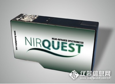
海洋光学高分辨率近红外光谱仪扩展了波长测量范围新款小型近红外光谱仪NIRQuest512-1.9 。这款高分辨率近红外光谱仪NIRQuest512-1.9的响应范围可达1100-1900纳米,从粮食生产和化学处理的变化监测到为半导体装配和医疗进行激光特征分析,该光谱仪可应用于各种领域。http://ng1.17img.cn/bbsfiles/images/2012/12/201212191311_413838_2432394_3.jpgNIRQuest512-1.9配置具有很高的稳定性,512像素Hamamatsu InGaAs线阵探测器,适用于多种光栅和光具座,用以优化1100至1900纳米之间的性能。标准的NIRQuest512-1.9光栅常数为150线/毫米,25微米的入射狭缝,以及一个非荧光长波通滤光器配置,可传输1000纳米以上的波长。该滤光器有助于缓和二阶效应。NIRQuest512-1.9外部配有一个硬件,通过该硬件,在出现外部情况时,用户可以通过外部触发获取相应数据信息,或者在数据获得之后再次引起触发。光谱仪操作通过SpectraSuite软件来控制,该软件是一个基于Java的模块化光谱学平台。NIRQuest的低沉噪声让其具备集成光谱仪的潜力(或者将光谱仪中的探测器暴露在光线下),从而延长使用时间,这在光线暗的环境中非常有用。满信号条件下的信噪比在每100毫秒积分时间内大于15000:1。因此,在对敏感性要求极高的应用环境中可以实现高效操作模式。
看书上说:不能通过增加光栅刻线密度来提高分辨率,为啥???
请教:在ICR质谱中分辨率与离子质量数的关系在离子回旋共振(ICR)质谱仪中,如果仪器状态较好时,在一般情况下小质量的离子可以得到超高分辨率(如m/z=100时分辨率可达1000000),但是在分辨能力会随质量数的增加线性下降,如m/z=10000时分辨率将会降低至10000。这是为什么?谢谢!


 我要推广仪器
 下载APP
下载APP