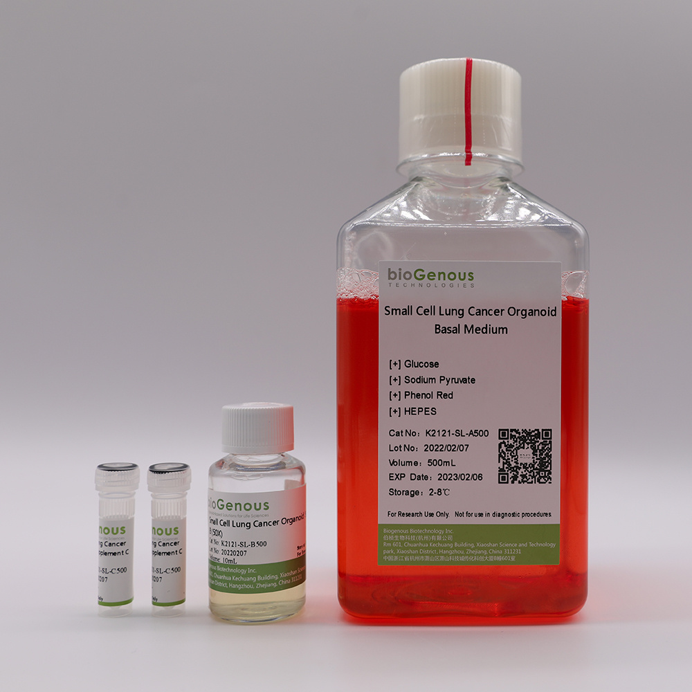
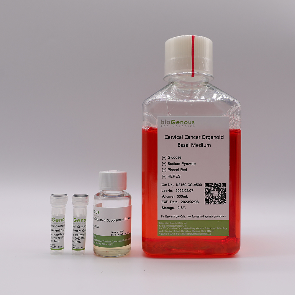
类器官Gastric Cancer Organoid Kit(胃癌)
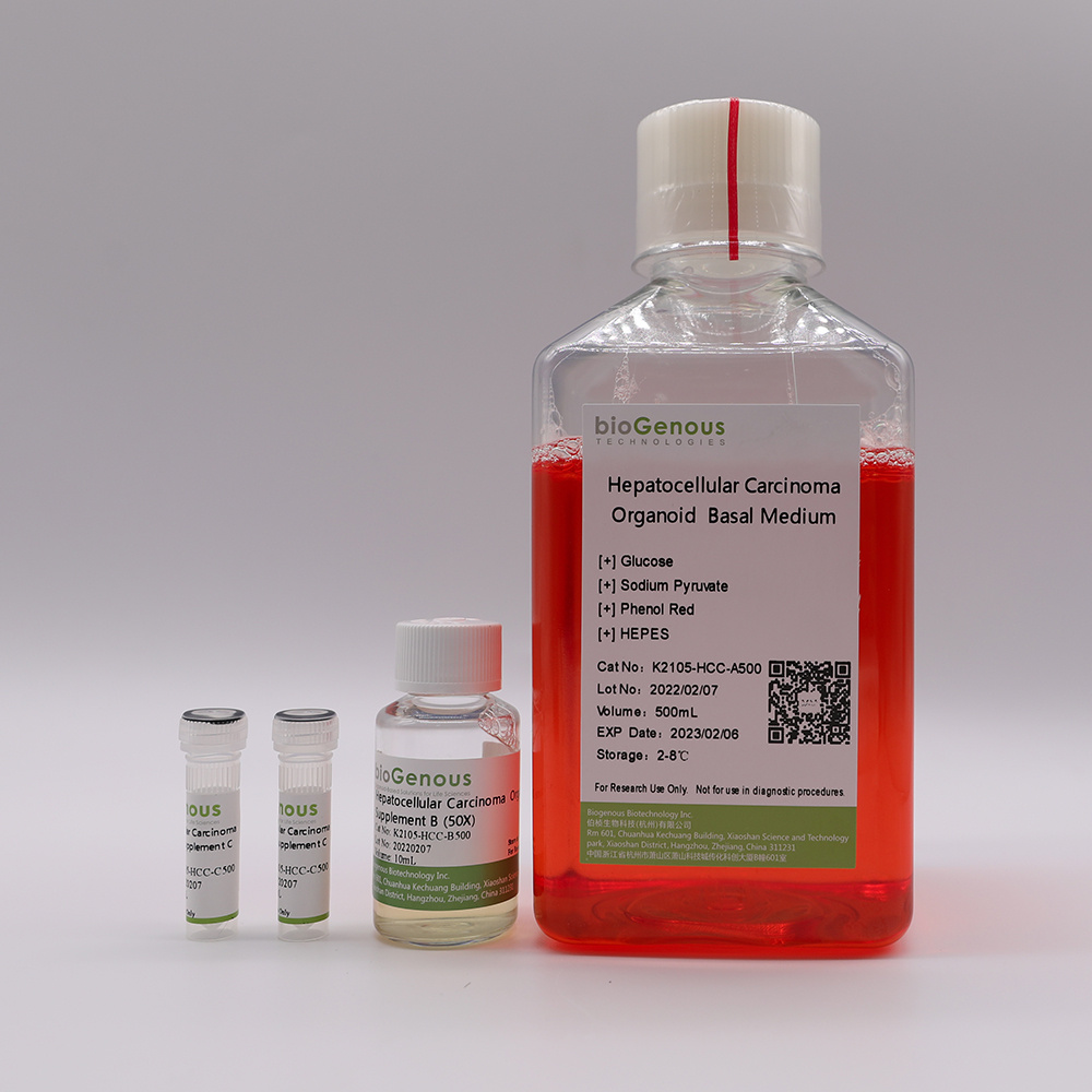
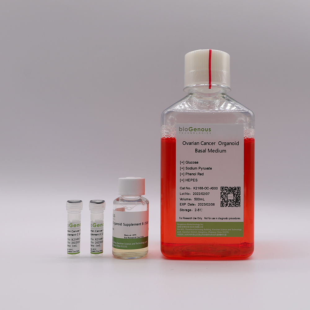
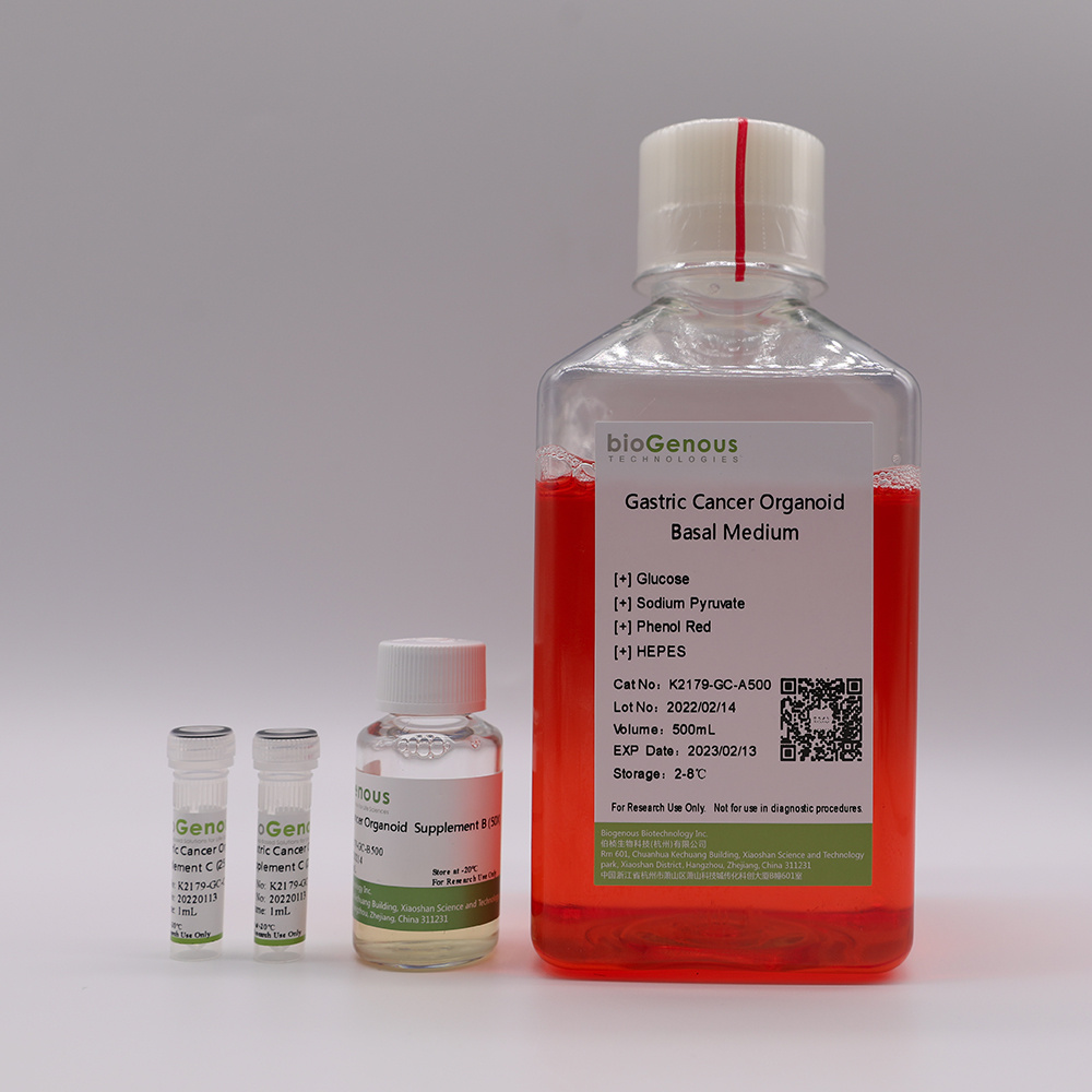
类器官(Organoids)是指将成体干细胞或多能干细胞在体外三维培养形成的具有一定空间结构的组织类似物。类器官在组织结构、细胞类型、自我更新能力和功能等方面与来源组织高度一致,从而在发育生物学、疾病造模、精准医学、药物研发、基因和细胞疗法、感染和免疫以及再生医学等生物医学的多个领域展现出独特的优势。产品介绍Product Description:bioGenousTMGastric Cancer Organoid Kit is a chemically defined cell culture medium for the establishment and maintenance of human gastric cancer organoid s. Patient-derived cancer organoids recapitulate the genomic and pathological features of original tumors and therefore hold great promise for medical research and precision medicine.技术参数Product Information:ComponentComponent Cat#VolumeStorage & StabilitybioGenousTMGastric Cancer Organoid Basal MediumK2179-GC-A100/A500100mL/500mL4℃,12 monthsbioGenousTMGastric Cancer Organoid Supplement B (50x)K2179-GC-B100/B5002mL/10mL-20℃,avoid repeated freeze-thaw cycles, 12 monthsbioGenousTMGastric Cancer Organoid Supplement C (250x)K2179-GC-C100/C5000.4mL/2mL-20℃, avoid repeated freeze-thaw cycles, 12 monthsPreparation of Gastric Cancer Organoid Complete MediumUse a sterile technique to prepare the gastric cancer organoid complete medium. The following example is for preparing 10mL complete medium. If preparing other volumes, adjust accordingly.1.Thaw Gastric Cancer Organoid Supplement B(50x) and Gastric Cancer Organoid Supplement C(250x) on ice. Mix thoroughly.NOTE:Once thawed, use immediately or aliquot and store at -20°C for not more than 10 months. After thawing the aliquots, use immediately. Do not re-freeze.2.Add 200μL Gastric Cancer Organoid Supplement B(50x) and 40μL Gastric Cancer Organoid Supplement C(250x) to 9.76mL Gastric Cancer Organoid Basal Medium. Mix thoroughly.NOTE:If not used immediately, store the complete medium at 2-8°C for not more than 2 weeks. The Gastric Cancer Organoid Supplement B contains fungicide and antibiotics (50x).Protocol for Establishment ofPatient-Derived Gastric Cancer OrganoidsCAUTIONStudies involving primary human tissue material must follow all relevant institutional and government regulations. Informed consent must be obtained from all subjects before the collection of the primary human tissue material.Establishment of Organoids from Primary Tissue1.Collect primary human gastric cancer tissue pieces in ice-cold Primary Tissue Storage Solution (K601005) with conical tubes. Keep tissue samples at 4°C until the start of the isolation.2.Assess whether the obtained tissue pieces consist purely of epithelium. If fat or muscle tissues are present, remove these non-epithelial components as much as possible using surgical scissors or scalpels and forceps under a dissection microscope. If no fat or muscle tissues are present, continue to the next step immediately.3.Rinse the tissuewith Cancer Organoid Basal Medium (B213152) or DPBS twice.4.Mince the tissue into small fragments of 1-3 mm3in a cell culture dish using surgical scissors or scalpels.5.Digest the tissue fragments with 10mL of Tumor Tissue Digestion Solution (K601003) in a 15mL conical tube at 37°C, with variable incubation times ranging from 30 min to 1 h. Carefully monitor the digestion process, mixing the content of the tube every 5-10 min by shaking vigorously or pipetting the mixture up and down. The digestion process could be finished when most of tissue fragments are able to pass through the 1mL pipette tips.6.Add FBS to the tissuedigestionmixture to a final concentration of 2%, and filter using a 100 μm cell strainer.7.Collect and centrifuge the filtered cells at 250g for 3 min at 4 °C. In case of a visible red pellet, aspirate the supernatant, and resuspend the pellet using 2mL of Red Blood Cell Lysis Solution (E238010) to lyse the erythrocytes at room temperature for 1 min and centrifuge at 250g for 3 min at 4°C.8.Aspirate the supernatant and resuspend the pellet in Cancer Organoid Basal Medium, centrifuge at 250g for 3 min at 4°C,repeat this step once more time.9.Aspirate the supernatant and resuspend the pellet in Matrigel. The Matrigel should be kept on ice to prevent it from solidifying. Perform the process as quickly as possible. The amount of Matrigel used depends on the size of the pellet. Approximately 10,000 cells should be plated in 25 μL of Matrigel.CRITICAL:Do not overly dilute the Matrigel (Matrigel should be 70% (Matrigel vol/Total vol)), as this may inhibit the proper formation of the solid droplets.10.Plate the Matrigel containing organoids at the bottom of 24-well cell culture plates in droplets of ~30μL each around the center of the well..CRITICAL:Once the organoids are resuspended in Matrigel, proceed with plating as quickly as possible, as the Matrigel may solidify in the tube or pipette tip. Do not let the Matrigel touch the wall of wells.11.Place the culture plate into a humidified incubator at 37 °C and 5% (vol/vol) CO2for15-25 minto let the Matrigel solidify.12.Prepare the required amount of gastric cancer organoid complete medium.13.Once the Matrigel droplets have solidified (15-25 min), open the plate and carefully add 500 μL of organoid complete medium to each well.CRITICAL:Do not add the medium directly on top of the Matrigel droplets, as this might disrupt the droplets.14.Place the culture plate in a humidified incubator at 37 °C and 5% (vol/vol) CO2.15.Change the medium every 3-4 d by carefully aspirating the medium from the wells and replacing it with a fresh, pre-warmed organoid complete medium.16.Closely monitor the organoid formation. Ideally, patient-derived gastric cancer organoids should be passaged for the first time between 7 and 10 d after the initial plating.Splitting and Passaging of Organoids17.Pipette up and down to disrupt the Matrigel and transfer the organoid suspension to a 1.5 mL conical tube.18.Pipette the organoid suspension up and down to mix thoroughly by pipetting against the bottom of the tube to create pressure, which will aid the removal of Matrigel.19.Centrifuge organoids at 250g for 3 min at room temperature.20.Aspirate the supernatant and split organoids using either Organoid Dissociation Solution (E238001) or by mechanical disruption. For Organoid Dissociation Solution-based cell dissociation, resuspend the pellet in 0.2 mL of Organoid Dissociation Solution, pipette up and down and incubate at 37 °C until organoids fall apart. Pipette up and down with a filter tip for ≥8 times every 2 min to aid in the disruption of the organoids. Closely monitor the digestion to keep the incubation time inthe Organoid Dissociation Solution to a minimum. In case of mechanical disruption, resuspend the pellet in 1.5 mL ofCancer Organoid Basal Medium. Carefully pipette the organoid suspension up and down 30 times by pipetting against the bottom of the tube to create pressure, which will aid organoid disruption.CRITICAL:Do not dissociate in Organoid Dissociation Solution for 5 min, as this may result in poor organoid outgrowth or even loss of the culture. As a rule of thumb, digestion is complete if a mixture of small lumps of cells (consisting of 10–50 cells) can be observed.21.After shearing is complete, wash once by topping up with 1 mL ofCancer Organoid Basal Medium, and centrifuge at 250g for 3 min at room temperature.22.Aspirate the supernatant and resuspend the organoid pellet in 70% (vol/vol) Matrigel, and plate organoids in droplets at the bottom of a culture plate as described in Steps 10. After plating is complete, transfer the plate into a humidified incubator at 37 °C and 5% (vol/vol) CO2for 15–25 min.23.Pre-warm gastric cancer organoid complete medium at 37 °C.24.After the Matrigel droplets have solidified (15–25 min), carefully pipette pre-warmed medium into the wells.25.Place culture plates in a humidified incubator at 37 °C and 5% (vol/vol) CO2until the organoids are needed for further experiments.


 我要推广仪器
 下载APP
下载APP