
 关注
关注
已关注

![]() 已认证
已认证
粉丝量 0
400-860-5168转4668
仪器信息网认证电话,请放心拨打
Anti-DAP Kinase 1 antibody
种属反应性Human,Mouse,Rat
验证应用WB,IHC-P,ICC,FC
抗体类型小鼠单抗
免疫原Recombinant protein within Human DAPK1 aa 400-670.
偶联Non-conjugated
Anti-DAP Kinase 1 antibody性能
形态Liquid
浓度2mg/ml.
存放说明Store at +4℃ after thawing. Aliquot store at -20℃. Avoid repeated freeze / thaw cycles.
存储缓冲液1*PBS (pH7.4), 0.2% BSA, 50% Glycerol. Preservative: 0.05% Sodium Azide.
亚型IgG1
纯化方式Protein A purified.
亚细胞定位Cytoplasm, Cytoskeleton.
其它名称
moreDAK1 antibody
DAP K1 antibody
DAP kinase 1 antibody
Anti-DAP Kinase 1 antibody应用
WB: 1:500-1:2,000
IHC-P: 1:50-1:200
ICC: 1:50-1:100
FC: 1:50-1:100
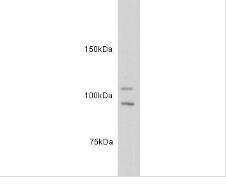
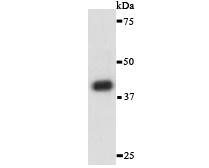
Fig1: Western blot analysis of DAP Kinase 1 on different lysates. Proteins were transferred to a PVDF membrane and blocked with 5% BSA in PBS for 1 hour at room temperature. The primary antibody was used in 5% BSA at room temperature for 2 hours. Goat Anti-Mouse IgG - HRP Secondary Antibody (HA1006) at 1:5,000 dilution was used for 1 hour at room temperature.
Positive control:
Lane 1: Human skin tissue lysate
Lane 2: Human skeletal muscle tissue lysate
Fig2: ICC staining of DAP Kinase 1 in MCF-7 cells (green). Formalin fixed cells were permeabilized with 0.1% Triton X-100 in TBS for 10 minutes at room temperature and blocked with 1% Blocker BSA for 15 minutes at room temperature. Cells were probed with the primary antibody for 1 hour at room temperature, washed with PBS. Alexa Fluor®488 Goat anti-Mouse IgG was used as the secondary antibody at 1/100 dilution. The nuclear counter stain is DAPI (blue).

Fig3: Immunohistochemical analysis of paraffin-embedded rat lung tissue using anti-DAP Kinase 1 antibody. The section was pre-treated using heat mediated antigen retrieval with Tris-EDTA buffer (pH 8.0-8.4) for 20 minutes.The tissues were blocked in 5% BSA for 30 minutes at room temperature, washed with ddH2O and PBS, and then probed with the primary antibody for 30 minutes at room temperature. The detection was performed using an HRP conjugated compact polymer system. DAB was used as the chromogen. Tissues were counterstained with hematoxylin and mounted with DPX.

Fig4: Immunohistochemical analysis of paraffin-embedded human lung cancer tissue using anti-DAP Kinase 1 antibody. The section was pre-treated using heat mediated antigen retrieval with Tris-EDTA buffer (pH 8.0-8.4) for 20 minutes.The tissues were blocked in 5% BSA for 30 minutes at room temperature, washed with ddH2O and PBS, and then probed with the primary antibody for 30 minutes at room temperature. The detection was performed using an HRP conjugated compact polymer system. DAB was used as the chromogen. Tissues were counterstained with hematoxylin and mounted with DPX.

Fig5: Immunohistochemical analysis of paraffin-embedded mouse liver tissue using anti-DAP Kinase 1 antibody. The section was pre-treated using heat mediated antigen retrieval with Tris-EDTA buffer (pH 8.0-8.4) for 20 minutes.The tissues were blocked in 5% BSA for 30 minutes at room temperature, washed with ddH2O and PBS, and then probed with the primary antibodfor 30 minutes at room temperature. The detection was performed using an HRP conjugated compact polymer system. DAB was used as the chromogen. Tissues were counterstained with hematoxylin and mounted with DPX.

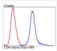
Fig6: Flow cytometric analysis of DAP Kinase 1 was done on MCF-7 cells. The cells were fixed, permeabilized and stained with the primary antibody ( (red). After incubation of the primary antibody at room temperature for an hour, the cells were stained with a Alexa Fluor 488-conjugated goat anti-mouse IgG Secondary antibody at 1/500 dilution for 30 minutes.Unlabelled sample was used as a control (cells without incubation with primary antibody; black).
特别提示:本公司的所有产品仅可用于科研实验,严禁用于临床医疗及其他非科研用途!
更多![]()
企业名称
上海泽叶生物科技有限公司
企业信息已认证
企业类型
信用代码
91310116MAJ8N934E
成立日期
2016-08-09
注册资本
100
经营范围
生物科技
上海泽叶生物科技有限公司
公司地址
上海市松江区国家经济开发区北松公路5629号聚科生物松江园区
客服电话
