
 关注
关注
已关注

![]() 已认证
已认证
粉丝量 0
400-860-5168转4668
仪器信息网认证电话,请放心拨打
Anti-TNFRSF19 antibody
种属反应性Human
验证应用WB,FC
抗体类型小鼠单抗
免疫原Purified recombinant fragment of human TNFRSF19 (AA: extra 30-170) expressed in E. Coli.
偶联Non-conjugated
Anti-TNFRSF19 antibody性能
形态Liquid
浓度1 mg/mL
存放说明Store at +4℃ after thawing. Aliquot store at -20℃. Avoid repeated freeze / thaw cycles.
存储缓冲液1*PBS with 0.05% sodium azide.
亚型IgG1
纯化方式Protein G purified.
亚细胞定位Cell Membrane. Single-pass type I membrane protein.
其它名称
moreTAJ alpha antibody
TAJ antibody
TNFRSF 19 antibody
Anti-TNFRSF19 antibody应用
WB: 1:500-1:2,000
FC: 1:100-1:200
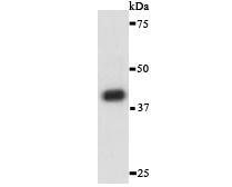
Fig1: Western blot analysis of TNFRSF19 against human TNFRSF19 (AA: extra 30-170) recombinant protein. Proteins were transferred to a PVDF membrane and blocked with 5% BSA in PBS for 1 hour at room temperature. The primary antibody was used in 5% BSA at room temperature for 2 hours. Goat Anti-Mouse IgG - HRP Secondary Antibody at 1:5,000 dilution was used for 1 hour at room temperature.

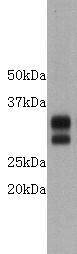
Fig2: Western blot analysis of against HEK293 (1) and TNFRSF19 (AA: extra 30-170)-hIgGFc transfected HEK293 (2) cell lysate.Proteins were transferred to a PVDF membrane and blocked with 5% BSA in PBS for 1 hour at room temperature. The primary antibodywas used in 5% BSA at room temperature for 2 hours. Goat Anti-Mouse IgG - HRP Secondary Antibody at 1:5,000 dilution was used for 1 hour at room temperature.

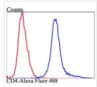
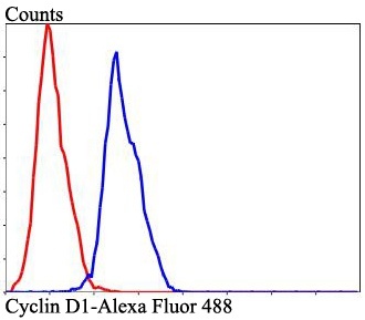
Fig3: Flow cytometric analysis of TNFRSF19 was done on Jurkat cells. The cells were fixed, permeabilized and stained with the primary antibody ( (green). After incubation of the primary antibody at room temperature for an hour, the cells were stained with a Alexa Fluor 488-conjugated goat anti-Mouse IgG Secondary antibody at 1/500 dilution for 30 minutes. Unlabelled sample was used as a control (cells without incubation with primary antibody; red).
特别提示:本公司的所有产品仅可用于科研实验,严禁用于临床医疗及其他非科研用途!
更多![]()

上海泽叶生物科技有限公司
公司地址
上海市松江区国家经济开发区北松公路5629号聚科生物松江园区
客服电话
