
 关注
关注
已关注

![]() 已认证
已认证
粉丝量 0
400-860-5168转3779
仪器信息网认证电话,请放心拨打
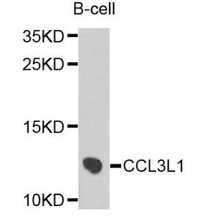
趋化因子C-C-基元配体3样蛋白1(CCL3L1)重组蛋白
实验方法双抗夹心法
反应时长3h
检测范围0.625-10ng/mL
灵敏度最小可检测剂量小于等于0.231ng/mL.
样本类型serum, plasma, tissue homogenates, cell lysates, cell culture supernates 趋化因子C-C-基元配体3样蛋白1(CCL3L1)重组蛋白and other biological fluids
Enzyme-linked Immunosorbent Assay Kit
For Chemokine C-C-Motif Ligand 3 Like Protein 1 (CCL3L1)
Organism Species: Homo sapiens (Human)
Instruction manual
FOR IN VITRO AND RESEARCH USE ONLY
NOT FOR USE IN CLINICAL DIAGNOSTIC PROCEDURES
11th Edition (Revised in July, 2013)
[ INTENDED USE ]
The kit is a sandwich enzyme immunoassay for in vitro quantitative measurement of CCL3L1 in human serum,
plasma, tissue homogenates, cell lysates, cell culture supernates and other biological fluids.
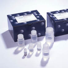
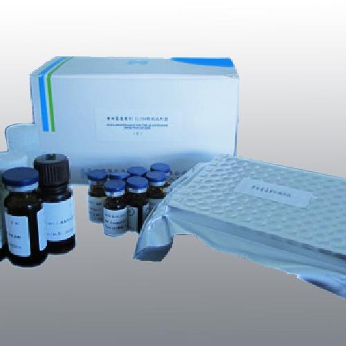
[ REAGENTS AND MATERIALS PROVIDED ]
Reagents Quantity Reagents Quantity
Pre-coated, ready to use 96-well strip plate 1 Plate sealer for 96 wells 4
Standard 2 Standard Diluent 1×20mL
Detection Reagent A 1×120μL Assay Diluent A 1×12mL
Detection Reagent B 1×120μL Assay Diluent B 1×12mL
TMB Substrate 1×9mL Stop Solution 1×6mL
Wash Buffer (30 × concentrate) 1×20mL Instruction manual 1
趋化因子C-C-基元配体3样蛋白1(CCL3L1)重组蛋白[ MATERIALS REQUIRED BUT NOT SUPPLIED ]
1. Microplate reader with 450 ± 10nm filter.
2. Precision single or multi-channel pipettes and disposable tips.
3. Eppendorf Tubes for diluting samples.
4. Deionized or distilled water.
5. Absorbent paper for blotting the microtiter plate.
6. Container for Wash Solution
[ STORAGE OF THE KITS ]
1. For unopened kit: All the reagents should be kept according to the labels on vials. The Standard, Detection
Reagent A, Detection Reagent B and the 96-well strip plate should be stored at -20oC upon receipt while
the others should be at 4
oC.
2. For opened kit: When the kit is opened, the remaining reagents still need to be stored according to the
above storage condition. Besides, please return the unused wells to the foil pouch containing the desiccant
pack, and reseal along entire edge of zip-seal.
Note:
It is highly recommended to use the remaining reagents within 1 month provided this is within the expiration date
of the kit. For the expiration date of the kit, please refer to the label on the kit box. All components are stable until
this expiration date. [ SAMPLE COLLECTION AND STORAGE ]
Serum - Use a serum separator tube and allow samples to clot for two hours at room temperature or overnight at
4
oC before centrifugation for 20 minutes at approximately 1000×g. Assay freshly prepared serum
immediately or store samples in aliquot at -20oC or -80oC for later use. Avoid repeated freeze/thaw cycles.
Plasma - Collect plasma using EDTA or heparin as an anticoagulant. Centrifuge samples for 15 minutes at
1000×g at 2 - 8
oC within 30 minutes of collection. Remove plasma and assay immediately or store samples
in aliquot at -20oC or -80oC for later use. Avoid repeated freeze/thaw cycles.
Tissue homogenates - The preparation of tissue homogenates will vary depending upon tissue type. For this
assay, tissues were rinsed in ice-cold PBS(0.01mol/L,pH 7.0-7.2) to remove excess blood thoroughly and
weighed before homogenization. Minced the tissues to small pieces and homogenized them in 5-10mL of
PBS with a glass homogenizer on ice(Micro Tissue Grinders woks, too). The resulting suspension was
sonicated with an ultrasonic cell disrupter or subjected to two freeze-thaw cycles to further break the cell
membranes. After that, the homogenates were centrifugated for 5 minutes at 5000×g. Remove the supernate
and assay immediately or aliquot and store at ≤-20oC. Cell Lysates - Cells must be lysed before assaying according to the following directions.
1. Adherent cells should be detached with trypsin and then collected by centrifugation (suspension cells can
be collected by centrifugation directly).
2. Wash cells three times in cold PBS.
3. Resuspend cells in PBS (1×) and the cells was subject to ultrasonication for 4 times (or Freeze cells at ≤
-20oC. Thaw cells with gentle mixing. Repeat the freeze/thaw cycle for 3 times.)
4. Centrifuge at 1500×g for 10 minutes at 2 - 8
oC to remove cellular debris.
Cell culture supernates and other biological fluids - Centrifuge samples for 20 minutes at 1000×g. Remove
particulates and assay immediately or store samples in aliquot at -20oC or -80oC for later use. Avoid repeated
freeze/thaw cycles.
Note:
1. Samples to be used within 5 days may be stored at 4
oC, otherwise samples must be stored at -20oC (≤1
month) or -80oC (≤2 months) to avoid loss of bioactivity and contamination.
2. Sample hemolysis will influence the result, so hemolytic specimen should not be detected.
3. When performing the assay, bring samples to room temperature.
趋化因子C-C-基元配体3样蛋白1(CCL3L1)重组蛋白[ REAGENT PREPARATION ]
1. Bring all kit components and samples to room temperature (18-25oC) before use.
2. Standard - Reconstitute the Standard with 0.5mL of Standard Diluent, kept for 10 minutes at room
temperature, shake gently(not to foam). The concentration of the standard in the stock solution is 10ng/mL. Please prepare 5 tubes containing 0.25mL Standard Diluent and produce a double dilution series according
to the picture shown below. Mix each tube thoroughly before the next transfer. Set up 5 points of diluted
standard such as 10ng/mL, 5ng/mL, 2.5ng/mL, 1.25ng/mL, 0.625ng/mL, and the last EP tubes with
Standard Diluent is the blank as 0ng/mL.
Tube 1 2 3 4 5 6
ng/mL 10 5 2.5 1.25 0.625 0
3. Detection Reagent A and Detection Reagent B - Briefly spin or centrifuge the stock Detection A and
Detection B before use. Dilute to the working concentration with Assay Diluent A and B, respectively
(1:100).
4. Wash Solution - Dilute 20mL of Wash Solution concentrate (30×) with 580mL of deionized or distilled water
to prepare 600mL of Wash Solution (1×).
5. TMB substrate - Aspirate the needed dosage of the solution with sterilized tips and do not dump the residual
solution into the vial again.
Note:
1. Making serial dilution in the wells directly is not permitted.
2. Prepare standard within 15 minutes before assay. Please do not dissolve the reagents at 37oC directly.
3. Please carefully reconstitute Standards or working Detection Reagent A and B according to the instruction,
and avoid foaming and mix gently until the crystals are completely dissolved. To minimize imprecision caused
by pipetting, use small volumes and ensure that pipettors are calibrated. It is recommended to suck more
than 10μL for once pipetting.
4. The reconstituted Standards, Detection Reagent A and Detection Reagent B can be used only once. 5. If crystals have formed in the Wash Solution concentrate (30×), warm to room temperature and mix gently
until the crystals are completely dissolved.
6. Contaminated water or container for reagent preparation will influence the detection result.
[ SAMPLE PREPARATION ]
1. We are only responsible for the kit itself, but not for the samples consumed during the assay. The user should
calculate the possible amount of the samples used in the whole test. Please reserve sufficient samples in
advance.
2. Please predict the concentration before assaying. If values for these are not within the range of the standard
curve, users must determine the optimal sample dilutions for their particular experiments. Sample should be
diluted by 0.01mol/L PBS(PH=7.0-7.2).
3. If the samples are not indicated in the manual, a preliminary experiment to determine the validity of the kit is
necessary.
4. Tissue or cell extraction samples prepared by chemical lysis buffer may cause unexpected ELISA results due
to the impacts from certain chemicals.
5. Due to the possibility of mismatching between antigen from other origin and antibody used in our kits (e.g.,
antibody targets conformational epitope rather than linear epitope), some native or recombinant proteins
from other manufacturers may not be recognized by our products.
6. Influenced by the factors including cell viability, cell number or sampling time, samples from cell culture
supernatant may not be detected by the kit.
7. Fresh samples without long time storage is recommended for the test. Otherwise, protein degradation and
denaturalization may occur in those samples and finally lead to wrong results.
[ ASSAY PROCEDURE ]
1. Determine wells for diluted standard, blank and sample. Prepare 5 wells for standard, 1 well for blank.
Add 100μL each of dilutions of standard (read Reagent Preparation), blank and samples into the appropriate
wells. Cover with the Plate sealer. Incubate for 2 hours at 37oC. 2. Remove the liquid of each well, don’t wash.
3. Add 100μL of Detection Reagent A working solution to each well. Incubate for 1 hour at 37oC after covering
it with the Plate sealer.
4. Aspirate the solution and wash with 350μL of 1× Wash Solution to each well using a squirt bottle,
multi-channel pipette, manifold dispenser or autowasher, and let it sit for 1~2 minutes. Remove the remaining
liquid from all wells completely by snapping the plate onto absorbent paper. Totally wash 3 times. After the
last wash, remove any remaining Wash Buffer by aspirating or decanting. Invert the plate and blot it against
absorbent paper. 5. Add 100μL of Detection Reagent B working solution to each well. Incubate for 30 minutes at 37oC after
covering it with the Plate sealer.
6. Repeat the aspiration/wash process for total 5 times as conducted in step 4.
7. Add 90μL of Substrate Solution to each well. Cover with a new Plate sealer. Incubate for 10 - 20 minutes at
37oC (Don't exceed 30 minutes). Protect from light. The liquid will turn blue by the addition of Substrate
Solution.
8. Add 50μL of Stop Solution to each well. The liquid will turn yellow by the addition of Stop solution. Mix the
liquid by tapping the side of the plate. If color change does not appear uniform, gently tap the plate to ensure
thorough mixing.
9. Remove any drop of water and fingerprint on the bottom of the plate and confirm there is no bubble on the
surface of the liquid. Then, run the microplate reader and conduct measurement at 450nm immediately.
Note:
1. Assay preparation: Keep appropriate numbers of wells for each experiment and remove extra wells from
microplate. Rest wells should be resealed and stored at -20oC. 2. Samples or reagents addition:Please use the freshly prepared Standard. Please carefully add samples
to wells and mix gently to avoid foaming. Do not touch the well wall. For each step in the procedure, total
dispensing time for addition of reagents or samples to the assay plate should not exceed 10 minutes. This
will ensure equal elapsed time for each pipetting step, without interruption. Duplication of all standards and
specimens, although not required, is recommended. To avoid cross-contamination, change pipette tips
between additions of standards, samples, and reagents. Also, use separated reservoirs for each reagent.
3. Incubation: To ensure accurate results, proper adhesion of plate sealers during incubation steps is
necessary. Do not allow wells to sit uncovered for extended periods between incubation steps. Once
reagents are added to the well strips, DO NOT let the strips DRY at any time during the assay. Incubation
time and temperature must be controlled.
4. Washing: The wash procedure is critical. Complete removal of liquid at each step is essential for good
performance. After the last wash, remove any remaining Wash Solution by aspirating or decanting and
remove any drop of water and fingerprint on the bottom of the plate. Insufficient washing will result in poor
precision and false elevated absorbance reading.
5. Controlling of reaction time: Observe the change of color after adding TMB Substrate (e.g. observation
once every 10 minutes), if the color is too deep, add Stop Solution in advance to avoid excessively strong
reaction which will result in inaccurate absorbance reading. 6. TMB Substrate is easily contaminated. Please protect it from light.
7. The environment humidity which is less than 60% might have some effects on the final performance,
therefore, a humidifier is recommended to be used at that condition.
[ TEST PRINCIPLE ]
The microtiter plate provided in this kit has been pre-coated with an antibody specific to CCL3L1. Standards or
samples are then added to the appropriate microtiter plate wells with a biotin-conjugated antibody specific to
CCL3L1. Next, Avidin conjugated to Horseradish Peroxidase (HRP) is added to each microplate well and
incubated. After TMB substrate solution is added, only those wells that contain CCL3L1, biotin-conjugated
antibody and enzyme-conjugated Avidin will exhibit a change in color. The enzyme-substrate reaction is
terminated by the addition of sulphuric acid solution and the color change is measured spectrophotometrically at
a wavelength of 450nm ± 10nm. The concentration of CCL3L1 in the samples is then determined by comparing
the O.D. of the samples to the standard curve.
[ CALCULATION OF RESULTS ]
Average the duplicate readings for each standard, control, and samples and subtract the average zero standard
optical density. Construct a standard curve by plotting the mean O.D. and concentration for each standard and
draw a best fit curve through the points on the graph or create a standard curve on log-log graph paper with
CCL3L1 concentration on the y-axis and absorbance on the x-axis. Using some plot software, for instance, curve
expert 1.30, is also recommended. If samples have been diluted, the concentration read from the standard curve
must be multiplied by the dilution factor.
[ TYPICAL DATA ]
In order to make the calculation easier, we plot the O.D. value of the standard (X-axis) against the known
concentration of the standard (Y-axis), although concentration is the independent variable and O.D. value is the
dependent variable. However, the O.D. values of the standard curve may vary according to the conditions of
assay performance (e.g. operator, pipetting technique, washing technique or temperature effects), plotting log of
the data to establish standard curve for each test is recommended. Typical standard curve below is provided for
reference only.
Typical Standard Curve for CCL3L1, Human ELISA.
[ DETECTION RANGE ]
0.625-10ng/mL. The standard curve concentrations used for the ELISA’s were 10ng/mL, 5ng/mL, 2.5ng/mL,
1.25ng/mL, 0.625ng/mL.
[ SENSITIVITY ]
The minimum detectable dose of CCL3L1 is typically less than 0.231ng/mL.
The sensitivity of this assay, or Lower Limit of Detection (LLD) was defined as the lowest protein concentration
that could be differentiated from zero. It was determined by adding two standard deviations to the mean optical
density value of twenty zero standard replicates and calculating the corresponding concentration.
[ SPECIFICITY ]
This assay has high sensitivity and excellent specificity for detection of CCL3L1. No significant cross-reactivity or interference between CCL3L1 and analogues was observed.
Note:
Limited by current skills and knowledge, it is impossible for us to complete the cross- reactivity detection between
CCL3L1 and all the analogues, therefore, cross reaction may still exist.
[ RECOVERY ]
Matrices listed below were spiked with certain level of recombinant CCL3L1 and the recovery rates were
calculated by comparing the measured value to the expected amount of CCL3L1 in samples.
Matrix Recovery range (%) Average(%)
serum(n=5) 84-98 93
EDTA plasma(n=5) 81-95 87
heparin plasma(n=5) 93-105 101
[ LINEARITY ]
The linearity of the kit was assayed by testing samples spiked with appropriate concentration of CCL3L1 and their
serial dilutions. The results were demonstrated by the percentage of calculated concentration to the expected.
Sample 1:2 1:4 1:8 1:16
serum(n=5) 87-94% 79-89% 96-104% 82-93%
EDTA plasma(n=5) 80-92% 89-103% 83-101% 88-102%
heparin plasma(n=5) 85-95% 81-96% 99-105% 78-90%
[ PRECISION ]
Intra-assay Precision (Precision within an assay): 3 samples with low, middle and high level CCL3L1 were tested
20 times on one plate, respectively.
Inter-assay Precision (Precision between assays): 3 samples with low, middle and high level CCL3L1 were tested
on 3 different plates, 8 replicates in each plate. CV(%) = SD/meanX100
Intra-Assay: CV<10%
Inter-Assay: CV<12%
[ STABILITY ]
The stability of ELISA kit is determined by the loss rate of activity. The loss rate of this kit is less than 5% within
the expiration date under appropriate storage condition.
To minimize extra influence on the performance, operation procedures and lab conditions, especially room
temperature, air humidity, incubator temperature should be strictly controlled. It is also strongly suggested that the
whole assay is performed by the same operator from the beginning to the end.
[ ASSAY PROCEDURE SUMMARY ]
1. Prepare all reagents, samples and standards;
2. Add 100μL standard or sample to each well. Incubate 2 hours at 37oC;
3. Aspirate and add 100μL prepared Detection Reagent A. Incubate 1 hour at 37oC;
4. Aspirate and wash 3 times;
5. Add 100μL prepared Detection Reagent B. Incubate 30 minutes at 37oC;
6. Aspirate and wash 5 times;
7. Add 90μL Substrate Solution. Incubate 10-20 minutes at 37oC;
8. Add 50μL Stop Solution. Read at 450nm immediately.
[ IMPORTANT NOTE ]
1. Limited by the current condition and scientific technology, we can't completely conduct the comprehensive
identification and analysis on the raw material provided by suppliers. So there might be some qualitative and
technical risks to use the kit.
2. The final experimental results will be closely related to validity of the products, operation skills of the end
users and the experimental environments. Please make sure that sufficient samples are available.
3. Kits from different batches may be a little different in detection range, sensitivity and color developing time.
Please perform the experiment exactly according to the instruction attached in kit while electronic ones from
our website is only for information.
4. Do not mix or substitute reagents from one kit lot to another. Use only the reagents supplied by manufacturer.
5. Protect all reagents from strong light during storage and incubation. All the bottle caps of reagents should be
covered tightly to prevent the evaporation and contamination of microorganism.
6. There may be some foggy substance in the wells when the plate is opened at the first time. It will not have
any effect on the final assay results. Do not remove microtiter plate from the storage bag until needed.
7. Wrong operations during the reagents preparation and loading, as well as incorrect parameter setting for the
plate reader may lead to incorrect results. A microplate plate reader with a bandwidth of 10nm or less and an
optical density range of 0-3 O.D. or greater at 450 ± 10nm wavelength is acceptable for use in absorbance
measurement. Please read the instruction carefully and adjust the instrument prior to the experiment.
8. Even the same operator might get different results in two separate experiments. In order to get better
reproducible results, the operation of every step in the assay should be controlled. Furthermore, a
preliminary experiment before assay for each batch is recommended.
9. Each kit has been strictly passed Q.C test. However, results from end users might be inconsistent with our
in-house data due to some unexpected transportation conditions or different lab equipments. Intra-assay
variance among kits from different batches might arise from above factors, too.
10. Kits from different manufacturers with the same item might produce different results, since we haven’t
compared our products with other manufacturers.
11. The instruction manual also suits for the kit of 48T, but all reagents of 48T kit are reduced by half.
[ PRECAUTION ]
The Stop Solution suggested for use with this kit is an acid solution. Wear eye, hand, face, and clothing protection
when using this material.
[ TROUBLE SHOOTING ]
Problem Possible Source Correction Action
Poor
Standard
Curve
Improper standard curve preparation Ensure accurate operation of the dilution
Incomplete washing and aspiration Adequate washing and adequate aspiration
Inaccurate Pipetting Check and Calibrate pipettes
Poor
Precision
Incomplete washing of wells Ensure sufficient washing
Inadequate mixing and aspiration reagents Adequate aspiration and mixing reagents
Reused pipette tips, containers and sealers Change and use new pipette tips, containers and sealers
Inaccurate Pipetting Check and Calibrate pipettes
Low
O.D
Values
Inadequate reagent volumes added to wells Calibrate pipettes and Add adequate reagents
Incorrect incubation times Ensure sufficient incubation times
Incorrect incubation temperature Reagents balanced to room temperature
Conjugate or substrate reagent failure Mix conjugate &substrate,color should develop immediately
No stop solution added Follow the assay protocol in the kit manual
Read beyond suggested reading time Read within the time recommended in the manual
Sample
Values
Improper Sample Storage Store the sample properly and use the fresh sample
Improper sample collection and preparation Take proper sample collection and preparation method
Low quantity of analyte in samples Use new sample and repeat assay
更多![]()


企业名称
上海康朗生物科技有限公司
企业信息已认证
企业类型
信用代码
310112001510759
成立日期
2015-05-29
注册资本
100
经营范围
从事生物科技技术领域内技术开发、技术咨询、技术服务、技术转让,实验分析仪器、化工产品及原料(除危险化学品、监控化学品、烟花爆竹、民用爆竹物品、易制毒化学品)、仪器仪表、环保设备、酒店用品、皮革制品、床上用品、电子产品的销售。【依法须经批准的项目,经相关部门批准后方可开展经营活动】
上海康朗生物科技有限公司
公司地址
上海市闵行区闵北路88弄1-30号第22栋BV145室
客服电话
企业名称: 上海康朗生物科技有限公司
企业地址: 上海市闵行区闵北路88弄1-30号第22栋BV145室 联系人: 蔡盼 邮编: 200612 联系电话: 400-860-5168转3779
仪器信息网APP

展位手机站