尊敬的会员请选择进入的厂商展位
开通仪会通服务,请联系客服人员
刘老师 13717560883(微信同号)

企业微信二维码

 0
0
 1
1
 投诉
投诉
 分享
分享
 17
2023-12-07
17
2023-12-07
 下载APP,观看精彩内容
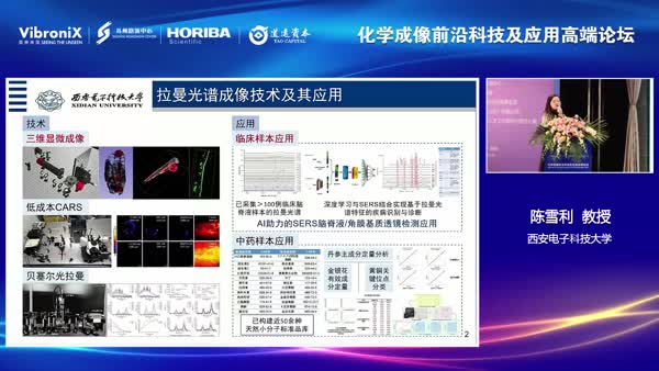
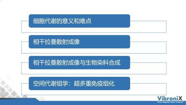 基于相干拉曼技术的空间代谢组学新进展
 187次观看
 20年受激拉曼成像20年人类基因组引发的医学变革
90次观看
20年受激拉曼成像20年人类基因组引发的医学变革
90次观看
 计算拉曼光谱与成像
187次观看
计算拉曼光谱与成像
187次观看
 领导致辞
133次观看
领导致辞
133次观看
 振电科技新品发布会
158次观看
振电科技新品发布会
158次观看

扫描二维码联系我

关注微服务 参会不迷路
下载app 回看更便捷